Low Toxin Diet Grant Genereux's Theory Of Vitamin A Toxicity

Blossom

Moderator
Forum Supporter
Joined
Nov 23, 2013
Messages
11,072
Location
Indiana USA
How about liver, will you ever eat that again?
I don’t think so. It’s hard to imagine but maybe if I became severely anemic for some reason I might consider it. The thought isn’t the least bit appealing. :/
I’m probably not going to make the amount of cream and butter I indulged in for Thanksgiving a regular habit anytime soon but it’s nice that I seem to tolerate them both okay now.
 

Blossom

Moderator
Forum Supporter
Joined
Nov 23, 2013
Messages
11,072
Location
Indiana USA
I don’t think so. It’s hard to imagine but maybe if I became severely anemic for some reason I might consider it.
Honestly in that unlikely scenario I’d probably exhaust my other options before resorting to liver.
 

Blossom

Moderator
Forum Supporter
Joined
Nov 23, 2013
Messages
11,072
Location
Indiana USA
@Janelle525, how’s it going with you? Are you noticing any improvement with your rash?
I noticed this morning I had a slight itch where my dermatitis herpetiformis rash used to be but thankfully thus far nothing has developed and the itching has gone away.
 

InChristAlone

Member
Joined
Sep 13, 2012
Messages
5,955
Location
USA
@Janelle525, how’s it going with you? Are you noticing any improvement with your rash?
I noticed this morning I had a slight itch where my dermatitis herpetiformis rash used to be but thankfully thus far nothing has developed and the itching has gone away.
Yeah I doubt I will ever eat liver again barring cancer or something.

I think I was going through detox as every few days I go through some symptoms. No change in butt rash :( still have some cracking today, doesnt seem as itchy but it was last night. It comes and goes.

Glad things are going well for you.
 

md_a

Member
Joined
Aug 31, 2015
Messages
468
Zinc and vitamin A interact in several ways. Zinc is a component of retinol-binding protein, a protein necessary for transporting vitamin A in the blood. Zinc is also required for the enzyme that converts retinol (vitamin A) to retinal. This latter form of vitamin A is necessary for the synthesis of rhodopsin, a protein in the eye that absorbs light and thus is involved in dark adaptation. Zinc deficiency is associated with decreased release of vitamin A from the liver, which may contribute to symptoms of night blindness that are seen with zinc deficiency (16, 17)….

Serum zinc levels in this study increased after zinc supplementation in the trial group was not accompanied by an increase in serum retinol levels. Adequate protein intake is needed as a tool for zinc transportation in circulation. Albumin is the main tool of zinc transportation. Albumin in plasma is a major determinant of zinc absorption. Zinc absorption decreases when the blood albumin decreased …Lack of protein intake for a long time caused low levels of albumin. Zinc is mainly requires albumin (70%) to circulate in the systemic circulation to body tissues and the remainder binds to other proteins such as transferrin, ceruloplasmin and amino acids, especially histidine and cystine. With the low protein intake (under the RDA) can cause low levels of albumin in the blood. Consume less protein for a long time can cause low albumin levels, so zinc intake from food and supplements are not absorbed optimally….

Consume enough protein and zinc needed to produce normal RBP. Therefore, zinc deficiency or protein malnutrition would interfere vitamin A function by preventing the release of vitamin A normally from its storage in the liver [21]. Thus low serum retinol levels in this study and the absence of increased levels of serum retinol average after zinc supplementation and high-dose vitamin A supplementation showed that albumin has an important role in the vitamin A metabolism. Zinc supplementation without being followed with normal albumin levels in serum cannot reach other networks with optimal. Similarly, highdose vitamin A supplementation in this study did not increase retinol serum levels though found a significant difference in serum zinc levels after supplementation because RBP synthesis which needed to mobilize vitamin A from the storage in the liver requiring zinc and protein. So in malnutrition pregnant women with low serum albumin levels, required supplementary feeding mainly containing protein and energy also zinc supplementation to optimize high-dose vitamin A supplementation program which is a government program. Besides, the increase in nutrition counseling is also needed to improve nutritional knowledge of pregnant women so although there is a limitation in the economy, pregnant women still able to choose good food, both in quality and quantity….

https://www.omicsonline.org/open-ac...women-in-third-trimester-2168-9652-S5-001.pdf

Zinc
 

Blossom

Moderator
Forum Supporter
Joined
Nov 23, 2013
Messages
11,072
Location
Indiana USA
Yeah I doubt I will ever eat liver again barring cancer or something.

I think I was going through detox as every few days I go through some symptoms. No change in butt rash :( still have some cracking today, doesnt seem as itchy but it was last night. It comes and goes.

Glad things are going well for you.
Thank you! I hope you’re butt rash improves soon.
 

InChristAlone

Member
Joined
Sep 13, 2012
Messages
5,955
Location
USA
@freyasam aren't you one who took a lot of vitamin A and developed sun sensitivity? Have you ever looked in to the vitamin A detox?
 

Amazoniac

Member
Joined
Sep 10, 2014
Messages
8,583
Location
Not Uganda
The problem in switching to carotenes-only is that some people just can't convert it, it will build up and it's not good. This is what happens in diabetes and hypopboydism, which are the only two cases that have elevated stores of vitamin A; all others have normal or low..


Median reserves from accidental death was used for normal reference and cancer was used for low.

"To establish the extent of the vitamin A reserves in health it is necessary to resort to data obtained on cases of accidental death. No assurance can be given that all the cases were in perfect health, but the accident group appears to afford a reasonable crosssection of subjects in "ordinary health". Since the reserve varies over a wide range (even in this group) (lowest individual value 10, highest 1500) it is not permissible to speak of a "normal " reserve.1 The median (220) of this group may, however, be taken as the "typical" vitamin A reserve in health."

"In almost all diseases the vitamin A median was lower than in accidental death. For the purpose of distinguishing those diseases showing outstandingly low reserves the median of the cancer group (110 i.U. per g.), and not the accident median, has been taken as a basis of comparison."​

The reserves last longer than expected because the body starts to sacrifice some of its functions to conserve it:

"Davies & Moore [1935] have estimated the physiological requirements of the rat at 2 B.U., or 1-2 i.u. per g. of liver per day. By analogy with this admittedly rough estimate the typical human reserve, assuming equal expenditure per g. of liver, should be sufficient to support life for about 6 months. The prediction of the time of survival is made difficult, however, by the fact that the vitamin A reserve, especially if high, may sometimes be dispersed at a rate greater than that demanded by apparent physiological requirements."
And the levels on each condition:

"Vitamin A reserves above the "level in health" were found in diabetes (ages 15-59, 300, over 60, 540 i.u. per g.). High values in this disease have been reported by other workers and appear to be mainly due to the adoption of vegetable dietaries. Whether this simple explanation represents the whole story is not certain. According to Ralli et al. [1935] the ingestion of carotene by diabetics results in a more intense carotenaemia than administration of the same amount of carotene to a normal subject. The observation of high vitamin A reserves in the livers of subjects dying from diabetes must rule out the suggestion that there is any serious impairment of the power of the liver to convert carotene into vitamin A. It is possible, however, that a general slowing down of the metabolism of both carotene and vitamin A takes place, with consequent accumulation of both substances in the organism."


"High reserves were also found in thyroid diseases (all types), although the small number of cases available (9) makes the authenticity of this result more questionable. It may be noted, however, that Wolff found a high average for 5 cases of exophthalmic goitre, while Breusch & Scalabrino observed the highest value in their 72 cases in a man 98 years old suffering from a swollen thyroid."

"From the above evidence it is apparent that low vitamin A reserves occur frequently in a number of quite common diseases. As already emphasized the significance of this finding is open to question. On the one hand, it may be argued that since vitamin A reserves are lowered in almost all diseases the extent of the subnormality will depend merely on the duration and severity of the disease. Thus chronic nephritis and valvular disease of the heart, in which low reserves are observed, are diseases which run a very long course and in which there is marked dyspepsia. In support of this view Wolff has found the mean reserve for a large group of cases of various chronic diseases to be slightly lower than the mean for a group of acute diseases."

"Approaching the problem from a slightly different angle it appears certain that in some diseases the power of the liver to store vitamin A must be impaired."

"Green [1932] has reported instances of puerperal cases, treated unsuccessfully with large doses of vitamin A concentrate, which showed only meagre stores of the vitamin in the liver at autopsy. On the basis of this evidence one might be tempted to assume that the explanation of low reserves observed in all diseases not recognized as being specifically due to vitamin A deficiency might lie in the reduced power of the liver to absorb the vitamin, or to effect its synthesis from carotene. It may be mentioned, in support of this latter explanation in the case of chronic nephritis, that the extracts from many of the specimens were deeply yellow in colour, suggesting a preponderance of unconverted carotene."

"On the other hand, it may be mentioned in favour of the aetiological significance of low vitamin A reserves, that many of the diseases showing low reserves are also among the most common terminations to experimental vitamin A deficiency in animals. No one would suggest that all cases of pneumonia, sepsis, kidney disease etc. are due to vitamin A deficiency, and this self-evident conclusion is confirmed by the observation of cases with adequate reserves even in those groups giving the lowest median values. The question to be answered, however, is not whether all cases of these diseases are due to vitamin A deficiency, but whether in a significant proportion of cases (e.g. even 5-10%) vitamin A deficiency, or the general nutritional subnormality which it may imply, is a contributory cause of the disease."​

- Bioavailability of vitamin A - Jody B. Duggan, Baking Soda

"The presence of nitrite in the diet has been shown to increase vitamin A disappearance in ruminants (Reddy and Thomas 1962, Mitchell et al. 1967, Keating et al. 1964, Roberts and Sell 1963, Goodrich et al. 1964) and reduce the utilization of vitamin A in chicks (Sell and Roberts 1963, Roberts and Sell 1963) and rats (O'Dell et al. 1960, Emerick and Olson 1962, Phillips 1966) when it is fed at various levels. Ascarelli (1969) showed strong decreases in vitamin A storage in chicks when potassium nitrite was fed at levels as low as .2% of the diet. He suggested the possibility of impairnent of vitamin A storage by nitrite could occur without the appearance of nitrite toxicity symptoms. Sell and Roberts (1963) also saw reduced storage of orally administered vitamin A and b-carotene when nitrite was fed to chicks. This reduced storage was accounted to the destruction of vitamin A and b-carotene by nitrite, the interference in the absorption process of vitamin A and b-carotene by nitrite, or a combination of the two."

"O'Dell et al. (1960) indicated that in addition to increasing vitamin A depletion nitrite also precipitated a vitamin E deficiency in an normally adequate diet. Phillips (1966) stated that the reduction in vitamin A storage was a direct result of oxidation of the vitamin by nitrite rather than interference in absorption, conversion of b-carotene to vitamin A, or liver function. Although nitrite has been shown to reduce liver storage of vitamin A, nitrate does not affect vitamin A utilization in the chick (Ascarelli 1969) or the rat (Emerick and Olson 1960). This is not the case in ruminants, however."

"Roberts and Sell (1963) reported that the destruction of vitamin A and carotene by nitrite is dependent upon pH. Destruction is almost complete and rapid in the abomasum and ventriculus where the pH is low (1 to 3). There is some destruction in the rumen (pH~6) and very little destruction in the intestine (pH ~7). Pugh and Garner (1963) also reported destruction by nitrite to be dependent on pH and stated that retinol was more susceptible to destruction than b-carotene. Mitchell et al. (1967) suggested that the destructive mechanism in the ruminant may not be oxidation of vitamin A but may be caused by reduction of vitamin A compounds due to the highly reduced environment of the rumen."

Supramolecular materials: Longer and safer gastric residence
upload_2018-11-25_20-34-38.png

@Wagner83

"Several essential nutrients can also have an effect on the metabolism of vitamin A. These include inorganic minerals, protein level, fat quality, high dietary energy levels, and other factors that could possibly affect vitamin A status through the influence of the thyroid hormones (Mitchel 1967). The thyroid status of the animal has been shown to influence vitamin A metabolism (Smith et al., 1964). Johnson and Bauman (1947) showed that a functioning thyroid is necessary for the conversion of b-carotene to vitamin A. Their results indicated minor effects of thyroxine on preformed vitamin A. However, there was a greater production of vitamin A from carotene in the presence of the thyroid gland than in its absence. They also indicated that this increase in production of the vitamin was not due to basal metabolism changes but resulted from some other physiological action of the thyroid gland. Smith (1961) stated that the conversion of b-carotene to vitamin A could also be affected by extensive periods of hot weather or other factors which inhibit the function of the thyroid. Moore (1957) stated that thyroxine accelerates the current flow of vitamin A metabolism, regardless of direction, and that thyroid inhibitors have opposite effects. Mitchell (1967) agreed that thyroid stimulation increased the requirements of vitamin A and accelerated depletion of stores. Vitamin A also has a considerable influence on the secretion of thyroxine. The rate of thyroxine secretion was lowered when vitamin A was fed at both insufficient and excessive levels to young pigs (Frape et al., 1959)."

"Phosphorus and zinc are two of the inorganic minerals that have been shown to influence the status of vitamin A."

"Several papers have shown a relationship between protein metabolism and vitamin A utilization. Vitamin A status can be affected by both the quality and quantity of protein in the diet. Rechcigl et al. (1962) showed that depletion and utilization of total vitamin A stores were lowest in rats fed a protein-free diet and increased as both the quality and quantity of protein in the diet increased. Rats which were fed casein as the protein source expressed lower liver vitamin A stores as compared to rats fed gluten or zein as the protein source. Supplementation of the gluten and zein diets resulted in a slight decrease of liver vitamin A stores. Surelda Rao et al. (1974) also showed liver storage and utilization in rats to be affected by a low protein diet. They showed that a protein deficiency impedes the absorption of vitamin A through the intestinal wall and reduces the mobilization of vitamin A from the liver. They concluded that these adverse effects on vitamin A transport in protein malnutrition resulted from the lowered synthesis of the albumin protein fraction."

"In addition to the influence of protein level on vitamin A metabolism, Arroyave (1969) indicated that protein metabolism is affected by vitamin A deficiency causing reduced utilization of ingested protein. His review stated that the lower hepatic vitamin A stores in rats fed low protein diets (3 and 6%) were possibly due to poor intestinal absorption and that the reduced stores in rats fed high protein diets (30%) were due to increased utilization of vitamin A in protein metabolism. He suggested a trend in inverse proportion of liver vitamin A stores to the protein level of the diet. A functional deficiency of vitamin A could result from a protein deficiency even when adequate amounts of the vitamin are available in the diet. A protein deficiency in combination with a vitamin A deficiency could impair development of symptoms of the vitamin deficiency due to a decrease in the vitamin A required for protein metabolism."

"Several papers have discussed the relationship between vitamin A and vitamin E and the influence of vitamin E on the metabolism of vitamin A. Bauernfeind et al. (1974) described four ways in which vitamin E may enhance vitamin A metabolism. They are (1) protection of vitamin A from oxidation in the digestive tract, (2) increase in the absorption of vitamin A, (3) increase in the utilization of vitamin A and, (4) increase in the storage of vitamin A."

"Kohlmeier and Burroughs (1962) also showed an interrelationship between vitamin A and vitamin E plus vitamin K in beef cattle. Supplementation of vitamin A alone improved weight gains and feed conversion, but the improvements were doubled when supplemental vitamin E plus vitamin K was fed in addition to the vitamin A."

"Tocopherol supplementation also increased the time necessary to deplete the vitamin A stores of the calves. Since the effect of the tocopherol supplementation was not consistent, the Connecticut workers suggest that there may be a given level or range of tocopherol intake for maximum utilization of vitamin A. This same study also showed a decrease in tissue concentrations of tocopherol when vitamin A was supplemented and suggested that these decreases may result from the influence of one vitamin on the biological activity of the other. Smith (1961) stated in his review that in addition to a vitamin E deficiency accelerating vitamin A depletion, an excess of vitamin E could inhibit the conversion of b-carotene to vitamin A due to its antioxidant properties. Bauernfeind et al. (1974) made similar remarks in their review. They stated that signs of vitamin A deficiency were accelerated in animals with low tocopherol reserves and that the utilization of both oral and intramuscular administrations of vitamin A were greatly impaired in vitamin E deficient animals. They also reported that vitamin E can protect against the toxic effects of high levels of vitamin A. Vitamin E appears to have a reverse effect on vitamin A in regards to cell membrane stability and subdued the growth depressing effects of excess vitamin A in rats."

"[..]aqueous form [of poison A] resulted in the highest liver storage followed by an emulsified dispersion and finally the oil solution."​

I mean yes, but I'd think it would still do the job no? Vitamin A is also unsaturated.
I used to think this way as well. I've tried to find an 'iodine value' for retinoids, 'peroxidation potential' or 'stability' without much success.

With all-trans retinoids, the chain is straight; it is unstable but when it changes its shape after excitation it's only bent at one point of its tail and it doesn't look coiled as DHA; such conformation is similar after being subjected to various stressors. But it's difficult to know how retinoids and DHA are affected once in the body because they must be protected in different ways.

- Carotenoids and Their Isomers: Color Pigments in Fruits and Vegetables (!)
- Factors Influencing the Chemical Stability of Carotenoids in Foods

--
From a link that you posted:
The Retinoid X Receptors and Their Ligands
"Polyunsaturated fatty acids (PUFAs) such as docosahexaenoic acid (DHA) and a saturated metabolite of chlorophyll, phytanic acid (Fig. 1A), were also identified as RXR ligands."
 

InChristAlone

Member
Joined
Sep 13, 2012
Messages
5,955
Location
USA
Matthias Nelke (the guy in the upcoming On the Back of a Tiger docu) found this study:

"Vitamin A is likely pro-estrogenic at higher doses. This can be seen in a large randomized trial in pregnant women in Bangladesh. They received either placebo, retinol (23.331 IE per week equalling 3333 IE retinol per day - much less than many Peatarians eat per day!) or beta-carotene (42mg per week). Women in the retinol and beta-carotene group died almost double as often from preeclampsia/eeclampsia than women in the placebo group (6 deaths placebo, 13 deaths retinol, 14 deaths beta-carotene). Eclampsia is a classical high-estrogen disease according to Peat. Bleedings were also increased by retinol and beta-carotene, also supporting the pro-estrogen effect of vitamin A. "
Effects of vitamin A or beta carotene supplementation on pregnancy-related mortality and infant mortality in rural Bangladesh: a cluster randomized... - PubMed - NCBI
 

md_a

Member
Joined
Aug 31, 2015
Messages
468
Transthyretin: Thyroid and Vitamin A Carrier Protein

Transthyretin is a prealbumin protein of 127 amino acids and is a primary transport protein for thyroxine and retinol (vitamin A).

Serum Prealbumin (Transthyretin)

Serum prealbumin is the carrier protein for retinol-binding protein and thyroxin. It has certain advantages for nutritional assessment as compared to serum albumin, as it is less abundant than serum albumin in the body and its half life is 2–3 days, making serum prealbumin a more sensitive test to detect subtle changes in visceral protein pools [48–50]. Other advantages of serum prealbumin are that it can be easily measured; it is less affected by liver disease than many other serum proteins, and it has a high ratio of essential to nonessential amino acids, making it a distinct marker for protein synthesis [51,52]. In addition, the serum prealbumin concentration is not affected by hydration status [53]. Low nutrient intake decreases serum prealbumin levels, which can be restored by re-feeding [54,55] which is a characteristic of a useful tool to monitor nutritional supplementation [48]. Similar to serum albumin, serum prealbumin has been reported to be a reliable outcome predictor in many patient populations, including patients with kidney disease [50, 56–60]. Limitations of its use as a marker to monitor nutritional status and intervention include the possible increase of serum prealbumin in acute alcohol intoxication [61], during prednisone therapy and while using progestational agents [62]. Serum prealbumin levels may decrease during zinc deficiency [63] and infections [64,65] as well as in response to cytokine and hormone infusions [66,67], suggesting a similar profile as serum albumin with regard to inflammatory response.


Moreover, because the protein components of prealbumin are degraded by the kidney, serum prealbumin levels tend to rise as glomerular filtration rate (GFR) falls, and it may not be a reliable tool to assess protein stores in patients with progressive kidney disease. However, in ESRD patients, in whom GFR is relatively stable and has essentially disappeared, serum prealbumin may be a useful tool. A serum level below 29 mg/dL has been recommended to indicate PEW [2,68]. Of note, this value is within the normal range (i.e. 10–40 mg/dL) for populations with normal GFR.

https://www.sciencedirect.com/topics/biochemistry-genetics-and-molecular-biology/transthyretin


The thyroid hormones associate with three types of simple proteins in the serum: Transthyretin (prealbumin), thyroid binding globulin, and albumin. -Ray Peat, PhD


Because the actions of T3 can be inhibited by many factors, including polyunsaturated fatty acids, reverse T3, and excess thyroxine, the absolute level of T3 can’t be used by itself for diagnosis. “Free T3” or “free T4” is a laboratory concept, and the biological activity of T3 doesn’t necessarily correspond to its “freedom” in the test. T3 bound to its transport proteins can be demonstrated to enter cells, mitochondria, and nuclei.

Transthyretin, which carries both vitamin A and thyroid hormones, is sharply decreased by stress, and should probably be regularly measured as part of the thyroid examination. -Ray Peat, PhD


Clin Chem Lab Med. 2002 Dec;40(12):1344-8.

Transthyretin: its response to malnutrition and stress injury. clinical usefulness and economic implications.

Bernstein LH, Ingenbleek Y.

Serum transthyretin is an ideal marker for monitoring patients who are malnourished or have metabolic consequences of acute stress injury because it has a short half-life, it measures the level of metabolic deficit, the response to nutritional metabolic support, and because it is a prognostic indicator. Mounting clinical evidence indicates that the use of transthyretin to assess and monitor a patient’s nutritional status results in improved treatment outcomes and lower overall healthcare costs.


Transthyretin, carrying the thyroid hormone, enters the cell’s mitochondria and nucleus (Azimova, et al., 1984, 1985). In the nucleus, it immediately causes generalized changes in the structure of chromosomes, as if preparing the cell for major adaptive changes. Respiratory activation is immediate in the mitochondria, but as respiration is stimulated, everything in the cell responds, including the genes that support respiratory metabolism. –Ray Peat, PhD


Biokhimiia. 1984 Sep;49(9):1478-85.

[The nature of thyroid hormone receptors. Thyroxine- and triiodothyronine-binding proteins of mitochondria].

[Article in Russian]

Azimova ShS, Umarova GD, Petrova OS, Tukhtaev KR, Abdukarimov A.

T4- and T3-binding proteins of rat liver were studied. It was found that the external mitochondrial membranes and matrix contain a protein whose electrophoretic mobility is similar to that of thyroxine-binding blood serum prealbumin (TBPA) and which binds either T4 or T3. This protein is precipitated by monospecific antibodies against TBPA. The internal mitochondrial membrane has two proteins able to bind thyroid hormones, one of which is localized in the cathode part of the gel and binds only T3, while the second one capable of binding T4 rather than T3 and possessing the electrophoretic mobility similar to that of TBPA. Radioimmunoprecipitation with monospecific antibodies against TBPA revealed that this protein also the antigenic determinants common with those of TBPA. The in vivo translocation of 125I-TBPA into submitochondrial fractions was studied. The analysis of densitograms of submitochondrial protein fraction showed that both TBPA and hormones are localized in the same protein fractions. Electron microscopic autoradiography demonstrated that 125I-TBPA enters the cytoplasm through the external membrane and is localized on the internal mitochondrial membrane and matrix.


Biokhimiia. 1984 Aug;49(8):1350-6.

[The nature of thyroid hormone receptors. Translocation of thyroid hormones through plasma membranes].

[Article in Russian]

Azimova ShS, Umarova GD, Petrova OS, Tukhtaev KR, Abdukarimov A.

The in vivo translocation of thyroxine-binding blood serum prealbumin (TBPA) was studied. It was found that the TBPA-hormone complex penetrates-through the plasma membrane into the cytoplasm of target cells. Electron microscopic autoradiography revealed that blood serum TBPA is localized in ribosomes of target cells as well as in mitochondria, lipid droplets and Golgi complex. Negligible amounts of the translocated TBPA is localized in lysosomes of the cells insensitive to thyroid hormones (spleen macrophages). Study of T4- and T3-binding proteins from rat liver cytoplasm demonstrated that one of them has the antigenic determinants common with those of TBPA. It was shown autoimmunoradiographically that the structure of TBPA is not altered during its translocation.


Biokhimiia. 1985 Jan;50(1):114-121.

[The nature of thyroid hormone receptors. The role of serum thyroxine binding prealbumin in the realization of the hormonal effect].

[Article in Russian]

Azimova ShS, Petrova OS, Abdukarimov A.

Data from determination of molecular weight and competitive displacement suggest that T3 and T4 are bound to the same protein in chromatin. It was shown that the antigenic determinants of T3 and T4 for the chromatin-binding protein coincide with those for blood serum thyroxine-binding prealbumin (TBPA). It was found also that the binding either to T3 and T4 decreases proportionally to the amount of the TBPA removed from the subcellular fractions. It may thus be concluded that blood serum TBPA is responsible for the binding to T3 and T4 as well as for the realization of the hormonal response.


Tagged with T3, T4, thyroid, thyroxine, thyroxine-binding blood serum prealbumin, Transthyretin, triiodothyronine, Vitamin A.


Transthyretin: Thyroid and Vitamin A Carrier Protein – Functional Performance Systems (FPS)


The great difference in water/oil solubility affects the strength of binding between a fatty acid and the lipophilic, oil-like, parts of proteins. When a protein has a region with a high affinity for lipids that contain double bonds, polyunsaturated fatty acids will displace saturated fats, and they can sometimes displace hormones containing multiple double bonds, such as thyroxine and estrogen, from the proteins that have a high specificity for those hormones. Transthyretin (also called prealbumin) is important as a carrier of the thyroid hormone and vitamin A. The unsaturation of vitamin A and of thyroxin allow them to bind firmly with transthyretin and certain other proteins, but the unsaturated fatty acids are able to displace them, with an efficiency that increases with the number of double bonds, from linoleic (with two double bonds) through DHA (with six double bonds).

The large amount of albumin in the blood is important in normal fatty acid binding and transport, but it is also an important part of our detoxifying system, since it can carry absorbed toxins from the intestine, lungs, or skin to the liver, for detoxification. Albumin facilitates the uptake of saturated fatty acids by cells of various types (Paris, et al., 1978), and its ability to bind fatty acids can protect cells to some extent from the unsaturated fatty acids (e.g., Rhoads, et al., 1983). The liver's detoxification system processes some polyunsaturated fats for excretion, along with hormones and environmental toxins…..


Albumin and transthyretin are two proteins that are sometimes found in large quantities inside cells, and their primary importance is that they bind and transport biologically active oily molecules.

While the competition by PUFA for protein binding sites blocks the effects of thyroid hormone and vitamin A, the action of PUFA on the sex steroid binding protein (SBP, or SSBG, for sex steroid binding globulin) increases the activity of estrogen. That's because the SSBG neutralizes estrogen by binding it, keeping it out of cells; free PUFA keep it from binding estrogen (Reed, et al., 1986). People with low SSBG/estrogen ratio have an increased risk of cancer. When the SSBG protein is free of estrogen, it is able to enter cells, and in that estrogen-free state it probably serves a similar protective function, capturing estrogen molecules that enter cells before they can act on other proteins or chromosomes. Transthyretin, the main transporter of thyroid and vitamin A, and albumin (which can also transport thyroid hormone) are both able to enter cells, while loaded with thyroid hormone and vitamin A. Albumin becomes more lipophilic as it binds more lipid molecules, so its tendency to enter cells increases in proportion to its fat burden. Albumin in the urine is a problem associated with diabetes and kidney disease; albumin loaded with fatty acids passes from the blood into the urine more easily than unloaded albumin, and it is the fatty acids, not the albumin, which causes the kidney damage (Kamijo, et al., 2002). It's possible that SSBG's opposite behavior, entering cells only when it carries no hormones, is the result of becoming less lipophilic when it's loaded with estrogen….


Fats, functions and malfunctions.


In Alzheimer’s disease, the commonly recognized features are tangles, amyloid deposits, hypometabolism, and evidence of inflammatory processes. Cells related to inflammation can produce amyloid, as well as remove it. Glycation, the attachment of sugar molecules to proteins, can happen quickly, and can occur either with or without enzyme catalysis. The failure of glucose consumption and of carbon dioxide production in Alzheimer’s disease predisposes to glycation.


Glycation imitates mutated forms of proteins, for example normal transthyretin behaves like the prion protein, forming amyloid. Transthyretin, the protein that carries thyroid hormone and vitamin A, is normally taken up along with cholesterol under the influence of thyroid hormone. Abnormal cholesterol metabolism is one of the traits associated with Alzheimer’s disease. In the absence of thyroid-supported respiration, carbon dioxide and other respiration-associated molecules (e.g., acetate) are replaced by lactate and unused sugar, causing abnormal modifications of proteins such as tau, which regulates microtubule assembly. Glycation of collagen in the extracellular matrix alters the properties of the matrix. The glycated matrix would become a preferred site for glycated prion-like proteins.


It is possible that the altered transthyretin makes vitamin A less available to cells. Vitamin A deficiency creates major disruption of the framework proteins. Fragments of starch molecules inhibit the enzymes that remove inappropriately bound sugar molecules from proteins, and the inability to metabolize sugar into carbon dioxide increases that binding. Starches and unsaturated fats cooperate in this process of inappropriate sugar binding, while thyroid hormone, and the carbon dioxide it produces, tend to prevent the binding…

Energy, structure, and carbon dioxide: A realistic view of the organism – Functional Performance Systems (FPS)

Physical signs (seen at autopsy) of Alzheimer's disease:


1) Death of neurons (increase of glial cells),

2) Amyloid plaques (extracellular), associated with a particular variant of apolipoprotein E, the epsilon 4 allele,

3) Fibrillary tangles (intracellular, or remaining after the rest of the cell has disappeared),

4) Amyloid in blood vessels.


Functional and biochemical observations:

1) The mitochondrial energy problem, cytochrome oxidase and its regulation; body temperature/pulse-rate cycle disturbance; lipid peroxidation; respiratory defect; altered amino acid uptake; memory impairment; dominance of the excitatory systems vs. the inhibitory adenosine/GABA/progesterone/pregnenolone system. Increased calcium uptake, which is associated with lipid peroxidation and cell death. Increased cortisol and DHEA.

2) Deposit of abnormal proteins, such as transthyretin-amyloid; albumin binding of PUFA, vs. transport of thyroid and retinol. Beta-glucuronidase increases, depositing estrogen in cells. (A. J. Cross, et al., "Cortical neurochemistry in Alzheimer-type dementia," Chapter 10, pages 153-170 in Aging of the Brain and Alzheimer's Disease, Prog. in Brain Res. 70, edited by D. F. Swaab, et al., Elsevier, N.Y., 1986.)

3) Abnormally phosphorylated (tau) proteins; association with the variant form of Apo E; tau microtubule organizing proteins, microtubules are involved in transporting cholesterol; phosphorylation, by the kinase systems, regulated by PUFA; the intermediate filaments are generally stress-associated.

4) ApoE, in cytoplasm, involved in cholesterol delivery for pregnenolone synthesis, as in the adrenal; its expression regulated by thyroid. Regulation of the side-chain cleaving enzymes; regulation of the cholesterol intake and conversion to pregnenolone by the endozepine receptor/GABA receptor, modified by progesterone.


AN EXAMPLE OF A REGULATORY PROBLEM

Vegetable oil suppresses the thyroid, increasing estrogen. Estrogen and calcium depolymerize microtubules. Microtubule transport for Apo E, transthyretin, thyroid, and cholesterol for pregnenolone synthesis is disrupted. Transthyretin and Apo E accumulate unused, and deposit in blood vessels, around nerves, and in cytoplasm. Pregnenolone and progesterone deficiency (aggravating thyroid deficiency) causes memory loss, destabilization of nerve cells, failure of myelin formation, and excess cortisol synthesis. Free radicals and calcium cause multiple cell injuries including nerve-death. Estrogen is released by elevated beta-glucuronidase. Imbalances of other steroids, including cortisol and DHEA, develop as cells compensate for pregnenolone deficiency, causing shifts in balance of glial cells. Hypothyroidism, estrogen excess, free unsaturated fats cause increased vascular permeability and brain edema, protein leakage, and alteration of the matrix..

The problem of Alzheimer's disease as a clue to immortality Part 2


ANDREW MURRAY: …. Okay. So, Dr Peat, you mentioned that there was a single protein, which carries vitamin A and cholesterol. If you know the name of the protein, great. But, otherwise, do you know if there’s anything that would be supportive to that protein as it seems like a fairly key intermediary in that process.

RAY PEAT: It’s the low-density lipoprotein that delivers cholesterol and then prealbumin or transthyretin which transports the thyroid and vitamin A.

SARAH JOHANNESEN MURRAY: Which is why it’d be so dangerous to have a low LDL which is what most doctors call the bad cholesterol, which is…

RAY PEAT: It’s a good one because it brings us stuff to make progesterone out of.

Herb Doctors: Mitochondria, GABA, Herbs, And More - Ray Peat


SARAH JOHANNESEN MURRAY: So these polyunsaturated fatty acids are found in very high levels that are mainly consisting of vegetable oils, corn oil, soy oil, sesame seed oil, safflower, canola – rapeseed and canola are the same – and fish hemp and flax seed oil. So a lot of these oils that are purported to be good for our health are actually quite thyroid toxic and long-term use could lead to conditions that are common in low thyroid and this is detrimental as cancer.

RAY PEAT: Yeah. And there is one which isn’t really a fatty acid, but it’s a highly unsaturated molecule, keratin, which is the precursor to vitamin A. It not only blocks the cellular sites that use vitamin A, but as a polyunsaturated molecule it also blocks the thyroid function every place that the vegetable oils do.

ANDREW MURRAY: So this would be just basically ingesting lots of cooked carrots, that would be the…

RAY PEAT: Yeah.

ANDREW MURRAY: Yeah. SARAH JOHANNESEN MURRAY: And cooked pumpkin, anything that had high levels of keratin.

ANDREW MURRAY: Okay. And that’s that yellow pigment or the orange pigment.

RAY PEAT: Yeah. Some of the studies confused people because they knew that vitamin A was protective against cancer, but they saw that some types of cancer increased with supplementation of keratin.

ANDREW MURRAY: Okay.

SARAH JOHANNESEN MURRAY: Right. So it’s that the keratin blocks the receptors that your body can’t use the vitamin A that’s in your diet. And so, it sits in the receptor, but it doesn’t stimulate the receptor, it sits somewhere in the cell that doesn’t stimulate the vitamin – they effect the vitamin A, the protective – cancer-protecting effects of vitamin A.

RAY PEAT: And vitamin A and thyroid work so closely together biologically that the protein that transports them is a single protein. It’s called transthyretin for retinol and thyroid transport. And in the 1930s, one of the ways of confirming that a person had died from hypothyroidism was that the steroid-forming tissues turned red because of the accumulated keratin because you can’t use vitamin A if you don’t have thyroid. And so, the keratin accumulates in the steroid-forming tissue. It makes them red.

ANDREW MURRAY: Would this be any reason behind the basis of people with yellow calluses being very apparent on their soles or their palms?

RAY PEAT: Yeah. That’s one of the old ways to diagnose hypothyroidism. ANDREW MURRAY: Okay.

SARAH JOHANNESEN MURRAY: No, I think you’ve told this out before…

ANDREW MURRAY: Okay. The list is pretty exhaustive. It may seem a little extreme, but pretty much all of these will be apparent in some people. So the things like the obvious ones, less stamina than others given that the metabolic rate helps us produce energy and gives us kind of – gives us life.

Less energy than others. A long recovery period after any activity. This is also the inability to fight infection, sort of low-grade chronic infections. Cold hands and feet are very kind of symptomatic of low thyroid. And then high – usually high or rising cholesterol in low thyroid patients does seem to be fairly common. And then things like dry hair, dry skin, hair loss, dry cracking skin.

SARAH JOHANNESEN MURRAY: Also, the other thing that seems to be contrary to dry skin is that you can have acne on the face, the shoulders, the chest, and the back. Dr. Peat, why would symptoms such as like dry skin and dry hair and acne and oily skin both be symptoms of low thyroid?

RAY PEAT: Partly it’s the close connection between vitamin A and thyroid. The skin needs vitamin A to differentiate properly and mucous membranes require vitamin A too, so that in an extreme deficiency the surface of the eye becomes scaly and like snakeskin. But the lack of both thyroid and vitamin A can cause lots of skin problems, including plugging the pores and allowing infection to set in because the thyroid doesn’t allow the immune cells to function properly. And thinning of the skin just because it isn’t growing fast enough. Estrogen is contrary to vitamin A’s effect. Progesterone and vitamin A are closely connected, so that when you have enough vitamin A and thyroid even your skin can produce progesterone and other steroids. And when they are lacking, then estrogen takes effect and it tends to prematurely harden or keratinize the skin cells. They’re called keratinized because they become horny that the juicy cell collapses and becomes just a bit of leathery scale-like material, like it makes a pore in our hair. SARAH JOHANNESEN MURRAY: Wow.

RAY PEAT: That’s accelerated by estrogen and retarded and the cells are allowed to stay vital and moist longer when there is enough thyroid, vitamin A, and progesterone.

Herb Doctors: Thyroid, Polyunsaturated Fats And Oils
 

md_a

Member
Joined
Aug 31, 2015
Messages
468
Hypervitaminosis A

Ray Peat:

"Vitamin A oxidizes easily and an excess can create symptoms of a deficiency, so vitamin E is the most important thing for correcting it; excess vitamin A, like PUFA, interferes with thyroid hormone transport, so it’s important to balance the two."

vitamin A is the main problem of a vegetarian because carotene can so easily get in the way of vitamin A functions. I had learned that by a young man who was extremely sick and his doctors had found that he had practically no vitamin A in his blood but extremely high carotene which was blocking all of his hormones

In his case, all it took was one dose of vitamin B12, which is needed to convert carotene to vitamin A and within a week his symptoms had gone and his vitamin A level was normal and he was able to convert the carotene to vitamin A easily.

any little source of vitamin B12 can keep a vegetarian in good health as long as they avoid too many of the toxins

I constantly warn people about [juicing carrots]. There are good chemicals in the juice, good nutrition, but unless your thyroid is pretty active, it’s going to accumulate the carotene, because you need to convert carotene to vitamin A, if you have vitamin B12 in proportion to how active your thyroid hormone is and if for some reason you are getting more carotene than your vitamin B12 and thyroid can handle, then it turns off the thyroid function. I experienced that myself about 40 years ago, eating so many carrots that I suppressed my thyroid. But, if your thyroid is good, then having a whole carrot every day has some very important anti-stress functions.

"The carrot is rich in carotene. When you eat it as a whole vegetable, raw, the fibre keeps you from absorbing, almost all the carotene passes through you. When you juice, you get quite a bit of good sugar and minerals, but you also get a tremendous amount of carotene. And carotene works like unsaturated fats in blocking thyroid function and progesterone function. In the early years of treating thyroid patients, they noticed that infertile women often turn out to have a red spot in the ovary in place of the yellow spot, which is called the corpus luteum. And it was red because such a tremendous amount of carotene had accumulated in it, that instead of being yellow it was a red spot. And that indicates both that the thyroid is unable to convert the cholesterol into progesterone and that the vitamin A, which should be used in the conversion, is blocked by the carotene accumulating in the tissue. It's the same with the adrenals. The excess carotene blocks adrenal steroid production."


"If a person has a very vigorous thyroid function and plenty of vitamin B12, they can take care of a lot of carotene. But if you notice that the calluses on your hands and feet are starting to get an orange tint to them, that means you've got too much carotene."


Vitamin A

Yes, it's definitely hard to get them coordinated when there's an imbalance in one direction or the other. For several years, when I had an extremely high metabolic rate, I needed 100,000 units per day during sunny weather to prevent acne and ingrown whiskers, but when I moved to a cloudy climate, suddenly that much was too much, and suppressed my thyroid. The average person is likely to be hypothyroid, and to need only 5,000 units per day. Avoiding large amounts of carotene, and getting plenty of vitamin B12 to be able to convert any carotene that's in your food, helps to use vitamin A efficiently.


Yes, vitamin A and estrogen are antagonistic, and while estrogen promotes keratinization (shedding of skin cells), vitamin A opposes it. Since vitamin A is highly unsaturated, in excess it suppresses the thyroid, so it has to be balanced with the thyroid; the combination is effective for increasing progesterone and decreasing estrogen, slowing the turnover of skin cells, and making the skin cells function longer before flaking off. Plugged pores, combined with a local shift toward synthesizing inflammatory substances, foster bacterial infection. Bright light stimulates the production of steroids, and consumes vitamin A very quickly, but when the balance is right, the acne clears up in just a day or two. Cream, butter, eggs, and liver are good sources of vitamin A. When people supplement thyroid and eat liver once or twice a week, their acne and dandruff (and many other problems) usually clear up very quickly. It was acne and dandruff that led me into studying the steroids and thyroid, and in the process I found that they were related to constipation and food sensitivity.


I found that I had an extremely high vitamin A requirement, increased by stress or bright light, and that it related to thyroid function. Usually, thyroid and vitamin A are the supplements that stop acne.


I avoid carotene, because it blocks thyroid and steroid production, and very large, excessive, amounts of vitamin A, retinol, can do the same. I use halibut liver oil-derived vitamin A, or retinyl palmitate.


Estrogen causes the oil glands to atrophy, so the skin doesn't support bacterial growth so well. Topical sulfur's germicidal effect can help, and topical aspirin and caffeine are antiseptic as well as antiinflammatory. One function of vitamin A is to increase progesterone in the skin, and it has to be in balance with thyroid to do that. Another function is to differentiate the skin cells, reducing keratin plugging of the glands.


For several years, I had a similar need to take 100,000 i.u. daily to prevent acne and ingrown whiskers, so I read a lot about its effects. The toxic effects of extremely big doses, such as 500,000 to a million i.u., seem to be from either oxidative processes (rancidity) that are prevented by adequate vitamin E, or by antithyroid effects. I found that when my need for vitamin A began to decrease I tended to accumulate carotene in my calluses; that happens when the thyroid function is lower, reducing the need for vitamin A. Since you are eating foods with carotene, the calluses on your palms or soles should serve as an indicator of when your tissues are saturated with vitamin A. About 100 i.u. of vitamin E would help to keep the vitamin A from being wasted by oxidation, and possibly could reduce your requirement for it.


[Are vitamin A supplements inherently allergenic or are they allergenic just because of modern production processes?] It's something in the manufactured product that's not in the natural.


[Do you know of a good product by the way] I use Nutrisorb-A on my skin.


[On this study] I think it would have been possible for any one of those six co-authors to write an equally worthless paper. The amount of "water-miscible, emulsified, and solid preparations" would have been about 40,000 i.u. per day for an average sized person, but there was no clear definition of what they mean by chronic hypervitaminosis A.


[Eye trouble and very dry skin] Vitamin A oxidizes easily and an excess can create symptoms of a deficiency, so vitamin E is the most important thing for correcting it; excess vitamin A, like PUFA, interferes with thyroid hormone transport, so it’s important to balance the two."
 

Tarmander

Member
Joined
Apr 30, 2015
Messages
3,772
These massive walls of text are probably skipped by most people. Try synthesizing it down to a few paragraphs and you will have contributed more then CTRL +V
 

sunraiser

Member
Joined
Feb 21, 2017
Messages
549
I took K2 for like a year by itself and started to get lower D symptoms as well.

What kind of k2 did you take and how much?

While the supplement level megadoses are not healthy day in day out I still feel k2 in small amounts is an important piece of the puzzle for a lot of c section, sedentry, indoor and antibiotic driven metabolic dysfunction in the modern day.
 

Tarmander

Member
Joined
Apr 30, 2015
Messages
3,772
What kind of k2 did you take and how much?

While the supplement level megadoses are not healthy day in day out I still feel k2 in small amounts is an important piece of the puzzle for a lot of c section, sedentry, indoor and antibiotic driven metabolic dysfunction in the modern day.
Yeah I like it still, just backed off on it. I took Haidut's K2, rubbed it on my legs
 

somuch4food

Member
Joined
Aug 23, 2018
Messages
1,281
There might be a way to test for vitamin A toxicity:

This study evaluated serum REs as a biomarker of VA status against TLRs (gold standard), and abnormal histology suggested that 7.5% total serum VA as REs [Retinyl esters] is diagnostic for toxicity at the individual level in adults. The long-term impact of VA supplements and fortificants on VA status is currently unknown. Considering the high prevalence of hypervitaminotic TLRs in this cohort, and given that many countries are adding preformed VA to processed products, population biomarkers diagnosing hypervitaminosis before toxicity are urgently needed.

Serum retinyl esters are positively correlated with analyzed total liver vitamin A reserves collected from US adults at time of death
 

Amazoniac

Member
Joined
Sep 10, 2014
Messages
8,583
Location
Not Uganda
The studies on liver reserves posted above were doned around 1930-40. I thoughteded that fish liver oils were used back then, but that results could perhaps be different if the publication was more recent. And they is.

- A survey of human liver reserves of retinol in London

This was doned around 1980, the median reserve of all people was 650 IU/g; for comparison, this is way above the median of the diabetic group over 60 y (which is already the highest of all) from the 1940 one: 540 IU/g.

upload_2018-11-29_19-35-37.png


upload_2018-11-29_19-35-46.png

The highestest level found back then was 1500 IU/g of liver; in the more recent, it was 4000 IU/g (which is insane).

They commented that it's difficult to stipulate a low value mark for liver reserves because animals are still capable of surviving for months with the content that some researchers considered inadequate. When it's low, an ingested dose is rapidly consumed in the body and isn't enough to be deposited as reserve, therefore it's only after a certain amount of vitamin A that the body starts to save it. Low level mark is before clear signs of deficiency.

Their normal range was 330-1000 IU/g, but this is also a reflect of the population as a whole consuming more.

upload_2018-11-29_19-35-58.png

Infants had low levels but rose 3x in childhood and stayed that way until 30 years. With age levels tended to decrease to eventually match those of infants. They commented that this is different in other places; in North and America the levels also increase in childhood (multivitamins), decline later and increase as life goes on; in the Asias, it keeps increasing and there are no early peaks (supplementation is rarer).​

There is no doubt that levels are increasing (and the authors acknowledged this), largely as a result of supplementation.

I remember Paul writting about it:

- Food Fortification: A Risky Experiment? - Perfect Health Diet | Perfect Health Diet

Slide5.jpg


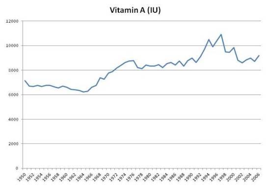
"In the book we noted studies showing that people whose intake of vitamin A was above 10,000 IU/day tended to have higher mortality. This was most commonly observed in people taking multivitamins.

There was a period of enthusiasm for vitamin A supplementation between the 1960s and 2000. Multivitamins had more vitamin A in that period. After studies showed negative results, the vitamin A content of multivitamins was reduced.

It is possible that the source of problems may not be vitamin A per se, but degradation products of vitamin A. I’ve previously blogged about how vitamin A plus DHA (a fatty acid in fish oil) plus oxidative stress can produce highly toxic degradation products (see DHA and Angiogenesis: The Bottom Line, May 4, 2011; Omega-3s, Angiogenesis and Cancer: Part II, Apr 29, 2011; Omega-3 Fats, Angiogenesis, and Cancer: Part I, Apr 26, 2011)."

- Omega-3s, Angiogenesis and Cancer: Part II - Perfect Health Diet | Perfect Health Diet

"DHA is oxidized to a compound called HOHA which then combines with a protein, generally a retinyl (vitamin A-derived) protein to form CEP.

Cancers generate lots of CEP from DHA, and perhaps one way they do that is by generating lots of retinyl proteins. Cancers are known to have disturbed vitamin A biology with lots of retinyl:

Disturbance in vitamin A metabolism seems to be an important attribute of cancer cells. Retinoids, particularly retinoic acid, have critical regulatory functions and appear to modulate tumor development and progression. The key step of vitamin A metabolism is the esterification of all-trans retinol, catalyzed by lecithin/retinol acyltransferase. In this work we show that malignant melanoma cells are able to esterify all-trans retinol and subsequently isomerise all-trans retinyl esters into 11-cis retinol, whereas their benign counterparts – melanocytes are not able to catalyze these reactions. Besides, melanoma cell lines express lecithin/retinol acyltranseferase both at the mRNA and protein levels. In contrast, melanocytes do not express this enzyme … [8]
"​

- Vitamin A hepatotoxicity: A cautionary note regarding 25,000 IU supplements
 
Last edited:

Brother John

Member
Joined
Feb 10, 2016
Messages
101
The studies on liver reserves posted above were doned around 1930-40. I thoughteded that fish liver oils were used back then, but that results could perhaps be different if the publication was more recent. And they is.

- A survey of human liver reserves of retinol in London

This was doned around 1980, the median reserve of all people was 650 IU/g; for comparison, this is way above the median of the diabetic group over 60 y (which is already the highest of all) from the 1940 one: 540 IU/g.


The highestest level found back then was 1500 IU/g of liver; in the more recent, it was 4000 IU/g (which is insane).

They commented that it's difficult to stipulate a low value mark for liver reserves because animals are still capable of surviving for months with the content that some researchers considered inadequate. When it's low, an ingested dose is rapidly consumed in the body and isn't enough to be deposited as reserve, therefore it's only after a certain amount of vitamin A that the body starts to save it. Low level mark is before clear signs of deficiency.

Their normal range was 330-1000 IU/g, but this is also a reflect of the population as a whole consuming more.


Infants had low levels but rose 3x in childhood and stayed that way until 30 years. With age levels tended to decrease to eventually match those of infants. They commented that this is different in other places; in North and America the levels also increase in childhood (multivitamins), decline later and increase as life goes on; in the Asias, it keeps increasing and there are no early peaks (supplementation is rarer).​
There is no doubt that levels are increasing (and the authors acknowledged this), largely as a result of supplementation.

I remember Paul writting about it:

- Food Fortification: A Risky Experiment? - Perfect Health Diet | Perfect Health Diet

Slide5.jpg


"In the book we noted studies showing that people whose intake of vitamin A was above 10,000 IU/day tended to have higher mortality. This was most commonly observed in people taking multivitamins.

There was a period of enthusiasm for vitamin A supplementation between the 1960s and 2000. Multivitamins had more vitamin A in that period. After studies showed negative results, the vitamin A content of multivitamins was reduced.

It is possible that the source of problems may not be vitamin A per se, but degradation products of vitamin A. I’ve previously blogged about how vitamin A plus DHA (a fatty acid in fish oil) plus oxidative stress can produce highly toxic degradation products (see DHA and Angiogenesis: The Bottom Line, May 4, 2011; Omega-3s, Angiogenesis and Cancer: Part II, Apr 29, 2011; Omega-3 Fats, Angiogenesis, and Cancer: Part I, Apr 26, 2011)."

- Omega-3s, Angiogenesis and Cancer: Part II - Perfect Health Diet | Perfect Health Diet

"DHA is oxidized to a compound called HOHA which then combines with a protein, generally a retinyl (vitamin A-derived) protein to form CEP.

Cancers generate lots of CEP from DHA, and perhaps one way they do that is by generating lots of retinyl proteins. Cancers are known to have disturbed vitamin A biology with lots of retinyl:

"​

- Vitamin A hepatotoxicity: A cautionary note regarding 25,000 IU supplements
Amazoniac, That was interesting!!
Thanks,
Brother John
 

Amazoniac

Member
Joined
Sep 10, 2014
Messages
8,583
Location
Not Uganda
Matthias Nelke (the guy in the upcoming On the Back of a Tiger docu) found this study:

"Vitamin A is likely pro-estrogenic at higher doses. This can be seen in a large randomized trial in pregnant women in Bangladesh. They received either placebo, retinol (23.331 IE per week equalling 3333 IE retinol per day - much less than many Peatarians eat per day!) or beta-carotene (42mg per week). Women in the retinol and beta-carotene group died almost double as often from preeclampsia/eeclampsia than women in the placebo group (6 deaths placebo, 13 deaths retinol, 14 deaths beta-carotene). Eclampsia is a classical high-estrogen disease according to Peat. Bleedings were also increased by retinol and beta-carotene, also supporting the pro-estrogen effect of vitamin A. "
Effects of vitamin A or beta carotene supplementation on pregnancy-related mortality and infant mortality in rural Bangladesh: a cluster randomized... - PubMed - NCBI
When will people stop using estrogenic in place of detrimental? raypeatclips could turn into a meme factory with these.

Where the pimp says that "deaths doubled", there were 13 deaths in 19806. This difference is decreased when you consider death from all causes rather than just (pre)eclampsia: 41 in 19862 for placebo versus 47 in 19806 for vitamin A. The values for deaths in 100,000 pregnancies aren't reliable because they were just projections.
This complication can have multiple causes other than improper amount of vitamin A (which might affect those indirectly).

I've just read Makrosky's post on 'news', and to be fair I find it annoying whenever reporters imply that the extent of an accident can be measured by the number of deaths since a lot of bad stuff can happen without doing the dyings.
"The fat soluble vitamins appear to function as integral parts of cell membranes and variations in availability of, for example vitamins A or E may have a profound effect on the structure and properties of the membranes of cells and their contained organelles. The biochemical make-up of these membranes varies not only from one tissue to another, but also from one intracellular organelle to another. As a result, pathological effects resulting from deficiency or excess of vitamins are highly selective in their situation, whether in the tissues or within the cells themselves. The fat soluble vitamins in general are more akin to the steroid hormones, than to the vitamins which function as enzymic cofactors."​

If people already have trouble with safer nutrients when given alone, how can you expect that giving extra vitamin A won't increase your needs for various other nutrients? It isn't difficult to become malnourished, especially if you're pregnant and under a regimen that imposes you to take something regardless of how you react to it. Still, many of the negative effects could probably be prevented with a decent program to support the poison.

- Retinoid-induced hemorrhaging and bone toxicity in rats fed diets deficient in vitamin K
 
Last edited:

Amazoniac

Member
Joined
Sep 10, 2014
Messages
8,583
Location
Not Uganda
I forgot to comment: the vitamin A gaved in that trial had [. . . Brewing document again . . .] 5 IU of vitamin E for every (weekly) 23000 IU of vitamin A, so people were receiving less than 1 IU of vitamin E per day. :thumbsdown:

Considering that the requirements for vitamin E are based on mg, less than 1 IU of it is negligible.
 
Last edited:
EMF Mitigation - Flush Niacin - Big 5 Minerals

Similar threads

Back
Top Bottom