Water Structure, Osmolytes And Cancer

OP
LLight

LLight

Member
Joined
May 30, 2018
Messages
1,411
Could dehydration increase the "detoxification enzymes" aldehyde oxydase / xanthine dehydrogenase?

These enzymes both require the element molybdenum, which has been found to be helpful in case of dehydrating conditions in plants. Molybdenum seems to help with osmolytes synthesis or uptake.

Dehydration has been found to increase ROS in plants. The source of these ROS seem to be these same enzymes:
The plant Mo-hydroxylases aldehyde oxidase and xanthine dehydrogenase have distinct reactive oxygen species signatures and are induced by drought and abscisic acid - PubMed
"Plant ROS production and transcript levels of AO and XDH were rapidly upregulated by application of abscisic acid and in water-stressed leaves and roots. These results, supported by in vivo measurement of ROS accumulation, indicate that plant AO and XDH are possible novel sources for ROS increase during water stress."

"The ability of human xanthine oxido-reductase to generate O2 and/or H2O2 in the presence of hypoxanthine and xanthine is well established and has led to the proposed role of the enzyme as a source of ROS in a range of humanpathological and physiological situations. In this case, ROS activity is the result of specific post-translational processingof the normal enzyme. NADH oxidase activity of mammalian XDH has also been detected; however, the physiological function of this activity has yet to be established (Harrison,2002; Sanders et al., 1997). The high homology between animal and plant XDH led us to hypothesize that XDH can also be a source of ROS in plants, either as O2 or H2O2 products."
I believe it's known that hyperosmolarity also induces ROS in mammalian cells.

Abscisic acid which could be an homologue of retinoic acid, seems to regulate the production of these enzymes.

Some enzymes (CYP2E1, CYP3A4 and CYP26B1) participating in vitamin A metabolism (potential retinoic acid catabolism) seem to be upregulated by dehydration. Aldehyde oxidase is thought to participate in retinoic acid production (and to potentially "act in concert with the P450 system"):
The mammalian aldehyde oxidase gene family
"The enzymes are involved in the phase I metabolism of numerous compounds of both medical and toxicological relevance, potentially acting in concert with the microsomal cytochrome P450 system [33-39]."​

"Vitamin A metabolism is probably the endogenous pathway for which more stringent information on the involvement of aldehyde oxidases is available. The ability of mammalian aldehyde oxidases to catalyse the oxidation of retinaldehyde (a physiological precursor) into retinoic acid (the active metabolite of vitamin A) was discovered in rabbit liver cytosol [53] and confirmed using purified preparations of mouse liver AOX1 [28,54]. In our hands, retinaldehyde has been one of the best substrates not only of mouse AOX1, but also of AOX3, AOX4 and AOX3L1 [14,19,27]."​

Could it be consistent that, while water restriction could be inducing the catabolism of retinoic acid somewhere, it would also induce its production "elsewhere"?

Also, dry fasting tends to increase uric acid (Dry Fasting Physiology: Responses to Hypovolemia and Hypertonicity - PubMed), whose production can be catalized by xanthine oxidase.

Sorry for this mish-mash ;)
 

Collden

Member
Joined
Oct 6, 2012
Messages
630
This is an interesting thread. Intermittent dehydration could stimulate the immune system. Does this mean that much of the benefit of dry fasting could be achieved simply by restricting fluid intake without actually fasting?
 
OP
LLight

LLight

Member
Joined
May 30, 2018
Messages
1,411
Does this mean that much of the benefit of dry fasting could be achieved simply by restricting fluid intake without actually fasting?

I think so. Maybe eating high fat can allow drinking even less than if you are fasting. Fat is the macro that induce the most important creation of metabolic water. If you bring more fat than with lipolysis alone you may end with less water need (more fat burning than with fasting alone, thus more metabolic water). Metabolic water might be more important because it is relatively deuterium depleted (with respect to water that you would drink). Deuterium is thought to impair mitochondria.

You can check this website: Hydration dehydration cycles - One Cure for All Diseases

Epileptic children had good results with the "dehydration therapy" (which is not the protocol presented on the website above).
 
Last edited:
OP
LLight

LLight

Member
Joined
May 30, 2018
Messages
1,411
Osmotic Adaptation by Na+-Dependent Transporters and ACE2: Correlation with Hemostatic Crisis in COVID-19

"The characteristics of permanent unresolved osmotic stress in cells include initiation of reversible thrombosis increasing D-dimer levels in blood, necrosis in ATP-deprived cells, or cell rupture causing acute thrombosis and inflammation."

"The osmotic crisis resulting from hypokalemia and disruption of the ACE2-based complex is compatible with clinical findings in damaged endothelial cells, early manifestations of elevated D-dimer levels, and the progression to thrombosis in COVID-19 patients. Current clinical trials use fibrinolytic and anti-inflammatory therapies reviewed in [19] to combat thrombosis. To address osmotic crisis, adjuvant supplementation with neutral amino acids could be implemented to replenish organic osmolytes and stabilize intracellular osmolality when the ACE2 complex is disabled. This safe approach improves the rate of distress, executive functions, attention, and vigilance in patients with genetic disorders and malnutrition [203]. Normalization of hypokalemia, resulting from the hyperactivation of ENaC, could be achieved by a moderate dose of amiloride combined with potassium and neutral amino acids’ supplementation to address both the root problem and its consequences."​
 
OP
LLight

LLight

Member
Joined
May 30, 2018
Messages
1,411
Ramadan diurnal intermittent fasting modulates SOD2, TFAM, Nrf2, and sirtuins (SIRT1, SIRT3) gene expressions in subjects with overweight and obesity - PubMed

"Results showed that the relative gene expressions in obese subjects in comparison to counterpart expressions of controls for the antioxidant genes (TFAM, SOD2, and Nrf2) were significantly increased at the end of Ramadan, with percent increments of 90.5%, 54.1% and 411.5% for the three genes, respectively. However, the metabolism-controlling gene (SIRT3) showed a highly significant (P < 0.001) downregulation accompanied with a trend for reduction in SIRT1 gene at the end of Ramadan month, with percent decrements of 61.8% and 10.4%, respectively. Binary regression analysis revealed significant positive correlation (P < 0.05) between high energy intake (>2000 Kcal/day vs. <2000 Kcal/day) and expressions of SOD2 and TFAM (r = 0.84 and r = 0.9, respectively)."
While dry fasting probably increases ROS, it also seems to concomitantly increase endogenous antioxidants.

NRF2 activation could help with iron metabolism:
Nrf2 controls iron homeostasis in haemochromatosis and thalassaemia via Bmp6 and hepcidin
"Pharmacological activation of Nrf2 stimulates the Bmp6-hepcidin axis, improving iron homeostasis in haemochromatosis and counteracting the inhibition of Bmp6 by erythroferrone in beta-thalassaemia. We propose that Nrf2 links cellular sensing of excess toxic iron to control of systemic iron homeostasis and antioxidant responses, and may be a therapeutic target for iron-associated disorders."
NRF2 seems to also be linked to the detoxification pathway.
 
OP
LLight

LLight

Member
Joined
May 30, 2018
Messages
1,411
Farnesoid X receptor is essential for the survival of renal medullary collecting duct cells under hypertonic stress

"Given a ubiquitous distribution pattern of both FXR and TonEBP, they may act in concert in regulating many other biological processes than those involved in the response to hypertonicity in renal medulla.

Similar to TonEBP, FXR may also represent a hypertonicity-responsive gene."
The FXR is hypothesized to be triggered by hyperosmolarity like TonEBP/NFAT5.

This receptor seems to be linked (in this species of mice at least) to the detoxification pathway:
Alterations in xenobiotic metabolism in the long-lived Little mice
"Our results suggest instead that bile acids and the bile acid receptor Fxr (farnesoid × receptor) are likely mediators of the up-regulation of xenobiotic detoxification genes in Little mice. Bile acid levels are significantly elevated in the bile, serum and liver of Little mice. We found that treatment of wild-type animals with cholic acid (CA) mimics in large part the up-regulation of xenobiotic metabolism genes observed in Little mice. Additionally, the loss of Fxr had a major effect on the expression of xenobiotic detoxification genes up-regulated in Little mice."
Reminds me the upregulation (again in mice) of bile acids production by vasopressin receptor agonist.
 
OP
LLight

LLight

Member
Joined
May 30, 2018
Messages
1,411
Farnesoid X receptor (FXR) gene deficiency impairs urine concentration in mice
"It was previously reported that the nuclear receptor liver X receptors (LXRs), which have a “yin-yang” relationship with FXR in regulation of bile acid metabolism, also participate in water homeostasis regulation. LXRβ deficiency results in central diabetes insipidus and impaired renal proximal tubule aquaporin-1 expression in mice (37). This finding suggests that LXR and FXR may act in concert in maintaining renal water homeostasis."​

Bile acid signaling in renal water regulation
"Emerging evidence has shown that bile acids play important roles in renal physiology and diseases by activating two major receptors, the nuclear farnesoid X receptor (FXR) and the membrane G protein-coupled bile acid receptor-1 (Gpbar1; also known as TGR5). Both FXR and TGR5 have been identified in human and rodent kidneys, where they are deeply involved in renal water handling."​

T0901317 is a dual LXR/FXR agonist - PubMed
"We characterize the ability of the liver X receptor (LXRalpha [NR1H3] and LXRbeta [NR1H2]) agonist, T0901317, to activate the farnesoid X receptor (FXR [NR4H4]). Although T0901317 is a much more potent activator of LXR than FXR, this ligand actually activates FXR more potently than a natural bile acid FXR ligand, chenodeoxycholic acid. Thus, the FXR activity of T0901317 must be considered when utilizing this agonist as a pharmacological tool to investigate LXR function."​

Oxysterol 22(R)-Hydroxycholesterol Induces the Expression of the Bile Salt Export Pump through Nuclear Receptor Farsenoid X Receptor but Not Liver X Receptor
"In a microarray study using human hepatocytes, BSEP was markedly induced not only by chenodeoxycholic acid (CDCA) but also by oxysterol 22(R)-hydroxycholesterol [22(R)-OHC]. We hypothesized that the expression of BSEP was induced by oxysterols through activation of LXR. To test the hypothesis, human primary hepatocytes or hepatoma cells were treated with 22(R)-OHC, and expression of BSEP was determined. The level of BSEP mRNA was increased as much as 5-fold upon oxysterol induction. In contrast to our hypothesis, the oxysterol-induced up-regulation of BSEP is mediated through FXR but not LXR. BSEP promoter activity was markedly induced by 22(R)-OHC in the presence of FXR but not LXRs."

"The FXR signaling is functionally related to LXR signaling. Both signaling pathways are involved in regulating a number of target genes important in the synthesis, metabolism, and transport of cholesterol and bile acids. For example, cholesterol-7α-hydroxylase (CYP7A1), a rate-limiting enzyme in the conversion of cholesterol to bile acids in the classic pathway of bile acid synthesis, is directly up-regulated through LXR pathway in rodents (Chiang et al., 2001; Menke et al., 2002) but indirectly down-regulated through FXR pathway in both rodents and human (Chiang et al., 2000; Lu et al., 2000; Davis et al., 2002). The species difference in the regulation of human CYP7A1 by LXR is noted because of the lack of an LXR-responsive element in human CYP7A1 (Chiang et al., 2001). Conversion of cholesterol to bile acids represents a major pathway for cholesterol elimination from the body. On the other hand, secretion or adsorption of cholesterol requires bile acids for cholesterol solubilization.

In a microarray study using human primary hepatocytes to identify transcriptionally regulated target genes by bile acid CDCA and oxysterol 22(R)-OHC, it was unexpectedly observed that BSEP was markedly induced by 22(R)-OHC. The observation is consistent with a recent study that found a high-cholesterol diet results in an increase in bile acid pool size and fecal secretion (Tiemann et al., 2004), indicating an elevated canalicular bile acid secretion by BSEP. We hypothesized that the expression of BSEP was induced by oxysterols through activation of LXR. To test the hypothesis, a reporter harboring the BSEP promoter was constructed and characterized for its responsiveness to 22(R)-OHC in the presence of LXR or FXR. In contrast to our hypothesis, the activation of BSEP promoter by 22(R)-OHC was achieved through FXR but not LXR. Disruption of the IR1 element in the BSEP promoter significantly reduced its ability to respond to oxy-sterol induction. In addition, mutational analysis of the FXR ligand binding domain (LBD) revealed that different structural features of FXR were involved in CDCA- and 22(R)-OHC-mediated activation."

"Our finding raises a series of not only interesting but also important questions, such as whether other oxysterols serve as FXR ligands to regulate FXR target genes, how oxysterols interplay with bile acids to regulate FXR target genes, and how oxysterols behave as dual ligands in the presence of FXR and LXR in consideration of the fact that FXR, LXR, and oxysterols coexist in many tissues including liver, intestine, kidney, adrenal, and vascular epithelial cells (Bishop-Bailey et al., 2004). Those questions are currently under investigation."​

Some elements :
  • Bile acid receptors (FXR; TGR5) seem to be involved in water homeostasis for some reasons. It's the same for the LXR.
  • FXR and LXR also feature a "yin-yang" relationship regarding bile acid metabolism (at least in rodents, regarding the LXR).
  • A synthetic LXR agonist (T0901317) is able to activate the FXR more potently than a natural bile acid. The oxysterol 22(R)-OHC is also a ligand for the FXR but bile acids and this oxysterol do not seem to act on the FXR in the same way. Note: 22(R)-OHC is the product of the conversion of cholesterol by CYP11A1, the first step in the metabolism of the steroid hormone biosynthesis pathway.
  • FXR is known for its role in the downregulation of the CYP7A1 enzyme ("rate-limiting enzyme in the conversion of cholesterol to bile acids").
  • In humans, LXR doesn't upregulate the CYP7A1 like it does in mice.

If the LXR is not the receptor that senses oxysterols levels and upregulate the bile acids production via the CYP7A1 enzyme in humans, could oxysterols also be significant ligands for the FXR that, in contrast to the negative feedback effect of bile acids on their own production, would increase (instead of repressing) bile acids production?

LXR could be indirectly activated via the potential [hyperosmolarity->NFAT5->CYP3A4->4β-Hydroxycholesterol] pathway, while FXR could also indirectly be activated by oxysterols ([hyperosmolarity->NFAT5->CYP11A1->22(R)-OHC]?) or directly sensing osmolarity according to the previous post. Is fluid restriction/fasting a significant way to activate them and benefit from their activation for their other metabolic purposes (in the liver or elsewhere)?

Why are LXR and bile acids receptors so "deeply involved in renal water handling"?
 
OP
LLight

LLight

Member
Joined
May 30, 2018
Messages
1,411
Could LXR be associated with the osmotic stress response?

I) Osmotic stress could induce the production of oxysterols (4-beta-hydroxycholesterol and 25-hydroxycholesterol) in the liver (via upregulation of the CYP3A4 enzymes by NFAT5/TonEBP), which appear to be LXR ligands. Could these oxysterols elevate systemic LXR activation and this long-term (half-life could be 17 days for 4β-HC)?

II) LXR beta "represents a key regulator of body water homeostasis". It would be odd that this effect is not in direct response to water restriction.

Liver X receptor increases aquaporin 2 protein level via a posttranscriptional mechanism in renal collecting ducts
"These findings provide additional evidence that the two LXR isoforms may also play a distinct role in renal physiology. LXR beta rather than LXR alpha is critical in regulating urine concentration by inducing collecting duct AQP2 protein expression."​

III) In certain conditions (high fat diet + diabetes inducer substances), the transcription NFAT5/TonEBP transcription factor seem to be involved in the expression of the LXR in the liver, cf. figure (d) below.

TonEBP/NFAT5 haploinsufficiency attenuates hippocampal inflammation in high-fat diet/streptozotocin-induced diabetic mice
"We found that HFD/STZ treatment increased the hepatic expression of these factors in WT and TonEBP+/− mice, while the increases in LXRβ and PPARα were significantly attenuated by TonEBP haploinsufficiency (Fig. 2d–f)."​

upload_2020-12-3_13-48-0.png

IV) This one is a bit far-fetched but LXR seems to be involved in antimicrobial peptides production, like NFAT5 is in macrophages subjected to osmotic stress.

IL-36/LXR axis modulates cholesterol metabolism and immune defense to Mycobacterium tuberculosis
"Of note, we identified several predicted binding sites for LXRα in the proximity of several AP sequences (Supplementary Table 1)."

"In contrast, LXR inhibition did not block vitamin D-induced expression of APs, showing that vitamin D triggers AP production independent of the IL-36/LXR axis (Fig. 3C,D). Importantly, similar results were observed upon Mtb infection, triggering the LXR- and IL-36-dependent induction of APs (Fig. 3E,F). These results could also be validated at the protein level (Fig. 3G). Our findings demonstrate that LXR activation is necessary for IL-36-induced AP production and inhibition of Mtb growth."​

LXR seems to be related to macrophages defenses to intracellular pathogens, like NFAT5 is.

Liver X receptors contribute to the protective immune response against Mycobacterium tuberculosis in mice
"Treatment with an LXR agonist reduced bacterial replication in M. tuberculosis–infected mice. Inversely, Lxra–/–Lxrb–/– mice, deficient in both LXRα and LXRβ, exhibited increased susceptibility to infection and a concomitant defective innate and acquired immune response. Our results implicate LXR-dependent pathways in the protective immune response against this major pathogen."

The evolving role of TonEBP as an immunometabolic stress protein - PubMed
"In summary, TonEBP is a central regulator of macrophages, which are involved in both innate and adaptive immunity against infections caused by bacteria and other pathogens."​

V) NFAT5/TonEBP and LXR share the same target SREBP-1c

X-box binding protein 1 (XBP1): A key protein for renal osmotic adaptation. Its role in lipogenic program regulation - PubMed
"Together our data demonstrate that hyperosmolarity induces IRE1α → XBP1s activation; XBP1s drives the expression of SREBP1 and SREBP2 which in turn regulates the expression of the lipogenic enzymes lipin1 (LPIN1) and 2 (LPIN2) and DGAT1. We also demonstrated for the first time that tonicity-responsive enhancer binding protein (TonEBP), the master regulator of osmoprotective response, regulates XBP1 expression. Thus, XBP1 acts as an osmoprotective protein since it is activated by high osmolarity and upregulates lipid metabolism, membranes generation and the restoration of ER homeostasis."​

Regulation of mouse sterol regulatory element-binding protein-1c gene (SREBP-1c) by oxysterol receptors, LXRα and LXRβ
In this report, we describe a novel LXR target, the sterol regulatory element-binding protein-1c gene (SREBP-1c), which encodes a membrane-bound transcription factor of the basic helix-loop-helix-leucine zipper family. SREBP-1c expression was markedly increased in mouse tissues in an LXR-dependent manner by dietary cholesterol and synthetic agonists for both LXR and its heterodimer partner, the retinoid X receptor (RXR).​

Interestingly, SREBP1 seems to be involved in the expression of the NIS (sodium/iodide symporter) while tissues of "previously treated tuberculosis" patients seem to uptake iodine:
Radioiodine uptake in inactive pulmonary tuberculosis - PubMed
"Radioiodine may accumulate at sites of inflammation or infection. We have seen such accumulation in six thyroid cancer patients with a history of previously treated pulmonary tuberculosis."
Also interestingly, the LXR could also be involved in the immune response against HIV:
Stimulation of Liver X Receptor Has Potent Anti-HIV Effects in a Humanized Mouse Model of HIV Infection - PubMed
"In conclusion, these results reveal a novel link between LXR stimulation and cell resistance to HIV infection and suggest that LXR agonists may be good candidates for development as anti-HIV agents, in particular for pre-exposure prophylaxis of HIV infection."​

While on the contrary, NFAT5 would favor HIV proliferation:
NFAT5 Regulates HIV-1 in Primary Monocytes via a Highly Conserved Long Terminal Repeat Site
"Using small interfering RNA to ablate expression of endogenous NFAT5 protein, we show that the replication of three major HIV-1 viral subtypes (B, C, and E) is dependent upon NFAT5 in human primary differentiated macrophages."​
 
OP
LLight

LLight

Member
Joined
May 30, 2018
Messages
1,411
More intrications between osmolarity, transcription factors, bile acids and immune system:

X-box binding protein 1 (XBP1): A key protein for renal osmotic adaptation. Its role in lipogenic program regulation - PubMed
"We also demonstrated for the first time that tonicity-responsive enhancer binding protein (TonEBP), the master regulator of osmoprotective response, regulates XBP1 expression. Thus, XBP1 acts as an osmoprotective protein since it is activated by high osmolarity and upregulates lipid metabolism, membranes generation and the restoration of ER homeostasis."

Farnesoid X receptor signaling activates the hepatic X-box binding protein 1 pathway in vitro and in mice - PubMed
"FXR signaling activates the IRE1α/XBP1 pathway in vivo and in vitro. FXR pathway activation increases XBP1 splicing and enhances p-IRE1α expression. These effects are mediated, at least in part, by SHP. IRE1α/XBP1 pathway activation by bile acids and pharmacological FXR agonists may be protective during liver injury and may have therapeutic implications for liver diseases."

Hepatic deletion of X-box binding protein 1 impairs bile acid metabolism in mice
"The unfolded protein response (UPR) is an adaptive response to endoplasmic reticulum stress and the inositol-requiring enzyme 1α/X-box binding protein 1 (IRE1α/XBP1) pathway of the UPR is important in lipid metabolism. However, its role in bile acid metabolism remains unknown. We demonstrate that liver-specific Xbp1 knockout (LS-Xbp1−/−) mice had a 45% reduction in total bile acid pool."

"In conclusion, loss of hepatic XBP1 decreased the bile acid pool and CYP7A1 synthetic activity. Cholesterol feeding, but not induction of CYP7A1 with cholestyramine, increased CYP7A1 synthetic activity and corrected the genotype-specific total bile acid pools. These data demonstrate a novel role of IRE1α/XBP1 regulating bile acid metabolism."

Immunodeficiency and Autoimmune Enterocolopathy Linked to NFAT5 Haploinsufficiency
"Crohn's disease (CD) and ulcerative colitis (UC), which together comprise IBD, are believed to result from an aberrant immune response to commensal gut microbes, leading to chronic intestinal inflammation. Compared to healthy controls, we observed that NFAT5 mRNA expression was significantly reduced in patients with active UC and CD (Fig. 7), raising the possibility that NFAT5 and other components of the osmoadaptation pathway may be dysregulated in IBD."

Farnesoid X receptor (FXR) activation and FXR genetic variation in inflammatory bowel disease - PubMed
"FXR activation in the ileum is decreased in patients with Crohn's colitis. This may be secondary to altered enterohepatic circulation of bile salts or transrepression by inflammatory signals but does not seem to be caused by the studied SNPs in FXR. Increasing FXR activity by synthetic FXR agonists may have benefit in CD patients."

XBP1: the last two decades - PubMed
"In summary, the induction of ER stress in intestinal epithelium through genetic deletion of XBP1 leads to spontaneous IBD. XBP1 controls organ-specific inflammation through two major mechanisms. First, Paneth cell function, as evidenced by diminished antimicrobial peptide secretion, was strikingly impaired in XBP12/2 mice, compromising the response to pathogenic bacteria. Second, XBP1 deletion-induced ER stress led to a heightened pro-inflammatory tone of the epithelium, as revealed by increased JNK activation upon stimulation with known inducers (flagellin, TNFa) of IBD. These two mechanisms are probably co-dependent, interactive and co-amplifying because Paneth cell dysfunction would be predicted to promote intestinal inflammation by enhanced JNK signalling in hypersensitive intestinal epithelial cells upon increased exposure to bacterial antigens such as flagellin. XBP1 thus unifies two key aspects of IBD: regulation of the intestinal microbiota and the inflammatory state of the mucosal immune system. Future experiments will be directed at testing the hypothesis that one can extrapolate this scenario to other autoimmune/proinflammatory diseases"

Summary:
  • TonEBP/NFAT5 and FXR seem to activate the XBP1 which itself could stimulate bile acids production and increase the immune system response (production of antimicrobial peptides).
  • TonEBP/NFAT5 and FXR could be downregulated in IBD, and XBP1 could be the reason for this link.
 
OP
LLight

LLight

Member
Joined
May 30, 2018
Messages
1,411
Link between osmotic stress and immune system activation in C. elegans:

Previous studies of infection in C. elegans have noted that transcriptional responses to infection appear highly specific and exhibit little significant overlap with other forms of well-studied environmental stress, such as heat shock and ER stress [12], [13]. However, infection by diverse types of pathogens gives rise to common transcriptional signatures [26], the etiology of which is not clear. Here, we find that activation of the osmotic stress response in C. elegans, either through physiological or genetic mechanisms, can mimic common transcriptional responses to a diverse array of infection models in C. elegans. The mechanism(s) by which activation of osmotic stress responses mimic infection is currently unclear but one intriguing mechanism may relate to infection-induced disruptions in osmotic homeostasis. For example, many pathogens, via their production of pore-forming toxins and Type III secretion systems, create ion and water permeable pores in the plasma membrane [31]. Due to the standing electrochemical ion gradients present in all cells, the insertion of these relatively non-selective pores will allow such gradients to collapse, producing osmotic dyshomeostasis [32], [33]. In this regard, osmotic stress may mimic early pathophysiological responses to infection. Consistent with this hypothesis, there is also a substantial overlap between the ORGs identified in this study and genes regulated by exposure of C. elegans to a purified pore-forming toxin [8]. In the future, it would be interesting to test whether common transcriptional signatures of infection in C. elegans are reduced following infection with toxin-compromised pathogens.

Functional links between infection and osmotic stress have also been observed in other systems. For example, in mammals, T-cell activation in the thymus occurs in hypertonic environments, the maintenance of which requires the rel-type transcription factor NFAT5/TonEBP [34]. NFAT5/TonEBP is also an essential mediator of hypertonicity-induced transcriptional responses in diverse cell types, including the extremely hypertonic environment of the inner medulla of the mammalian kidney [35], [36]. In Drosophila, salt stress induces a broad transcriptional response that includes the induction of numerous infection regulated genes [37]. Along with the present studies in C. elegans, these data suggest the intriguing hypothesis that cellular osmotic stress may be an evolutionarily conserved and physiologically significant activator of immune responses. Interestingly, the C. elegans genome does not contain rel-type transcription factors similar to NFAT5/TonEBP, suggesting that the functional links between osmotic stress and immunity may have preceded the evolution of this family of transcription factors. C. elegans should provide an outstanding model system in which to better explore the functional, molecular, and evolutionary relationships between immunity and osmotic stress.

In conclusion, our findings show that both osmotic stress and pathogen infection regulate similar transcriptional targets in C. elegans. Tissue-specific GATA-type transcription factors elt-2 and elt-3 play a major role in coordinating both of these gene expression programs and are required for the response to osmotic stress. Our studies functionally link cellular osmotic stress responses with innate immunity and suggest that osmotic dyshomeostasis might represent a new and evolutionarily significant paradigm for the physiological basis of host cell pathogen sensing.
 

zarrin77

Member
Joined
May 12, 2015
Messages
173
Location
San Diego, CA
This is all super interesting. This might be the reason why MSM (methylsulfonylmethane) has been shown to have strong anti-cancer properties in essentially every study and type tested
 

Inaut

Member
Joined
Nov 29, 2017
Messages
3,620
This is all super interesting. This might be the reason why MSM (methylsulfonylmethane) has been shown to have strong anti-cancer properties in essentially every study and type tested

I forgot to tag you in the MSM revisited thread. I'm really interested in your posts on MSM. Do you currently take it? If so how much?
 
OP
LLight

LLight

Member
Joined
May 30, 2018
Messages
1,411
Interesting publication:

Aging as a consequence of intracellular water volume and density - ScienceDirect


Aging as a consequence of intracellular water volume and density
Author links open overlay panelDiegoBonattoaBruno CésarFeltesbJoice de FariaPolonib
RedirectingGet rights and content
Open Access funded by Brazilian Government
Under an Elsevier user license
open access

Abstract
Aging is the result of a gradual failure of physiological and/or biochemical pathways that culminates with the death of the organism. Until now, the causative factors of aging are elusive, despite the increasing number of theories that try to explain how aging initiates. Interestingly, aging cells show an increase in intracellular water volume, but this fact is barely explored in aging studies. All cells have a crowded cytoplasm, where the high concentration and proximity of macromolecules create an environment that excludes many small molecules, including water. In this crowded environment, water can be found in two states termed low density water (LDW), which shows low reactivity and has an ice-like structure, and high density water (HDW) that has a disorganized structure and is highly reactive. LDW predominates in a macromolecular crowded environment, while HDW is found only in microenvironments within cytoplasm. In this sense, we hypothesized that the failure in the water homeostasis mechanisms with time changes the equilibrium between LDW and HDW, increasing the concentration of intracellular HDW. Being reactive, HDW leads to the generation of reactive oxygen species and disturbs the crowded cytoplasm environment, resulting in a diminished efficiency of metabolic reactions. Noteworthy, the cell becomes less prone to repair damage when the concentration of HDW increases with time, resulting in aging and finally death. Interestingly, some biological mechanisms (e.g., anhydrobiosis) reduce the concentration of intracellular water and prolong the life of cells and/or organisms. In this sense, anhydrobiosis and related biological mechanisms could be used as a platform to study new anti-aging therapies.
 
OP
LLight

LLight

Member
Joined
May 30, 2018
Messages
1,411


Here is a short clip of an experiment I ran it with only 8 control rats and 2 water restricted rats. The control rats all grew old and died off normally with an average lifespan of 23 months and a maximum lifepsan of 29 months. The 2 water restricted rats were retained at the same maximum weight and were given water after they reached a minimum weight. eventualy the rats could go for 13 days or even more without water (I never pushed it to the limit) and then they would regain the weight they lost by being given water freely for about 1/2 hour or so. When they began to look a little like they weren't thriving, they were allowed to grow about 10 grams. This was similar to the protocol for Clive McCay's 1933 experiment (described in the Journal of Nutrition) were he first discovered that caloric restriction increased the lifespan of rats.
This led to one water restricted rat dying at about 29 months from a leg tumor (this rat of the two WR rats was the one that was most nervous about coming out of the cage to get water-the other long lived one seemed to be more carefree-I just mention this since I've seen studies where more stressed animals have shorter lifespans ), and the other rat lived 47 months and finally being killed by an eye infection.
The 47 months old rat broke a WORLD record for life span extension in rats of this type- Sprague Dawley Females from Taconic labs. Even including C.R. experiments in this rat type and sex for 1000's of rats! As far as I could find, looking through all caloric restriciton experiments using 1000's of these rats, the oldest one generated was about 44 months.

Maybe insignificant or fake, but interesting if true.
 

Amazoniac

Member
Joined
Sep 10, 2014
Messages
8,583
Location
Not Uganda

"Renal medullary cells contain the highest levels of organic osmolytes consequent to their being exposed to extremely high concentrations of NaCl and urea because of their roles in concentrating the urine. The principal organic osmolytes in renal medullary cells are sorbitol, betaine, inositol, taurine, and glycerophosphocholine (GPC). Cells in other tissues may also experience hyperosmolality, albeit to a lesser degree than in the renal medulla. Accordingly, they also accumulate organic osmolytes. The principal ones in brain are amino acids, choline, creatine, inositol, and taurine (20). Liver cells accumulate betaine, inositol, and taurine (21)."​
 
OP
LLight

LLight

Member
Joined
May 30, 2018
Messages
1,411
Betaine (trimethylglycine, an osmolyte) really seems like a wonder nutrients:

Investigation of betaine as a novel psychotherapeutic for schizophrenia
 
Last edited:
OP
LLight

LLight

Member
Joined
May 30, 2018
Messages
1,411

Solanum lycopersicum is an important vegetable crop, and its growth and productivity are strongly affected by abiotic stresses, such as salt or drought, worldwide (Duan et al. 2012, Sadashiva et al. 2013). Therefore, identifying novel genes involved in this process and understanding their functions can improve plant stress tolerance.
In this study, SlVKOR was determined to be induced at the transcriptional level by salt or drought stress.
Overexpression of SlVKOR in E. coli also increased the survival rate of cells under osmotic stress. The results suggested that SlVKOR played an important role in the response to osmotic stress.

[...]

As a homologue of mammalian VKOR, the up- and down-expression of plant VKOR may also be related to the ROS homeostasis, but more evidence is needed.

[...]

The results indicated that down- and up-regulation of SlVKOR in vivo changed the activities of antioxidant enzymes and resulted in differential accumulation of ROS. Our recent study also showed that the involvement of AtVKOR to osmotic stress was related to abscisic acid synthesis in Arabidopsis (Lu et al.
2015). In resistance to osmotic stress, SlVKOR may play an important role in facilitating cross-talk between ROS homeostasis and the abscisic acid signal pathway.
 
OP
LLight

LLight

Member
Joined
May 30, 2018
Messages
1,411

SlVKOR was determined to be induced at the transcriptional level by salt or drought stress.
Overexpression of SlVKOR in E. coli also increased the survival rate of cells under osmotic stress. The results suggested that SlVKOR played an important role in the response to osmotic stress.


It is known that K is not only distributed in the liver and bones but also abundantly distributed in the brain, kidney, and gonadal tissues. However, the role of K in these tissues is not well clarified. In this study, we used DNA microarray and identified the genes whose expression was affected in the testis under the K-deficient (K-def) state. The expression of genes involved in the biosynthesis of cholesterol and steroid hormones was decreased in the K-def group. The mRNA levels of Cyp11a - a rate-limiting enzyme in testosterone synthesis - positively correlated with the menaquinone-4 (MK-4) concentration in the testis.


....it also increases oxidative stress. Previous studies have shown potassium supplementation to decrease the oxidative stress occurring from high salt intake in rodents (via decreasing the increased NADPH oxidase activity). Furthermore, a diet abundant in vitamin e, anti-oxidative polyphenols, etc. would help negate the oxidative stress.

Also, one rodent study showed that sea salt is not as “detrimental” as refined salt:
Natural sea salt consumption confers protection against hypertension and kidney damage in Dahl salt-sensitive rats

Attached is the pic of the testosterone increase.
1619174571228.png

I'm wondering if the effect seen in these rats, with a doubling of the testosterone level, could correspond to (at least in part) to the in vivo effect of the in vitro results presented here:

It seems like the transcription factor NFAT5 could have a link with this enzyme, RNA-Seq analysis of high NaCl-induced gene expression:
  • "Categories of NFAT5 Target Genes Upregulated after Adaptation to High NaCl, but Not after as Little as 24 h of High NaCl.
  • Steroid hormones. Cyp11a1 protein localizes to the mitochondrial inner membrane and catalyzes the conversion of cholesterol to pregnenolone, the first and rate-limiting step in the synthesis of the steroid hormones."

Could vitamin K be necessary during osmotic stress, especially in the testes where it would be necessary for steroidogenesis which could be increased by high salt/NFAT5 activation?
 
OP
LLight

LLight

Member
Joined
May 30, 2018
Messages
1,411
As already posted previously, an enzyme "looking like" CYP11A1 could be upregulated during dehydrating conditions in insects:
Insect CYP Genes and P450 Enzymes

"The conversion of ecdysone to 20-hydroxyecdysone does not occur in the prothoracic glands, but does occur in many peripheral tissues, such as the fat body, midgut, and Malpighian tubules. The P450 nature of the enzyme catalyzing the 20-hydroxylation of ecdysone was well established [...] The agreement on the P450 nature of the reaction was accompanied by a lack of consensus on the subcellular localization of E20MO."

"The mitochondrial E20MO activity was reportedly inhibited by antibodies to vertebrate P450scc (CYP11A), P45011β (CYP11B), adrenodoxin, and adrenodoxin reductase (Chen et al., 1994), despite the considerable sequence divergence predicted between the vertebrate and insect proteins."
The enzyme that catalyses the production of 20-hydroxyecdysone could have similarities with the one metabolizing cholesterol in 20-hydroxyvitamin D3.

Secondly: Dehydration triggers ecdysone-mediated recognition-protein priming and elevated anti-bacterial immune responses in Drosophila Malpighian tubule renal cells

"Ecdysone titers measured from aged flies were also significantly elevated relative to young females in control (food) conditions, while desiccation increased 20E (20-hydroxyecdysone) in both young and old animals"
So water restriction increases the production of the 20-hydroxyecdysone hormone in drosophila. Which would remind the upregulation of CYP11A1 in human during dehydration.​

The 20-hydroxyecdysone insect hormone seems to be increased during dehydration while its metabolism relies on an enzyme that "was reportedly inhibited by antibodies to vertebrate P450scc (CYP11A)".

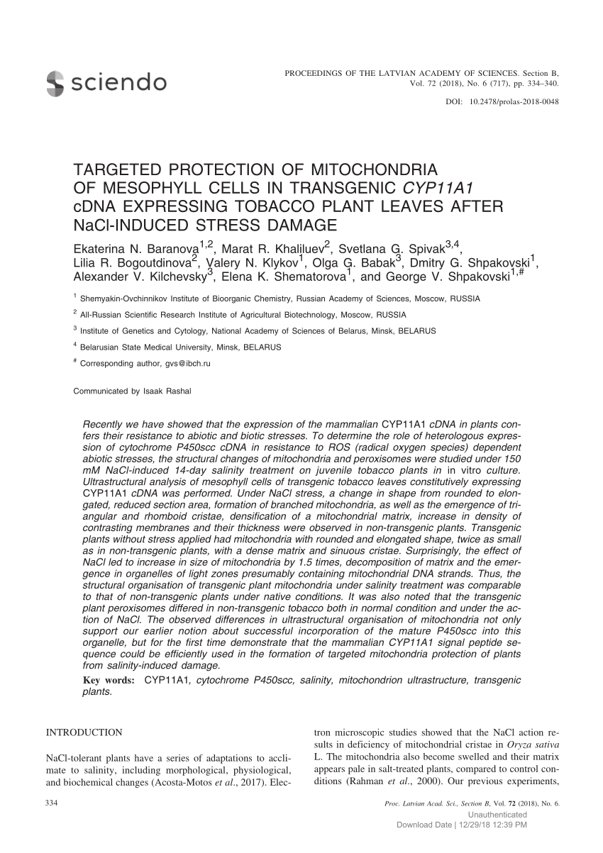
It seems like CYP11A1 enzyme (GMO) is protective against salinity-stress in plants as found in this study:
Recently we have showed that the expression of the mammalian CYP11A1 cDNA in plants confers their resistance to abiotic and biotic stresses. To determine the role of heterologous expression of cytochrome P450scc cDNA in resistance to ROS (radical oxygen species) dependent abiotic stresses, the structural changes of mitochondria and peroxisomes were studied under 150 mM NaCl-induced 14-day salinity treatment on juvenile tobacco plants in in vitro culture.
Ultrastructural analysis of mesophyll cells of transgenic tobacco leaves constitutively expressing CYP11A1 cDNA was performed. Under NaCl stress, a change in shape from rounded to elongated, reduced section area, formation of branched mitochondria, as well as the emergence of triangular and rhomboid cristae, densification of a mitochondrial matrix, increase in density of contrasting membranes and their thickness were observed in non-transgenic plants. Transgenic plants without stress applied had mitochondria with rounded and elongated shape, twice as small as in non-transgenic plants, with a dense matrix and sinuous cristae. Surprisingly, the effect of NaCl led to increase in size of mitochondria by 1.5 times, decomposition of matrix and the emergence in organelles of light zones presumably containing mitochondrial DNA strands. Thus, the structural organisation of transgenic plant mitochondria under salinity treatment was comparable to that of non-transgenic plants under native conditions. It was also noted that the transgenic plant peroxisomes differed in non-transgenic tobacco both in normal condition and under the action of NaCl. The observed differences in ultrastructural organisation of mitochondria not only support our earlier notion about successful incorporation of the mature P450scc into this organelle, but for the first time demonstrate that the mammalian CYP11A1 signal peptide sequence could be efficiently used in the formation of targeted mitochondria protection of plants from salinity-induced damage.​

In this one, CYP11A1 was protective regarding heat stress (in GMO plants again):​

Higher relative water content (by 13%) and superoxide dismutase (SOD) activity, lower electrolyte leakage (up 1.4-fold) and smaller increase in chlorophyll a and carotenoid contents in cyp11A1 canola leaves in comparison with wild-type plants under stress allowed to conclude cyp11A1 plants are more tolerant to high temperature than the control ones.​

Would hormesis be a real thing regarding steroid hormones production and heat stress / dehydration? If this is the case, what would be the best strategy to trigger CYP11A1 upregulation? Is this enzyme giving mammals a greater protection against dehydration (as seen in the modification of mitochondria appearance with salitinity stress that does not occur when there is CYP11A1)? Does vitamin K deficiency renders one more susceptible to dehydration stress?


Also, betaine is protective against osmotic / high glucose stress in ovarian cells:
Exposure of mice granulosa cells to high glucose concentration inhibited the steroidogenesis by decreasing estradiol and progesterone secretion and downregulation of steroidogenesis-related genes including 3βHSD, Cyp11a1, Cyp19a1, and StAR. Betaine treatment could ameliorate the steroidogenesis impairment at molecular and biochemical levels. High glucose concentration also enhanced apoptosis in mice granulosa cells that were characterized by elevation of caspase-3 activity, upregulation of bax gene and downregulation of bcl2 gene. Betaine treatment could attenuate the apoptotic-related changes induced by high glucose concentration in granulosa cells. According to the results of the present study, betaine could ameliorate the adverse effects of hyperglycemia on the physiological function of ovarian granulosa cells. The results highlight the potential role of betaine for the intervention of ovarian dysfunction in diabetic patients.​
 
EMF Mitigation - Flush Niacin - Big 5 Minerals

Similar threads

Back
Top Bottom