Cancer Cells Addicted To Fat And Use Fat Oxidation For Survival
Ray has written several times that while cancer cells use glucose for their normal activities they use fat to evade apoptosis and increase metastasis. Thus, according to Ray, using substances that block fat oxidation can be therapeutic. Niacinamide and aspirin are some of the most beneficial...
Forcing The Heart To Burn Glucose Instead Of Fat May Cure Heart Failure
One of the blockbuster studies I have seen over the last 12 months. It reads as if written by Peat himself and discusses tissue regeneration, Randle cycle, dietary control of metabolism, etc. AFAIK this is the first study brave enough to demonstrate that simply switching fuel types in an...
Dr. Stephen Hussey
To understand why cancer in the heart is extremely rare, we must first understand what cancer is. Cancer is a metabolic disease, which means that it occurs when there is a breakdown in normal metabolism that in turn causes shifts in the cell that result in it becoming cancerous. Way back in the 1920’s Otto Warburg and his team discovered that cancer cells stopped relying on what is called oxidative phosphorylation, the main way our cells make energy, and instead relied on what is called glycolysis and lactate fermentation.(r) This is called the Warburg Effect. It basically means that instead of using oxygen to make energy the cells found a way, or were forced to find a way, to make energy without having to use oxygen.
More recently, metabolic researcher Dominic D’Agostino and cancer researcher Dr. Thomas Seyfried have solidified the metabolic theory of cancer. (r,r) Let’s get a better understanding before we move on to the heart. If we look at cancer cells, they have some interesting characteristics. They are anaerobic, meaning they don’t use oxygen, they are acidic, they are rapidly dividing, and they are undifferentiated, meaning they are no particular type of cell but just a general cell. (r) So, we have cancer cells that display this Warburg Effect and have these unique characteristics. But why would the cell decide to have these characteristics and become cancerous? Let’s illustrate this another way.
When a human in conceived the egg and sperm come together to form a zygote cell. That cell implants itself on the side of the uterus. At this point, the cell has no blood supply and therefore no oxygen. Initially this cell grows into what is called a Morula, and then a blastocyst, by rapidly dividing to start the process of growing a fetus. Interestingly, these early dividing cells are anaerobic, (r) undifferentiated, and rapidly dividing. Sounds like cancer. Once the blood supply from the placenta develops are around the 2-week mark, the cells of the fetus start to use oxygen and become aerobic, they start to differentiate into different types of cells, and they have more controlled cell division. The key here is the presence of oxygen.
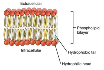
The question when it comes to cancer is why a cell stops using oxygen and become cancerous. Well, the structures in our cells that allow our cells to use oxygen to break the chemical bonds in our food to make energy for our bodies are called our mitochondria. These structures are very good at oxidative phosphorylation, provided they do not become damaged. Every time our mitochondria make energy we also make a waste product called a free radical, kind of like a car makes and exhaust when burning fuel. These free radicals can be damaging to our bodies if not taken care of. Normally, our bodies make what are called antioxidants that immediately take care of these free radicals.
However, if we are not careful how we live our lives then we can end up creating an abundance of these free radicals that can overwhelm the cells. Excess free radicals can be caused by toxin exposure, (r) relying on carbs for fuel, (r) inflammation, and high blood sugar. (r) When this happens, it can cause damage to the mitochondria. (r) Since the mitochondria are what allow us to us oxygen to make fuel now the cell cannot use oxygen anymore. If it can’t do this, it can’t survive. This triggers the cell to turn on oncogenes in the DNA of the cell that instruct the cell to become anaerobic, undifferentiated, and rapidly dividing cancer cells, the only thing it knows how to do to survive. When faced with the situation of not being able to use oxygen the cells must either die or become cancerous. It’s sort of a survival mechanism. The cancer solution keeps the tissue alive short-term, but it is obviously not a good long-term solution.
So, as I mentioned there are many thing that can cause excess free radicals in our bodies that damage our mitochondria. One is the large amount of toxins our bodies are exposed to on a daily basis. Everything from heavy metals, to plastics, to artificial fragrances. According to Herbert Needleman, who spent his life studying the effect of chemicals on children, at least 70,000 new chemical compounds have been invented and dispersed into our environment since 1950, and many of them can act as free radicals directly. But even worse than toxins, free radicals can increase dramatically when our cells become too reliant on burning carbohydrates for fuel instead of fatty acids and ketones. (r,r) This is where everything starts to relate to the heart.
The cardiac myocytes are the most mitochondrial dense cells in the body, (r) this is because of the massive amount on energy the heart needs and the large amount of oxygen it uses. Most of our organs prefer to burn fat for fuel, but especially the heart. (r) When other organs are forced to burn predominantly glucose for long periods of time then the sequence of events described above can start to happen and result in cancer. However, if the heart is forced to burn predominantly glucose then something far worse and potentially fatal can happen, a heart attack.
Because of this, the body has built in mechanisms that make sure the heart is never forced to burn predominantly glucose for fuel. One is the direct delivery of the fatty acid carrying chylomicrons from digestion through the lymphatic system to the veins that drain into the heart. (r) Secondly, the heart has a direct signaling pathway to fat cells (r) that I believe allows it to mobilize fats when glucose burning becomes to prevalent. Unfortunately, there is a situation where the heart could still be forced to burn predominantly glucose. It has to do with an imbalanced stress response signal to the heart cells.
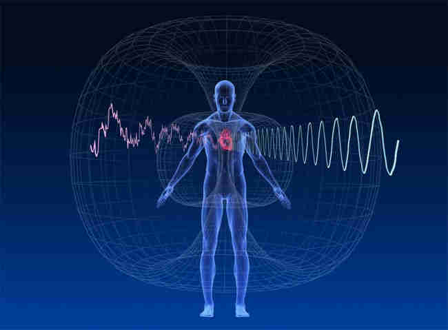
The control of the balance of the autonomic nervous system in cardiac cells, and many other cells, relies on two messenger molecules called cAMP and cGMP. cAMP levels rise in the heart cells when we have a stress response and cGMP levels rise when we are in a relaxation state. The only difference is that when it comes to cGMP, the relax molecule, something else is also needed to increase its levels. That something else is nitric oxide, NO, which is produced in the walls of arteries. These two molecules—cAMP and cGMP—keep each other in check within heart cells, they should always balance each other out. When we experience a stressful response and the nervous system causes spikes in cAMP within the heart then cGMP, provided there is enough NO, also has an increase just to keep the system more in balance.(r) This is depicted in the image below.
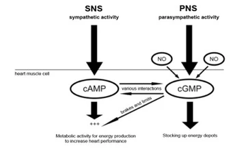
Sroka, K. (2013). What is the connection between oxidative stress and heart attacks? Retrieved from heartattacknew.com/faq/what-is-the-connection-between-oxidative-stress-and-heart-attacks/
But the system can become unbalanced. When we have prolonged periods in our life with many surges of stress responses that increase levels of cAMP and not enough stimulation of the relax response and CGMP, then we can lose the ability to effectively balance these two states. We can get stuck in our stress state. This is called decreased vagal tone because the vagus nerve is the nerve that carries these signals. When this happens the failsafe within the cardiac cells is that those consistently high levels of cAMP are balanced by also rising levels of cGMP. But remember that cGMP can only do this if NO is present. If NO gets depleted, it is really bad news.
We can get depletion of NO by having lots of free radicals in the body. Free radicals are damaging because they have an unpaired electron and they do not like to be unpaired. Because of this, the free radicals go running around the body, like the looney tunes Tasmanian devil, trying to find another electron to make a pair. It can steal them from tissues and cause damage to the tissue. But another place it can find an electron is NO. (r) If NO is uses to neutralize these free radicals too much it can decreases the NO available for cGMP simulation.
Now that we have set the stage, it is time for the big event. When humans experience decreased vagal tone for long periods of time while also experiencing decreases in NO levels, this can cause a surge in the stress response and subsequent elevation in cAMP in our heart cells without the balanced rise in cGMP. This is shown in the image below.
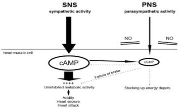
Sroka, K. (2013). What is the connection between oxidative stress and heart attacks? Retrieved from heartattacknew.com/faq/what-is-the-connection-between-oxidative-stress-and-heart-attacks/
When this happens the cascade of events that is a heart attack plays out. The sudden unchecked rise in adrenaline from the stress response has been shown to cause an increase in lactic acid production within cardiac cells. (r,r,r) This happens because the heart usually prefers to burn ketones—a product of burning fat—but in this situation the body thinks it needs to burn energy quicker to get away from a threat. Since it is quicker to burn glucose the heart cells revert to burning it rather than burning the more efficient and preferred energy source of ketones. (r,r) Burning glucose causes the build-up in lactic acid and hydrogen ions within the heart cells. This is similar to when you do a sprint or a hard, fast workout, lactic acid builds up in the muscles causing the muscle to have that burning feeling.
When this happens in a muscle in the legs or arms we can just stop moving it and the lactic acid and hydrogen ions will move along stopping the build-up an burning feeling. Since the heart can’t just stop contracting the lactic acid quickly builds up causing a major problem. The presence of acid in the heart tissue causes swelling in the area of tissue forced to burn glucose. (r) This swelling creates a higher pressure in the tissue than there is pressure from the blood flow into heart tissue and therefore prevents the blood from reaching the cells. This explains why nearly 100% of heart attacks happen in the left ventricle. The left ventricle is under the most pressure and is more suseptible to any changes in pressure casued by this edema. Without blood, this leads to dysfunctional cells walls and heart tissue death. In other words, a heart attack.
So, while cancer is a change in metabolism due to damage to our oxygen using mitochondria, which then forces the cell to become cancerous to survive short-term, a heart attack is a forced change in metabolism due to an imbalanced stress response and depletion of NO due to excess free radicals. They are both forced shifts in metabolism just in different tissues and using a slightly different mechanism. Because a forced change in metabolism in the heart immediately results in a heart attack, there is never a chance for the heart cells mitochondria to become damaged enough to force the cells to become cancerous. The heart attack beats them to the punch and kills the cells before that happens. That’s why we don’t see cancer in the heart, only heart attacks.