The mechanistic overview of SARS-CoV-2 using angiotensin-converting enzyme 2 to enter the cell for replication

md_a

Member
Joined
Aug 31, 2015
Messages
468
Aside from the anti-inflammatory stuff, I think two important methods as prevention / treatment against covid is to block the virus from attaching to ACE2 and to reduce the the angiotensin II type 1 receptor (AT1 receptor) in preventing the virus from entering the cell.
..........

The mechanistic overview of SARS-CoV-2 using angiotensin-converting enzyme 2 to enter the cell for replication: possible treatment options related to the renin–angiotensin system​

Abstract​

The SARS-CoV-2 pandemic is a healthcare crisis caused by insufficient knowledge applicable to effectively combat the virus. Therefore, different scientific discovery strategies need to be connected, to generate a rational treatment which can be made available as rapidly as possible. This relies on a solid theoretical understanding of the mechanisms of SARS-CoV-2 infection and host responses, which is coupled to the practical experience of clinicians that are treating patients. Because SARS-CoV-2 enters the cell by binding to angiotensin-converting enzyme 2 (ACE2), targeting ACE2 to prevent such binding seems an obvious strategy to combat infection. However, ACE2 performs its functions outside the cell and was found to enter the cell only by angiotensin II type 1 receptor (AT1R)-induced endocytosis, after which ACE2 is destroyed. This means that preventing uptake of ACE2 into the cell by blocking AT1R would be a more logical approach to limit entry of SARS-CoV-2 into the cell. Since ACE2 plays an important protective role in maintaining key biological processes, treatments should not disrupt the functional capacity of ACE2, to counterbalance the negative effects of the infection. Based on known mechanisms and knowledge of the characteristics of SARS-CoV we propose the hypothesis that the immune system facilitates SARS-CoV-2 replication which disrupts immune regulatory mechanisms. The proposed mechanism by which SARS-CoV-2 causes disease immediately suggests a possible treatment, since the AT1R is a key player in this whole process. AT1R antagonists appear to be the ideal candidate for the treatment of SARS-CoV-2 infection. AT1R antagonists counterbalance the negative consequences of angiotesnin II and, in addition, they might even be involved in preventing the cellular uptake of the virus without interfering with ACE2 function. AT1R antagonists are widely available, cheap, and safe. Therefore, we propose to consider using AT1R antagonists in the treatment of SARS-CoV-2.

Conclusion​

Because SARS-CoV-2 enters the cell bound to ACE2, which induces ACE2 deficiency at the cell membrane, AngII is persistently activated. Increased AngII induces activation of AT1R, causing more uptake of SARS-CoV-2 and increasing ACE2 deficiency, thus maintaining and exacerbating a non-specific immune response, consisting of cytokine-induced inflammation. This non-specific immune response is an attempt to reduce the viral load, while the specific immune response is mounted. Unrestrained AngII eventually causes death by respiratory distress induced by excessive inflammation and its deleterious effects on other organs. Therefore, SARS-CoV-2-induced mortality is promoted by three mechanisms: (i) increased AngII induces endocytosis of ACE2-bound SARS-CoV-2, leading to ACE2 deficiency and viral replication; (ii) ACE2 deficiency prevents the priming of an adaptive immune response by lack of NO; and (iii) Ang II induces an increase in viral load leading to an increased innate immune response and a further increase in AngII levels. Therefore, treatments should aim at preventing the AngII ‘storm’ in an early phase of the infection, restoring the modulation of NO, and preventing the entry of SARS-CoV-2 into the cell. All these mechanisms are targeted by AT1R antagonists. They may reduce morbid inflammatory distress and provide an environment to facilitate an effective, virus-specific adaptive immune response.

The mechanistic overview of SARS-CoV-2 using angiotensin-converting enzyme 2 to enter the cell for replication: possible treatment options related to the renin–angiotensin system

..........

Emodin blocks the SARS coronavirus spike protein and angiotensin-converting enzyme 2 interaction​

Abstract​

Severe acute respiratory syndrome (SARS) is an emerging infectious disease caused by a novel coronavirus (SARS-CoV). SARS-CoV spike (S) protein, a type I membrane-bound protein, is essential for the viral attachment to the host cell receptor angiotensin-converting enzyme 2 (ACE2). By screening 312 controlled Chinese medicinal herbs supervised by Committee on Chinese Medicine and Pharmacy at Taiwan, we identified that three widely used Chinese medicinal herbs of the family Polygonaceae inhibited the interaction of SARS-CoV S protein and ACE2. The IC(50) values for Radix et Rhizoma Rhei (the root tubers of Rheum officinale Baill.), Radix Polygoni multiflori (the root tubers of Polygonum multiflorum Thunb.), and Caulis Polygoni multiflori (the vines of P. multiflorum Thunb.) ranged from 1 to 10 microg/ml. Emodin, an anthraquinone compound derived from genus Rheum and Polygonum, significantly blocked the S protein and ACE2 interaction in a dose-dependent manner. It also inhibited the infectivity of S protein-pseudotyped retrovirus to Vero E6 cells. These findings suggested that emodin may be considered as a potential lead therapeutic agent in the treatment of SARS.

Emodin blocks the SARS coronavirus spike protein and angiotensin-converting enzyme 2 interaction - PubMed

.........

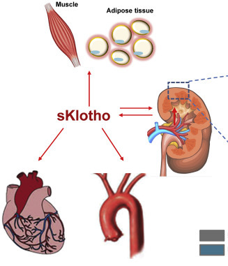
Angiotensin II blockade upregulates the expression of Klotho, the anti-ageing gene, in an experimental model of chronic cyclosporine nephropathy​

Abstract​

Background: The Klotho gene plays a role in suppressing ageing-related disorders. It is suggested that activation of renin-angiotensin system (RAS) or oxidative stress suppresses Klotho in the kidney. This study evaluated the association between Klotho expression and RAS in cyclosporine (CsA)-induced renal injury.
Methods: Chronic CsA nephropathy was induced by administering CsA (30 mg/kg) to mice on a low-salt diet (LSD) for 4 weeks. A normal-salt diet (NSD) was used as the control. Reverse transcription-polymerase chain reaction, western blot and immunohistochemistry were performed for Klotho and intrarenal RAS activity was measured using immunohistochemistry for angiotensinogen and renin. Oxidative stress was measured with urinary excretion of 8-hydroxy-2'-deoxyguanosine (8-OHdG).
Results: CsA treatment decreased Klotho mRNA and protein in mouse kidney in a dose-dependent and time-dependent manner, but a concurrent treatment with losartan, an angiotensin II type 1 (AT1) receptor blocker, reversed the decrease in Klotho expression with histological improvement. This finding was more marked in the LSD than the NSD. Klotho expression was correlated with angiotensinogen and renin expression, tubulointerstitial fibrosis score and urinary 8-OHdG excretion.
Conclusions: Angiotensin II may play a pivotal role in regulating Klotho expression in CsA-induced renal injury. AT1 receptor blocker may inhibit the ageing process by decreasing oxidative stress caused by CsA.

..............

Ray Peat klotho protein (anti-aging) similar to Vitamin D.​


View: https://www.youtube.com/watch?v=JvqgiMuRRRY
 

J.R.K

Member
Joined
Aug 4, 2020
Messages
1,837
Very @coolmd_a!
If my understanding is correct you have just shown a pathway as to how Vitamin D works to protect someone from COVID-19?
Do you believe that Vitamin D will also raise Klotho, or does it just mimic it?
 
OP
md_a

md_a

Member
Joined
Aug 31, 2015
Messages
468
Very @coolmd_a!
If my understanding is correct you have just shown a pathway as to how Vitamin D works to protect someone from COVID-19?
Do you believe that Vitamin D will also raise Klotho, or does it just mimic it?
"The aging suppressing gene discovered in 1997, named after the Greek life-promoting goddess Klotho, suppresses the reabsorption of phosphate by the kidney (which is also a function of the parathyroid hormone), and inhibits the formation of the activated form of vitamin D, opposing the effect of the parathyroid hormone. In the absence of the gene, serum phosphate is high, and the animal ages and dies prematurely. In humans, in recent years a very close association has been has been documented between increased phosphate levels, within the normal range, and increased risk of cardiovascular disease. Serum phosphate is increased in people with osteoporosis (Gallagher, et al., 1980), and various treatments that lower serum phosphate improve bone mineralization, with the retention of calcium phosphate (Ma and Fu, 2010; Batista, et al., 2010; Kelly, et al., 1967; Parfitt, 1965; Kim, et al., 2003)."RP

The Klotho gene and its related protein were identified as a putative aging factors in 1997, when the aging process was aggravated in a group of Klotho knockout mice (1). Klotho is expressed mainly in the kidneys, parathyroid glands, brain choroid plexus, and testes (2-4). Studies have confirmed Klotho expression in other tissues, including the aorta, colon, thyroid gland, and pancreas, but the kidney remains the strongest Klotho-producing organ (5).

There are two types of Klotho: circulating and membrane-bound. The latter functions as a co-receptor for fibroblast growth factor-23 (FGF23). The membrane-bound form, after losing its membrane domain, enters into the circulation as soluble Klotho (sKl), acting as a hormone with anti-aging and anti-oxidative stress properties; sKl can also be directly generated by alterative splicing of the Klotho transcript (2, 5). Klotho deficiency is an early biomarker for chronic kidney disease, and its upregulation could protect the kidney from fibrosis progression (6). The beneficial effect of physical activity in preventing premature mortality has been established by epidemiological studies showing that exercise may delay aging through various mechanisms. Exercise-induced Klotho upregulation could be one explanation. Klotho upregulates nitrous oxide (NO) production and inhibits angiotensin II-induced reactive oxygen species production within endothelial cells (7). In an epidemiological study, handgrip strength, an indicator of total body muscle strength, was correlated with plasma Klotho concentration (8).

Klotho production is affected by many physiological and non-physiological conditions. Angiotensin II downregulates renal Klotho protein expression (23), and AT1R blockade increases circulating Klotho. Conversely, oxidative stress downregulates Klotho production (23).
....

In chronic infectious diseases, low 25(OH)D is often found with elevated levels of the more active metabolite 1,25 dihydroxyvitamin D (1,25D).
1,25 (OH)2D is influenced by PTH and chronic infections, pathogens and aging.
The classic symptoms of vitamin D toxicity are entirely attributable to hypercalcemia. High 1,25 (OH)2D predispose to hypercalcemia.
Macrophages within the granuloma ↑ calcitriol (1,25-[OH]2 vitamin D3) activation → hypercalcemia
Granulomatous disorders (e.g., sarcoidosis): due to increased 1α-hydroxylase activation in epithelioid macrophages → increased 1,25-dihydroxyvitamin D synthesis
......

The majority of hypercalcemia cases in sarcoidosis are explained by the overproduction of 1,25(OH)2D3 (calcitriol) by activated macrophages. Despite quite convincing evidence supporting this hypothesis, some questions have yet to be completely answered. Moreover, some recent studies suggest that vitamin D supplementation may improve not only calcium homeostasis but also the course of sarcoidosis.
Conclusions
Sarcoidosis-associated hypercalcemia is quite a common problem as it affects about 6% of patients. It is also one of the indications to introduce pharmacotherapy with steroids. Its pathophysiology appears to be quite well explained. Although it seems logical that vitamin D metabolites should be good tools for assessing disease activity, clinically it is not that simple. Increased conversion of 25(OH)D3 to calcitriol suggests that perhaps the ratio of 25(OH)D3 to 1,25(OH)2D3 could be more adequate than absolute values. One study confirms this theory.
SAGE Journals: Your gateway to world-class research journals
......

In granulomatous disease hypercalcemia is produced because of the presence of 1α-hydroxylase enzyme in macrophages (6) and giant cells that form part of the granuloma. In the granuloma the 25 (OH) vitamin D is converted to 1,25-(OH)2 vitamin D without any type of homeostatic control.
......

Three types of the Klotho protein are distinguished, i.e., cell membrane-related, intracellular, and secretory forms. Klotho associated with the cell membrane (transmembrane Klotho) can be detected in the kidneys, pituitary gland, inner ear, brain, parathyroid glands, pancreas, large intestine, skeletal muscles, bladder, ovaries, testes, and epithelial breast cells [12]. The largest concentrations are detected in distal convoluted tubules, in the kidney, and in choroid plexus of the brain [1]. Klotho is involved in the renal metabolism of calcium, phosphates, and vitamin D. The membrane Klotho forms a complex with the fibroblast growth factor receptor (FGFR) and provides selective binding affinity to the fibroblast growth factor (FGF) [13]. This complex inhibits phosphate resorption in the proximal tubule of the kidney. In the distal tubule, it also regulates Ca2+ absorption by stabilizing the Ca2+ transient receptor potential vanilloid 5 (TRPV5) channel in the plasma membrane. It inhibits renal 1-alpha 25 hydroxylase activity, thus decreasing the levels of circulating calcitriol. Therefore, hyperphosphatemia, hypercalcemia, elevated plasma calcitriol, vascular calcification, and premature aging can be observed in Klotho-deficient mice [14]. FGF23 leads to the activation of extracellular signal-regulated kinase 1 and 2 (ERK1/2), which activates serum and glucocorticoid-induced kinase 1 (SGK1) in cortical renal tubular cells. SGK1 activates with-no-lysine kinase 4 (WNK 4), stimulating WNK4-TRPV5 complex formation [15].

Klotho has been shown to inhibit 1-α hydroxylase, an enzyme responsible for the production of 1,25-dihydroxyvitamin D3, which is an active form of vitamin D [16]. Lower levels of Klotho in mice induced the formation and toxicity of vitamin D. The toxicity was reduced by limiting the formation of vitamin D [17].

On the molecular level, permanent chronic inflammation, cell proliferation disorders, or cellular aging leads to the formation of a number of age-related chronic diseases, such as obesity, diabetes, atherosclerosis, Alzheimer’s disease (AD), cancer, renal diseases, or degenerative diseases [18]. The secretory Klotho results in the reduction in TNFα and IFNγ, which can show anti-inflammatory properties. The Wnt protein is a signaling molecule that regulates intercellular interactions in the developmental period and in adult tissues. Increased signaling (activity) of Wnt disrupts the function of stem and progenitor cells and leads to cellular aging. Liu et al. [19] demonstrated that Klotho can interact with Wnt, which results in the inhibition of Wnt pathway activity, thus inhibiting the aging process. Cellular aging is also activated by oxidative stress and mitochondrial dysfunction by stimulating the p53/p21 pathways. The p53 protein is a tumor suppressor and can be activated by the ataxia telangiectasia-mutated kinase that, in turn, activates p21, which effectively inhibits cell proliferation [20]. Klotho deficiency results in the overexpression of p53/p21 by inhibiting the formation of new cells and increasing the number of aging cells [21]. Therefore, Klotho supplementation reduces cellular aging by inhibiting the p53/p21 signaling pathway [22].
Klotho protein in neurodegenerative disorders
........

Androgens boost, estrogen inhibits, anti-aging gene Klotho​

...........

Policosanol is antiviral and a potent inducer of anti-aging gene Klotho​

...........

Klotho's Role In Longevity, Intelligence, Muscle Strength, And Cancer


J.R.K

Member
Joined
Aug 4, 2020
Messages
1,837
Klotho upregulates nitrous oxide (NO) production and inhibits angiotensin II-induced reactive oxygen species production within endothelial cells (7). In an epidemiological study, handgrip strength, an indicator of total body muscle strength, was correlated with plasma Klotho concentration (8).

Klotho production is affected by many physiological and non-physiological conditions. Angiotensin II downregulates renal Klotho protein expression (23), and AT1R blockade increases circulating Klotho. Conversely, oxidative stress downregulates Klotho production (23).

Wow! Thanks so much @md_a, you really know how to overdeliver!!
The quote above is one I am a little confused on. Specifically Klotho unregulates NO, I am under the impression that nitric oxide is one thing we needed to keep low and try to minimize, as it can be involved in the cancer metabolism, I am missing a piece of the puzzle there.
The other part was that Angiotensin 2 is if my understanding is correct to be anti inflammatory so I am unclear why Klotho and Angiotensin 2 would be on an opposing field with each other?
But this is why I am here to ask the simple questions that I do not know yet.
 
OP
md_a

md_a

Member
Joined
Aug 31, 2015
Messages
468
Klotho upregulates nitrous oxide (NO) production and inhibits angiotensin II-induced reactive oxygen species production within endothelial cells (7). In an epidemiological study, handgrip strength, an indicator of total body muscle strength, was correlated with plasma Klotho concentration (8).

Klotho production is affected by many physiological and non-physiological conditions. Angiotensin II downregulates renal Klotho protein expression (23), and AT1R blockade increases circulating Klotho. Conversely, oxidative stress downregulates Klotho production (23).

Wow! Thanks so much @md_a, you really know how to overdeliver!!
The quote above is one I am a little confused on. Specifically Klotho unregulates NO, I am under the impression that nitric oxide is one thing we needed to keep low and try to minimize, as it can be involved in the cancer metabolism, I am missing a piece of the puzzle there.
The other part was that Angiotensin 2 is if my understanding is correct to be anti inflammatory so I am unclear why Klotho and Angiotensin 2 would be on an opposing field with each other?
But this is why I am here to ask the simple questions that I do not know yet.
Most likely it's eNOS.

.......
Klotho protein protects against endothelial dysfunction
Abstract
Arteriosclerosis caused by aging is recognized to be a crucial risk factor of cardiovascular disease. We recently established klotho mouse which causes age-related disorders including arteriosclerosis. However, no information on endothelial function of klotho mouse or the physiological role of klotho protein as a circulating factor is available. In this report, we demonstrate that 50% effective dose of aortic relaxation in response to acetylcholine in heterozygous klotho mice is significantly greater (4 x 10(-5) M) than in wild-type mice (8 x 10(-6) M, n = 7, p < 0.05) and that the vasodilator response of arterioles to acetylcholine is significantly attenuated in heterozygous (20% effective dose; 2 x 10(-6) M) and homozygous klotho mice (>1 x 10(-5) M) as compared with wild-type mice (1 x 10(-7) M, n = 7, p < 0.05). Nitric oxide metabolites (NO-2 and NO-3) in urine are significantly lower in heterozygous klotho mice (142 +/- 16 nmol/day) than wild-type mice (241 +/- 28 nmol/day, n = 13, p < 0.05). Parabiosis between wild-type and heterozygous klotho mice results in restoration of endothelial function in heterozygous klotho mice. We conclude that the klotho protein protects the cardiovascular system through endothelium-derived NO production by humoral pathways.

..........
Klotho protein secreted in the blood could act as a hormone to regulate various target organs and have a protective effect on the cardiovascular system. Numerous studies had shown that Klotho protein had antioxidative stress, anti-inflammatory, and antiapoptotic effects on vascular endothelial cells. The purpose of this study was to investigate the protective mechanism of Klotho protein on oxidative damage of vascular endothelial cells induced by H2O2. Klotho protein significantly enhanced human umbilical vein endothelial cells viability and increased the activities of antioxidant enzymes (superoxide dismutase, catalase, and heme oxygenase-1 (HO-1)), scavenged reactive oxygen species, and inhibited tumor necrosis factor alpha and interleukin 6 secretion. Klotho protein also reduced the rate of apoptosis of cells and improved the function of vascular endothelial cells (increased nitric oxide secretion). Klotho protein activated nuclear translocation of Nrf2 and increased HO-1 expression. Klotho protein also activated phosphorylation of protein kinase B (AKT), whereas the addition of LY294002, a pharmacological inhibitor of phosphatidylinositol 3-kinase (PI3K), blocked Klotho-protein-induced Nrf2/HO-1 activation and cytoprotection. Klotho protein enhanced the antioxidant defense ability of the cells by activating the PI3K/AKT pathway, which upregulated the expression of Nrf2/HO-1, thereby inhibiting H2O2-induced oxidative damage.
......

Based on the previous studies proclaiming a potentially positive effect of Klotho in CVD, in 2016 Corsetti et al. assessed that Klotho is expressed in myocardial tissue and proved its association with the prevalence of CVD [2]. The right atrium biopsy samples of thirty patients at high and low risk for atherosclerotic cardiovascular disease (ASCVD) were examined. Research showed that Klotho, β-Klotho, FGF21, and FGF23 proteins are expressed in cardiomyocytes and subjects at higher cardiovascular risk had a reduced expression of cardiac Klotho and an elevated expression of cardiac FGFs [2]. The analysis of cardiomyocytes from patients at high risk for ASCVD also revealed an augmented expression of glucose-regulated protein 78 (GRP78), a marker of endoplasmic reticulum (ER) stress, and superoxide dismutase 1 (SOD1), an oxygen free radicals scavenger [2]. It is also known that high level of nitric oxide (NO) triggers an inflammation in cardiac tissue and disrupts its contractile function [71]. An expression of inducible nitric oxide synthase (iNOS) and subsequent NO formation can be induced by the agents such as inflammatory cytokines and endotoxins. It is also mediated through cytokine-inducible transcription factors like nuclear factor-kappa-B (NF-κB), which is accountable for the inflammatory responses to a variety of signals [72–75]. Therefore, an excessive activation of NF-κB and overexpression of iNOS in cardiomyocytes lead to the cellular dysfunction and damage and, hence, result in the cardiomyopathy, bradyarrhythmia, and sudden cardiac death [72–74]. Importantly, the immunoreactivity of these molecules was increased in cardiomyocytes obtained from subjects at high risk for ASCVD, confirming the active inflammation and apoptosis of these cells [2]. On the contrary, endothelial nitric oxide synthase (eNOS) is expressed constitutively and participates in the vasodilation, blood pressure, vasoprotection, and antiatherosclerotic action [72, 73]. An expression of cytoplasmic eNOS in cardiomyocytes of patients at high risk for ASCVD was significantly decreased and hence led to disruption of cellular energetic metabolisms and mitochondrial biogenesis [2]. Authors also emphasized higher expression of TGF-β1 intensified fibrosis [2]. On this basis, there is a deep suggestion that depletion of Klotho can promote prooxidative, proinflammatory, and proapoptotic activity in cardiomyocytes, leading to their damage in patients at higher risk of cardiovascular diseases. Thus, expression of cardiac Klotho might improve the ability of cells to withstand the stress conditions [2].

There is a link between Klotho deficiency and CKD, renal disorders, and kidney damage.
Several studies mentioned that Klotho can protect the kidneys against decline of their function [76, 77]. In the research including patients with CKD undergoing hemodialysis, an expression of Klotho was significantly reduced. It should be noted that high serum level of Klotho was associated with the low risk of cardiovascular events and death during chronic dialysis [78]. It has become clear that CKD is associated with the cardiovascular episodes, so the repletion of Klotho synthesis could provide renal protection and thus counteracts the accidents of cardiovascular mortality [78, 79].
.............

DISCUSSION
The present observations reveal that dehydration leads to marked downregulation of Klotho transcription and protein expression. Dehydration was followed by the expected increase in plasma osmolarity and stimulation of ADH and aldosterone release. More importantly, the present observations disclose that the two hormones are potent negative regulators of Klotho expression. The downregulation of Klotho transcript levels and protein abundance by ADH and aldosterone contributes to the decline of Klotho transcript levels and protein abundance during dehydration. In a recent study, angiotensin II has been shown to downregulate Klotho expression (24). As angiotensin II is increased during dehydration (4), enhanced release of angiotensin II could well contribute to the decline of Klotho expression following dehydration.

Klotho has previously been shown to be downregulated by reactive oxygen species and NF-κB (31). As formation of reactive oxygen species and the activation of NF-κB may both result from mineralocorticoid receptor activation (6, 8, 27), they could well contribute to or even account for the observed downregulation of Klotho.

Klotho expression is further downregulated by the soluble amyloid precursor protein (23) sirolimus (10), hyperparathyroidism (1), TNF, and INF-γ (39). TNF and IFN-γ are effective by stimulating the expression of inducible nitric oxide synthase (iNOS) with subsequent NO production (39). Klotho expression is upregulated by vitamin D (11), calcitonin gene-related peptide (44), peroxisome proliferator-activated receptor-γ (43), and epidermal growth factor
(2). Uremia has been reported to increase (12) or decrease (17, 42) Klotho expression.

Dehydration further leads to an increase in 1,25(OH)2D3 plasma concentrations, an effect which may be secondary to downregulation of Klotho. Klotho is known to inhibit 1α-hydroxylase and thus to decrease 1,25(OH)2D3 production (29, 40, 41). Accordingly, Klotho deficiency is followed by excessive 1,25(OH)2D3 formation (29, 40, 41). 1,25(OH)2D3 in turn stimulates Klotho expression (11). Thus enhanced 1,25(OH)2D3 plasma levels in dehydrated animals does not contribute to but rather attenuates the downregulation of Klotho expression.

The hormone 1,25(OH)2D3 stimulates intestinal and renal Ca2+ and phosphate transport (28, 30). Thus excess 1,25(OH)2D3 contributes to the increase in plasma Ca2+ (18) and phosphate (30) concentration in Klotho-deficient mice (29, 40, 41). Hyperphosphatemia is considered to be a decisive determinant of life span (19). For instance, CaHPO4 precipitations during hyperphosphatemia could contribute to vascular calcification.
Accordingly, vitamin D restriction reverses the untoward effect of Klotho deficiency on life span (40), growth deficit (40), and erythrocyte survival (16).

The present observations may shed new light on the impact of volume depletion and hyperaldosteronism in Klotho hypomorphic mice (7). The life span of these mice may be substantially extended by saline (7). In theory, hyperaldosteronism of these mice could have led to downregulation of residual Klotho expression, and the decline of plasma aldosterone levels following treatment with saline could have augmented residual Klotho expression in these mice. Alternatively, saline may have been effective more directly by counteracting extracellular volume depletion of the mice.

Dehydration is relatively common in the elderly (32, 36). Decreased water intake or plasma hyperosmolarity impacts body weight (33, 34), development of diabetes (35), and early frailty, incident disability, and mortality (37). The present observations may provide a mechanistic link between dehydration and development of frailty and reduced life span.

In conclusion, dehydration leads to downregulation of Klotho. The effect may accelerate the development of age-related disorders.
....


Nitric oxide: more than just a vasodilator
 

J.R.K

Member
Joined
Aug 4, 2020
Messages
1,837
Most likely it's eNOS.

.......
Klotho protein protects against endothelial dysfunction
Abstract
Arteriosclerosis caused by aging is recognized to be a crucial risk factor of cardiovascular disease. We recently established klotho mouse which causes age-related disorders including arteriosclerosis. However, no information on endothelial function of klotho mouse or the physiological role of klotho protein as a circulating factor is available. In this report, we demonstrate that 50% effective dose of aortic relaxation in response to acetylcholine in heterozygous klotho mice is significantly greater (4 x 10(-5) M) than in wild-type mice (8 x 10(-6) M, n = 7, p < 0.05) and that the vasodilator response of arterioles to acetylcholine is significantly attenuated in heterozygous (20% effective dose; 2 x 10(-6) M) and homozygous klotho mice (>1 x 10(-5) M) as compared with wild-type mice (1 x 10(-7) M, n = 7, p < 0.05). Nitric oxide metabolites (NO-2 and NO-3) in urine are significantly lower in heterozygous klotho mice (142 +/- 16 nmol/day) than wild-type mice (241 +/- 28 nmol/day, n = 13, p < 0.05). Parabiosis between wild-type and heterozygous klotho mice results in restoration of endothelial function in heterozygous klotho mice. We conclude that the klotho protein protects the cardiovascular system through endothelium-derived NO production by humoral pathways.

..........
Klotho protein secreted in the blood could act as a hormone to regulate various target organs and have a protective effect on the cardiovascular system. Numerous studies had shown that Klotho protein had antioxidative stress, anti-inflammatory, and antiapoptotic effects on vascular endothelial cells. The purpose of this study was to investigate the protective mechanism of Klotho protein on oxidative damage of vascular endothelial cells induced by H2O2. Klotho protein significantly enhanced human umbilical vein endothelial cells viability and increased the activities of antioxidant enzymes (superoxide dismutase, catalase, and heme oxygenase-1 (HO-1)), scavenged reactive oxygen species, and inhibited tumor necrosis factor alpha and interleukin 6 secretion. Klotho protein also reduced the rate of apoptosis of cells and improved the function of vascular endothelial cells (increased nitric oxide secretion). Klotho protein activated nuclear translocation of Nrf2 and increased HO-1 expression. Klotho protein also activated phosphorylation of protein kinase B (AKT), whereas the addition of LY294002, a pharmacological inhibitor of phosphatidylinositol 3-kinase (PI3K), blocked Klotho-protein-induced Nrf2/HO-1 activation and cytoprotection. Klotho protein enhanced the antioxidant defense ability of the cells by activating the PI3K/AKT pathway, which upregulated the expression of Nrf2/HO-1, thereby inhibiting H2O2-induced oxidative damage.
......

Based on the previous studies proclaiming a potentially positive effect of Klotho in CVD, in 2016 Corsetti et al. assessed that Klotho is expressed in myocardial tissue and proved its association with the prevalence of CVD [2]. The right atrium biopsy samples of thirty patients at high and low risk for atherosclerotic cardiovascular disease (ASCVD) were examined. Research showed that Klotho, β-Klotho, FGF21, and FGF23 proteins are expressed in cardiomyocytes and subjects at higher cardiovascular risk had a reduced expression of cardiac Klotho and an elevated expression of cardiac FGFs [2]. The analysis of cardiomyocytes from patients at high risk for ASCVD also revealed an augmented expression of glucose-regulated protein 78 (GRP78), a marker of endoplasmic reticulum (ER) stress, and superoxide dismutase 1 (SOD1), an oxygen free radicals scavenger [2]. It is also known that high level of nitric oxide (NO) triggers an inflammation in cardiac tissue and disrupts its contractile function [71]. An expression of inducible nitric oxide synthase (iNOS) and subsequent NO formation can be induced by the agents such as inflammatory cytokines and endotoxins. It is also mediated through cytokine-inducible transcription factors like nuclear factor-kappa-B (NF-κB), which is accountable for the inflammatory responses to a variety of signals [72–75]. Therefore, an excessive activation of NF-κB and overexpression of iNOS in cardiomyocytes lead to the cellular dysfunction and damage and, hence, result in the cardiomyopathy, bradyarrhythmia, and sudden cardiac death [72–74]. Importantly, the immunoreactivity of these molecules was increased in cardiomyocytes obtained from subjects at high risk for ASCVD, confirming the active inflammation and apoptosis of these cells [2]. On the contrary, endothelial nitric oxide synthase (eNOS) is expressed constitutively and participates in the vasodilation, blood pressure, vasoprotection, and antiatherosclerotic action [72, 73]. An expression of cytoplasmic eNOS in cardiomyocytes of patients at high risk for ASCVD was significantly decreased and hence led to disruption of cellular energetic metabolisms and mitochondrial biogenesis [2]. Authors also emphasized higher expression of TGF-β1 intensified fibrosis [2]. On this basis, there is a deep suggestion that depletion of Klotho can promote prooxidative, proinflammatory, and proapoptotic activity in cardiomyocytes, leading to their damage in patients at higher risk of cardiovascular diseases. Thus, expression of cardiac Klotho might improve the ability of cells to withstand the stress conditions [2].

There is a link between Klotho deficiency and CKD, renal disorders, and kidney damage.
Several studies mentioned that Klotho can protect the kidneys against decline of their function [76, 77]. In the research including patients with CKD undergoing hemodialysis, an expression of Klotho was significantly reduced. It should be noted that high serum level of Klotho was associated with the low risk of cardiovascular events and death during chronic dialysis [78]. It has become clear that CKD is associated with the cardiovascular episodes, so the repletion of Klotho synthesis could provide renal protection and thus counteracts the accidents of cardiovascular mortality [78, 79].
.............

DISCUSSION
The present observations reveal that dehydration leads to marked downregulation of Klotho transcription and protein expression. Dehydration was followed by the expected increase in plasma osmolarity and stimulation of ADH and aldosterone release. More importantly, the present observations disclose that the two hormones are potent negative regulators of Klotho expression. The downregulation of Klotho transcript levels and protein abundance by ADH and aldosterone contributes to the decline of Klotho transcript levels and protein abundance during dehydration. In a recent study, angiotensin II has been shown to downregulate Klotho expression (24). As angiotensin II is increased during dehydration (4), enhanced release of angiotensin II could well contribute to the decline of Klotho expression following dehydration.

Klotho has previously been shown to be downregulated by reactive oxygen species and NF-κB (31). As formation of reactive oxygen species and the activation of NF-κB may both result from mineralocorticoid receptor activation (6, 8, 27), they could well contribute to or even account for the observed downregulation of Klotho.

Klotho expression is further downregulated by the soluble amyloid precursor protein (23) sirolimus (10), hyperparathyroidism (1), TNF, and INF-γ (39). TNF and IFN-γ are effective by stimulating the expression of inducible nitric oxide synthase (iNOS) with subsequent NO production (39). Klotho expression is upregulated by vitamin D (11), calcitonin gene-related peptide (44), peroxisome proliferator-activated receptor-γ (43), and epidermal growth factor
(2). Uremia has been reported to increase (12) or decrease (17, 42) Klotho expression.

Dehydration further leads to an increase in 1,25(OH)2D3 plasma concentrations, an effect which may be secondary to downregulation of Klotho. Klotho is known to inhibit 1α-hydroxylase and thus to decrease 1,25(OH)2D3 production (29, 40, 41). Accordingly, Klotho deficiency is followed by excessive 1,25(OH)2D3 formation (29, 40, 41). 1,25(OH)2D3 in turn stimulates Klotho expression (11). Thus enhanced 1,25(OH)2D3 plasma levels in dehydrated animals does not contribute to but rather attenuates the downregulation of Klotho expression.

The hormone 1,25(OH)2D3 stimulates intestinal and renal Ca2+ and phosphate transport (28, 30). Thus excess 1,25(OH)2D3 contributes to the increase in plasma Ca2+ (18) and phosphate (30) concentration in Klotho-deficient mice (29, 40, 41). Hyperphosphatemia is considered to be a decisive determinant of life span (19). For instance, CaHPO4 precipitations during hyperphosphatemia could contribute to vascular calcification.
Accordingly, vitamin D restriction reverses the untoward effect of Klotho deficiency on life span (40), growth deficit (40), and erythrocyte survival (16).

The present observations may shed new light on the impact of volume depletion and hyperaldosteronism in Klotho hypomorphic mice (7). The life span of these mice may be substantially extended by saline (7). In theory, hyperaldosteronism of these mice could have led to downregulation of residual Klotho expression, and the decline of plasma aldosterone levels following treatment with saline could have augmented residual Klotho expression in these mice. Alternatively, saline may have been effective more directly by counteracting extracellular volume depletion of the mice.

Dehydration is relatively common in the elderly (32, 36). Decreased water intake or plasma hyperosmolarity impacts body weight (33, 34), development of diabetes (35), and early frailty, incident disability, and mortality (37). The present observations may provide a mechanistic link between dehydration and development of frailty and reduced life span.

In conclusion, dehydration leads to downregulation of Klotho. The effect may accelerate the development of age-related disorders.
....


Nitric oxide: more than just a vasodilator
This is great stuff @md_a, I cannot say that I grasp it completely yet, but it is a good thing that it should not make any sense until it makes complete sense. What I take away from this is that much of the things that raise Klotho are covered in Dr Peats thoughts, vitamin D3, salt to cut aldosterone, higher calcium than phosphate ratios, to keep parathyroid in check,Aspirin, vitamin E, progesterone, to keep the inflammatory mediators down, the B vitamins to help keep prolactin down as well as clearing ammonia, cutting inflammation. Stress trying to keep it to as low as life will allow to avoid activation of the RAS. All these things contribute to the raising of Klotho thereby contributing to longevity. If I am off on this please feel free to redirect my thought process to another pathway, no harm on my end I am happy to learn more.
 

J.R.K

Member
Joined
Aug 4, 2020
Messages
1,837
OP
md_a

md_a

Member
Joined
Aug 31, 2015
Messages
468
This is great stuff @md_a, I cannot say that I grasp it completely yet, but it is a good thing that it should not make any sense until it makes complete sense. What I take away from this is that much of the things that raise Klotho are covered in Dr Peats thoughts, vitamin D3, salt to cut aldosterone, higher calcium than phosphate ratios, to keep parathyroid in check,Aspirin, vitamin E, progesterone, to keep the inflammatory mediators down, the B vitamins to help keep prolactin down as well as clearing ammonia, cutting inflammation. Stress trying to keep it to as low as life will allow to avoid activation of the RAS. All these things contribute to the raising of Klotho thereby contributing to longevity. If I am off on this please feel free to redirect my thought process to another pathway, no harm on my end I am happy to learn more.
You have a very good summary. In theory it is simple but in practice it is more complicated due to pollution, plastic, pesticides, radiation, vaccination .... there are strong blockages for those who want to get a better quality of life. I think that the most natural and unprocessed food is basic, such as fruits, fruit juice, raw milk ..., potato juice (reduces ammonia), exposure of the body to sunlight and adequate movements to stimulate muscle mass. From what you have written, it is also important to limit PUFA as much as possible, through which I think it is the central mechanism for viruses / toxins to penetrate cells.

.....

COVID-19/SARS may be due simply to serum PUFA and its peroxidation

..........

RDS and linoleic acid
Adult/Acute Respiratory Distress Syndrome is topical at the moment. In the comments to the last post I wondered whether omega six fatty acids, especially linoleic acid, might be a driver of ARDS, which is one of the most intractable ITU problems in response to major infection/trauma/inflammatory insults.
Tucker came up with this abstract
Plasma fatty acid changes and increased lipid peroxidation in patients with adult respiratory distress syndrome
and I peeked at the related papers to find this gem:
An increase in serum C18 unsaturated free fatty acids as a predictor of the development of acute respiratory distress syndrome
Again, only an abstract and mostly describing a pilot study. But here is the critical statement:
"Increases in unsaturated serum acyl chain ratios differentiate between healthy and seriously iII patients, and identify those patients likely to develop ARDS".
That is, the more linoleic (and oleic) acid you have as FFAs in your bloodstream, relative to my beloved palmitic acid, the more likely you are to develop ARDS. Which carries a high risk of death.
That was 1996. The work will have been done before that, so we have known that linoileic acid is bad news for well over 20 years.
If you are a Standard American on the Standard American Diet, or anyone else in the world poisoned by a cardiologist-promoted PUFA based diet, any weight loss through illness will release significant amounts of linoleic acid from your adipocytes. That might just trigger ARDS in the aftermath of a viral pneumonia.
There's a lot of it about.
...........

Upon the analysis of the molecular structure of the spike protein, the team has found something interesting. They revealed the presence of a small molecule, called linoleic acid (LA), which was discovered in a customized pocket within the spike protein.
"We were truly puzzled by our discovery and its implications. So here we have LA, a molecule which is at the center of those functions that go haywire in COVID-19 patients, with terrible consequences. And the virus that is causing all this chaos, according to our data, grabs and holds on to exactly this molecule – basically disarming much of the body's defenses," Professor Berger said.
The researchers also explained that in other illnesses, interfering with the metabolic pathways of the linoleic acid can induce systemic inflammation, pneumonia, and acute respiratory distress syndrome (ARDS). All these health problems are seen in patients with COVID-19. The study findings provide the first direct link between LA, the health consequences, and the virus itself.
For instance, in the common cold virus, rhinovirus, scientists also discovered a similar pocket to develop molecules that attached firmly to the pocket to disrupt the structure, stemming from its infectivity.
...........

Role of arachidonic cascade in COVID-19 infection
Abstract
The World Health Organization has described the 2019 Coronavirus disease caused by an influenza-like virus called SARS-CoV-2 as a pandemic. Millions of people worldwide are already infected by this virus, and severe infection causes hyper inflammation, thus disrupting lung function, exacerbating breath difficulties, and death. Various inflammatory mediators bio-synthesized through the arachidonic acid pathway play roles in developing cytokine storms, injuring virus-infected cells. Since pro-inflammatory eicosanoids, including prostaglandins, and leukotrienes, are key brokers for physiological processes such as inflammation, fever, allergy, and pain but, their function in COVID-19 is not well defined. This study addresses eicosanoid's crucial role through the arachidonic pathway in inflammatory cascading and recommends using bioactive lipids, NSAIDs, steroids, cell phospholipase A2 (cPLA2) inhibitors, and specialized pro-resolving mediators (SPMs) to treat COVID-19 disease. The role of soluble epoxide hydrolase inhibitors (SEHIs) in promoting the activity of epoxyeicosatrienoic acids (EETs) and 17-hydroxide-docosahexaenoic acid (17-HDHA) is also discussed. Additional research that assesses the eicosanoid profile in COVID-19 patients or preclinical models generates novel insights into coronavirus-host interaction and inflammation regulation.
Conclusion
COVID-19 disease caused by the influenza virus is highly contagious and rapidly transmitted worldwide and has been declared by WHO as a pandemic. After insertion into the host alveolar epithelium, the SARS-CoV-2 virus can develop a severe inflammatory state that ultimately disrupts the alveolar function and develops the severe COVID-19 disease condition. The hyperinflammatory state develops through the hyperactivity of the biosynthetic pathway of synthesizing different inflammatory cytokines from AA. We can hypothesize different interventions for the treatment of COVID-19 disease by blocking or promoting different steps of the AA cascade. Some clinical studies on some NSAIDs and steroids found success in reducing the case severity and morbidity rate of COVID-19 disease. Developing specific small-molecule for inhibiting cPLA2 is another COVID-19 management strategy as it reduces viral RNA synthesis. SPM precursors, including 17-HDHA, and EETs, can consider managing the inflammatory condition in COVID-19 disease. Judging by the current study, further study can be started targeting specific molecules in the AA cascade to manage COVID-19 disease.
 

J.R.K

Member
Joined
Aug 4, 2020
Messages
1,837
You have a very good summary. In theory it is simple but in practice it is more complicated due to pollution, plastic, pesticides, radiation, vaccination .... there are strong blockages for those who want to get a better quality of life. I think that the most natural and unprocessed food is basic, such as fruits, fruit juice, raw milk ..., potato juice (reduces ammonia), exposure of the body to sunlight and adequate movements to stimulate muscle mass. From what you have written, it is also important to limit PUFA as much as possible, through which I think it is the central mechanism for viruses / toxins to penetrate cells.

.....

COVID-19/SARS may be due simply to serum PUFA and its peroxidation

..........

RDS and linoleic acid
Adult/Acute Respiratory Distress Syndrome is topical at the moment. In the comments to the last post I wondered whether omega six fatty acids, especially linoleic acid, might be a driver of ARDS, which is one of the most intractable ITU problems in response to major infection/trauma/inflammatory insults.
Tucker came up with this abstract
Plasma fatty acid changes and increased lipid peroxidation in patients with adult respiratory distress syndrome
and I peeked at the related papers to find this gem:
An increase in serum C18 unsaturated free fatty acids as a predictor of the development of acute respiratory distress syndrome
Again, only an abstract and mostly describing a pilot study. But here is the critical statement:
"Increases in unsaturated serum acyl chain ratios differentiate between healthy and seriously iII patients, and identify those patients likely to develop ARDS".
That is, the more linoleic (and oleic) acid you have as FFAs in your bloodstream, relative to my beloved palmitic acid, the more likely you are to develop ARDS. Which carries a high risk of death.
That was 1996. The work will have been done before that, so we have known that linoileic acid is bad news for well over 20 years.
If you are a Standard American on the Standard American Diet, or anyone else in the world poisoned by a cardiologist-promoted PUFA based diet, any weight loss through illness will release significant amounts of linoleic acid from your adipocytes. That might just trigger ARDS in the aftermath of a viral pneumonia.
There's a lot of it about.
...........

Upon the analysis of the molecular structure of the spike protein, the team has found something interesting. They revealed the presence of a small molecule, called linoleic acid (LA), which was discovered in a customized pocket within the spike protein.
"We were truly puzzled by our discovery and its implications. So here we have LA, a molecule which is at the center of those functions that go haywire in COVID-19 patients, with terrible consequences. And the virus that is causing all this chaos, according to our data, grabs and holds on to exactly this molecule – basically disarming much of the body's defenses," Professor Berger said.
The researchers also explained that in other illnesses, interfering with the metabolic pathways of the linoleic acid can induce systemic inflammation, pneumonia, and acute respiratory distress syndrome (ARDS). All these health problems are seen in patients with COVID-19. The study findings provide the first direct link between LA, the health consequences, and the virus itself.
For instance, in the common cold virus, rhinovirus, scientists also discovered a similar pocket to develop molecules that attached firmly to the pocket to disrupt the structure, stemming from its infectivity.
...........

Role of arachidonic cascade in COVID-19 infection
Abstract
The World Health Organization has described the 2019 Coronavirus disease caused by an influenza-like virus called SARS-CoV-2 as a pandemic. Millions of people worldwide are already infected by this virus, and severe infection causes hyper inflammation, thus disrupting lung function, exacerbating breath difficulties, and death. Various inflammatory mediators bio-synthesized through the arachidonic acid pathway play roles in developing cytokine storms, injuring virus-infected cells. Since pro-inflammatory eicosanoids, including prostaglandins, and leukotrienes, are key brokers for physiological processes such as inflammation, fever, allergy, and pain but, their function in COVID-19 is not well defined. This study addresses eicosanoid's crucial role through the arachidonic pathway in inflammatory cascading and recommends using bioactive lipids, NSAIDs, steroids, cell phospholipase A2 (cPLA2) inhibitors, and specialized pro-resolving mediators (SPMs) to treat COVID-19 disease. The role of soluble epoxide hydrolase inhibitors (SEHIs) in promoting the activity of epoxyeicosatrienoic acids (EETs) and 17-hydroxide-docosahexaenoic acid (17-HDHA) is also discussed. Additional research that assesses the eicosanoid profile in COVID-19 patients or preclinical models generates novel insights into coronavirus-host interaction and inflammation regulation.
Conclusion
COVID-19 disease caused by the influenza virus is highly contagious and rapidly transmitted worldwide and has been declared by WHO as a pandemic. After insertion into the host alveolar epithelium, the SARS-CoV-2 virus can develop a severe inflammatory state that ultimately disrupts the alveolar function and develops the severe COVID-19 disease condition. The hyperinflammatory state develops through the hyperactivity of the biosynthetic pathway of synthesizing different inflammatory cytokines from AA. We can hypothesize different interventions for the treatment of COVID-19 disease by blocking or promoting different steps of the AA cascade. Some clinical studies on some NSAIDs and steroids found success in reducing the case severity and morbidity rate of COVID-19 disease. Developing specific small-molecule for inhibiting cPLA2 is another COVID-19 management strategy as it reduces viral RNA synthesis. SPM precursors, including 17-HDHA, and EETs, can consider managing the inflammatory condition in COVID-19 disease. Judging by the current study, further study can be started targeting specific molecules in the AA cascade to manage COVID-19 disease.
That is something that I have been wondering about for quite awhile now. I personally have been trying to be vigilant on being aware and avoiding PUFA to the best of my abilities for five plus years now. But given the wide range of severity of the symptoms in COVID-19 and knowing a little about the chronic inflammatory diseases they can lead to anything from diabetes to cancer, I have thought what if it isn’t just the general health and comorbidities that are the issue, what if the levels of PUFA are one the main determining factors that drive the levels of severity of the symptoms? What if elimination or reducing PUFA would actually be a life saving intervention to help those most at risk of being a COVID-19 victim?
Thank you for all this information, my friend!
 
OP
md_a

md_a

Member
Joined
Aug 31, 2015
Messages
468
That is something that I have been wondering about for quite awhile now. I personally have been trying to be vigilant on being aware and avoiding PUFA to the best of my abilities for five plus years now. But given the wide range of severity of the symptoms in COVID-19 and knowing a little about the chronic inflammatory diseases they can lead to anything from diabetes to cancer, I have thought what if it isn’t just the general health and comorbidities that are the issue, what if the levels of PUFA are one the main determining factors that drive the levels of severity of the symptoms? What if elimination or reducing PUFA would actually be a life saving intervention to help those most at risk of being a COVID-19 victim?
Thank you for all this information, my friend!
From what I have read and experienced I can say for sure that PUFA is the basis of health problems.
FB_IMG_1619410474612.jpg
 

J.R.K

Member
Joined
Aug 4, 2020
Messages
1,837
From what I have read and experienced I can say for sure that PUFA is the basis of health problems.View attachment 23019
You almost would think that they are trying to reduce population on a global level wouldn’t you?
I am not much on the conjecture side of things but I do listen to all sides of the arguments, no matter how wild they might seem.
But I do agree that the PUFA intakes in the modern diet not just the western diet as I believe these seed based oils are something that are used world wide with the exception of a few indigenous tribes. These oils I am of the mind they play as equal of a role as the stress, pollution, compromised food, and the political establishment in the health of the human population.
Perhaps if there is a potential silver lining to this pandemic and the dangers of these products will be brought to the forefront of the medical establishment and the health advocates that promote them.
 
OP
md_a

md_a

Member
Joined
Aug 31, 2015
Messages
468
You almost would think that they are trying to reduce population on a global level wouldn’t you?
I am not much on the conjecture side of things but I do listen to all sides of the arguments, no matter how wild they might seem.
But I do agree that the PUFA intakes in the modern diet not just the western diet as I believe these seed based oils are something that are used world wide with the exception of a few indigenous tribes. These oils I am of the mind they play as equal of a role as the stress, pollution, compromised food, and the political establishment in the health of the human population.
Perhaps if there is a potential silver lining to this pandemic and the dangers of these products will be brought to the forefront of the medical establishment and the health advocates that promote them.
"...Michael Yeadon [former Pfizer vp]... says from what's known about the spike protein & the mRNA so-called vaccines; it's very probable that they could have long-range degenerative effects... One of the possibilities is that it's intended to depopulate the world." — Ray Peat

What is happening in society for many years and lately more accelerated is not normal, it is visible that it is part of a mafia plan, related to dehumanization, digital control, infertility, extreme experiments on children, depopulation, financial benefits. For me, these trends are visibly clear, I am unpleasantly surprised that the mob plan is easily accepted as the new normal by most people, especially by the young.
 

Regina

Member
Joined
Aug 17, 2016
Messages
6,511
Location
Chicago
"...Michael Yeadon [former Pfizer vp]... says from what's known about the spike protein & the mRNA so-called vaccines; it's very probable that they could have long-range degenerative effects... One of the possibilities is that it's intended to depopulate the world." — Ray Peat

What is happening in society for many years and lately more accelerated is not normal, it is visible that it is part of a mafia plan, related to dehumanization, digital control, infertility, extreme experiments on children, depopulation, financial benefits. For me, these trends are visibly clear, I am unpleasantly surprised that the mob plan is easily accepted as the new normal by most people, especially by the young.
+1
 
EMF Mitigation - Flush Niacin - Big 5 Minerals

Similar threads

Back
Top Bottom