How Coronavirus Kills: Acute Respiratory Distress Syndrome (ARDS)

OP
md_a

md_a

Member
Joined
Aug 31, 2015
Messages
468
Clove oil inhibited 97.3% lipid peroxidation of linoleic acid emulsion at 15 μg/mL concentration. However, under the same conditions, the standard antioxidant compounds such as butylated hydroxyanisole (BHA), butylated hydroxytoluene (BHT), α-tocopherol and trolox demonstrated inhibition of 95.4, 99.7, 84.6 and 95.6% on peroxidation of linoleic acid emulsion at 45 μg/mL concentration, respectively. In addition, clove oil had an effective DPPH scavenging, ABTS+ scavenging, superoxide anion radical scavenging, hydrogen peroxide scavenging, ferric ions (Fe3+) reducing power and ferrous ions (Fe2+) chelating activities.

Antioxidant activity of clove oil – A powerful antioxidant source - ScienceDirect


Eugenol: a dual inhibitor of platelet-activating factor and arachidonic acid metabolism.

Eugenol is an active principal and responsible for several pharmacological activities of clove oil. We studied the effects of eugenol on human platelet aggregation, arachidonic acid (AA) and platelet-activating factor (PAF) metabolism and in vivo effects on AA and PAF-induced shock in rabbits. Eugenol strongly inhibited PAF-induced platelet aggregation with lesser effect against AA and collegen. The IC(50) values were against AA: 31 ± 0.5; collagen: 64 ± 0.7 and PAF 7 ± 0.2 μM (n=9) respectively. In addition, eugenol stimulated PAF-acetylhydrolase activity suggesting that inhibition of PAF could be due to its inactivation to lyso-PAF. Pretreatment of rabbits with eugenol (50-100 mg/kg) prevented the lethal effects of intravenous PAF (11 μgg/kg) or AA (2 mg/kg) in a dose-dependent fashion. The protective effects of eugenol in the rabbits, however, were more pronounced against PAF-induced mortality (100% protection). In addition, eugenol also inhibited AA metabolism via cyclooxygenase and lipoxygenase pathways in human platelets. Both the production of thromboxane-A(2) and 12-hydroxy-eicosatetraenoic acid was inhibited by eugenol in a concentration-related manner (30-120 μM). In vivo, eugenol (50-100 mg/kg; i.p.) inhibited carrageenan-induced rat paw oedema (P < 0.001). In this test, eugenol was 5 times more potent than aspirin. These results provide evidence that eugenol acts as a dual antagonist of AA and PAF.

Eugenol: a dual inhibitor of platelet-activating factor and arachidonic acid metabolism. - PubMed - NCBI
 

RealNeat

Member
Joined
Jan 9, 2019
Messages
2,377
Location
HI
I think that there are situations in which mechanical ventilation is the only way to prevent certain death, but in pneumonia or COPD there are gentler treatments that should be tried first: T3, pregnenolone, progesterone, vitamin D and others work in a more coherent way.

Ray Peat talked about COPD in a KMUD interview.
Current Trends In Nitric Oxide, KMUD, 2015
Do you have a really good study or proof of vitamin D in supplement form actually benefitting this thing or any other pneumonia related condition?
 
Joined
Dec 18, 2018
Messages
2,206
Do you have a really good study or proof of vitamin D in supplement form actually benefitting this thing or any other pneumonia related condition?


Im dumping abstracts to this important question,you and me want to know if supplemental D works,
it will be about that and about natural occurence D.


Relationship between vitamin D levels and outcome of pneumonia in children
AO Oduwole, JK Renner, E Disu, E Ibitoye, E Emokpae
West African journal of medicine 29 (6), 2010
ABSTRACT BACKGROUND: Pneumonia, a common childhood infection in Nigerian children with a number of debilitating complications such as empyema thoracis, has been linked to vitamin D deficiency due to its ability to modulate the T lymphocyte of the immune system.
OBJECTIVE: To determine the relationship between vitamin D and outcome of pneumonia in children. METHODS: This was a case-control study involving 24 children, admitted for pneumonia as subjects and 10 children without pneumonia as controls. Pre-formatted questionnaire was utilized to obtain background information, anthropometric measurements were made to determine nutritional status and estimation of 25-hydroxy cholecalciferol (25OHD) done for all those studied. RESULTS: The mean (SD) serum 25OHD concentration was 104 (59) nmol/L and 130 (107) nmol/L for subjects and controls respectively. Amongst the subjects 15 (54%) had serum 25OHD less than 70nmol/L and 11 (46%) serum 25OHD greater than70nmol/L. Hypocalcaemia was present in 15 (54%) of the subjects. Further analysis of hypocalcaemia with regards to the concentration of serum 25OHD showed that 2 (13%) had levels below 40nmol/L, 10 (67%) had levels below 70nmol/L and 3 (20%) above 70nmol/L. Hypocalcaemia was more frequent among subjects with 25OHD below 70nmol/L compared with those above70nmol/L,(p= 0.01). Empyema thoracis and death occurred amongst the two subjects with 25OHD between 27.5 and 40nmol/L. Anaemia was more frequent among subjects with 25OHD below 70nmol/L compared with those above70nmol/L (p= 0.03). CONCLUSION: The study showed that Vitamin D insufficiency, and not solely its deficiency, may have an important role to play in the immune and haemopoetic system. It may therefore affect the response of a child to infections especially pneumonia. WAJM 2010; 29 (6): 373–378.


Effects of vitamin D supplementation to children diagnosed with pneumonia in Kabul: a randomised controlled trial
Semira Manaseki‐Holland, Ghulam Qader, Mohammad Isaq Masher, Jane Bruce, M Zulf Mughal, Daniel Chandramohan, Gijs Walraven
Tropical Medicine & International Health 15 (10), 1148-1155, 2010
Objectives  To determine whether (i) supplementation of oral 100 000 iu of vitamin D3 (cholecalciferol) along with antibiotics will reduce the duration of illness in children with pneumonia; (ii) supplementation will reduce the risk of repeat episodes.
Methods  Double‐blind individually randomised placebo‐controlled trial in an inner‐city hospital in Kabul, of 453 children aged 1–36 months, diagnosed with non‐severe or severe pneumonia at the outpatient clinic. Children with rickets, other concurrent severe diseases, very severe pneumonia or wheeze, were excluded. Children were given vitamin D3 or placebo drops additional to routine pneumonia treatment.
Results  Two hundred and twenty‐four children received vitamin D3; and 229 received placebo. There was no significant difference in the mean number of days to recovery between the vitamin D3 (4.74 days; SD 2.22) and placebo arms (4.98 days; SD 2.89; P  = 0.17). The risk of a repeat episode of pneumonia within 90 days of supplementation was lower in the intervention (92/204; 45%) than the placebo group [122/211; (58%; relative risk 0.78; 95% CI 0.64, 0.94; P = 0.01]. Children in the vitamin D3 group survived longer without experiencing a repeat episode (72 days vs. 59 days; HR 0.71; 95% CI 0.53–0.95; P = 0.02).
Conclusion  A single high‐dose oral vitamin D3 supplementation to young children along with antibiotic treatment for pneumonia could reduce the occurrence of repeat episodes of pneumonia.


Is ventilator-associated pneumonia in trauma patients an epiphenomenon or a cause of death?
Louis J Magnotti, Martin A Croce, Timothy C Fabian
Surgical infections 5 (3), 237-242, 2004
Background: Ventilator-associated pneumonia (VAP) is a common infection among patients in trauma intensive care units (ICUs). It has been suggested by different investigators that VAP is an indicator of injury severity and not necessarily associated with mortality. Crude mortality rates approximating 20% have been reported for trauma patients with VAP. Most studies have involved the most severely injured patients, making it difficult to determine the relative contribution of either VAP or injury severity to death. If VAP is independently associated with mortality, this relationship should be most evident in less severely injured patients. We studied patients with less severe injuries (Injury Severity Score, ISS < 25) to determine the impact of VAP on outcomes.
Methods: Patients admitted to the trauma ICU with ISS < 25 were identified from the trauma registry of a level I trauma center. Patients with penetrating injuries and those who died within 48 h of injury were excluded. Pneumonia was diagnosed using quantitative cultures of bronchoalveolar lavage effluent (≥ 105 colony forming units/mL). Risk factors for VAP, including age, transfusions with 24 h of admission, brain injury, and chest injury severity were analyzed. Logistic regression analysis was then performed to determine independent factors for death.Results: There were 15,492 blunt admissions over a 5.5 year study period who survived >48 h. Of these, 5,860 (38%) were admitted to the ICU, and 4,111 (70% of ICU admissions) had ISS < 25. The incidence of VAP in this group was 8%. Patients with VAP were older (47 vs 39 years), had more transfusions within 24 h (2.5 vs 0.9 units of red blood cell concentrates) and had greater injury severity by ISS (16.7 vs 12.6 points), GCS (Glasgow Coma Scale) score (11.8 vs. 13.7 points) and chest AIS (Abbreviated Injury Scale) (1.7 vs 0.9 points; all p < 0.001). Overall mortality was 4%. Mortality was 16% in patients with VAP compared to 3% in those without VAP (p < 0.0001). Logistic regression analysis identified transfusions, age, and VAP as independent predictors of mortality. Other descriptors of injury severity (ISS, GCS, or chest AIS) were not associated with death.
Results: There were 15,492 blunt admissions over a 5.5 year study period who survived . 48 h. Of these, 5,860 (38%) were admitted to the ICU, and 4,111 (70% of ICU admissions) had ISS , 25. The incidence of VAP in this group was 8%. Patients with VAP were older (47 vs 39 years), had more transfusions within 24 h (2.5 vs 0.9 units of red blood cell concentrates) and had greater injury severity by ISS (16.7 vs 12.6 points), GCS (Glasgow Coma Scale) score (11.8 vs. 13.7 points) and chest AIS (Abbreviated Injury Scale) (1.7 vs 0.9 points; all p , 0.001). Overall mortality was 4%. Mortality was 16% in patients with VAP compared to 3% in those without VAP (p , 0.0001). Logistic regression analysis identified transfusions, age, and VAP as independent predictors of mortality. Other descriptors of injury severity (ISS, GCS, or chest AIS) were not associated with death.
Conclusions: Ventilator-associated pneumonia is independently associated with death in less severely injured trauma patients. This demonstrates the need for effective diagnostic techniques so that adequate therapy may be initiated. Prevention of VAP in less severely injured trauma patients should increase survival.


Vitamin D Status and Community-Acquired Pneumonia: Results from the Third National Health and Nutrition Examination Survey
Abstract
Objective
To investigate the association between serum 25-hydroxyvitamin D [25(OH)D] level and history of community-acquired pneumonia (CAP).

Patients and Methods
We identified 16,975 individuals (≥17 years) from the third National Health and Nutrition Examination Survey (NHANES III) with documented 25(OH)D levels. To investigate the association of 25(OH)D with history of CAP in these participants, we developed a multivariable logistic regression model, adjusting for demographic factors (age, sex, race, poverty-to-income ratio, and geographic location), clinical data (body mass index, smoking status, asthma, chronic obstructive pulmonary disease, congestive heart failure, diabetes mellitus, stroke, chronic kidney disease, neutropenia, and alcohol consumption), and season. Locally weighted scatterplot smoothing (LOWESS) was used to depict the relationship between increasing 25(OH)D levels and the cumulative frequency of CAP in the study cohort.

Results
The median [interquartile range (IQR)] serum 25(OH)D level was 24 (IQR 18–32) ng/mL. 2.1% [95% confidence interval (CI): 1.9–2.3] of participants reported experiencing a CAP within one year of their participation in the national survey. After adjusting for demographic factors, clinical data, and season, 25(OH)D levels <30 ng/mL were associated with 56% higher odds of CAP [odds ratio 1.56; 95% confidence interval: 1.17–2.07] compared to levels ≥30 ng/mL. LOWESS analysis revealed a near linear relationship between vitamin D status and the cumulative frequency of CAP up to 25(OH)D levels around 30 ng/mL.

Conclusion
Among 16,975 participants in NHANES III, 25(OH)D levels were inversely associated with history of CAP. Randomized controlled trials are warranted to determine the effect of optimizing vitamin D status on the risk of CAP.


Vitamin D Status and Acute Respiratory Infection: Cross Sectional Results From the United States National Health and Nutrition Examination Survey, 2001-2006
Dominique J Monlezun 1 2 , Edward A Bittner 3 4 , Kenneth B Christopher 5 6 , Carlos A Camargo 7 8 9 , Sadeq A Quraishi 10 11
Affiliations

Abstract
Vitamin D is a promising, though under-explored, potential modifiable risk factor for acute respiratory infections (ARIs). We sought to investigate the association of vitamin D status with ARI in a large, nationally-representative sample of non-institutionalized individuals from the United States. We analyzed 14,108 individuals over 16 years of age in the National Health and Nutrition Survey (NHANES) 2001-2006 in this cross-sectional study. We used locally weighted scatterplot smoothing (LOWESS) to depict the relationship between increasing 25-hydroxyvitamin D (25OHD) levels and ARI. We then performed a multivariable regression analysis to investigate the association of 25OHD levels with ARI, while adjusting for known confounders. The median serum 25OHD level was 21 (IQR 15-27) ng/mL. Overall, 4.8% (95% CI: 4.5-5.2) of participants reported an ARI within 30 days before their participation in the national survey. LOWESS analysis revealed a near-linear relationship between vitamin D status and the cumulative frequency of ARI up to 25OHD levels around 30 ng/mL. After adjusting for season, demographic factors, and clinical data, 25OHD levels <30 ng/mL were associated with 58% higher odds of ARI (OR 1.58; 95% CI: 1.07-2.33) compared to levels ≥30 ng/mL. Among the 14,108 participants in NHANES 2001-2006, 25OHD levels were inversely associated with ARI. Carefully designed, randomized, controlled trials are warranted to determine the effect of optimizing vitamin D status on the risk of ARI


Effect of Vitamin D Supplementation on Procalcitonin as Prognostic Biomarker in Patients with Ventilator Associated Pneumonia Complicated with Vitamin D Deficiency
Abstract
Ventilator-associated pneumonia (VAP) is a common and serious problem that develops after more than 48 h of mechanical ventilation. Improving the activity of immune system with vitamin D, and its consequent impact on prognostic biomarkers of VAP was studied in the current study.

A randomized double blind placebo controlled clinical trial was designed. A total of 46 patients with VAP, who were suffering from vitamin D deficiency, were randomly allocated into the study groups of placebo (n=22) and treatment (n=24) The treatment group received 300,000 units of intramuscular vitamin D. Serum levels of procalcitonin and vitamin D along with SOFA and CPIS scores were determined at baseline and on day 7 after intervention. The mortality rate of patients was also monitored for the succeeding 28 days after the injection.

The administration of vitamin D significantly enhanced its levels (P<0.0001) in the treated patients (12.28 ± 8.26) in comparison to placebo group (1.15 ± 1.50). The levels of PCT were significantly decreased (p=0.001) in the treatment group (– 0.02 ± 0.59 ng/mL) compared to that of placebo group (0.68 ± 1.03 ng/mL). However, changes in (SOFA) and CPIS scores were not significantly different between study groups (p=0.63 and p=0.32, respectively). Interestingly, the mortality rate of patients in the treatment group (5/24) was significantly lower (p=0.04) than that of the placebo group (11/22).

In conclusion, our results indicate that vitamin D supplementation can significantly reduce the procalcitonin in (VAP) patients, and must be considered as a preventive and/or therapeutic strategy.

Key Words: Procalcitonin, VAP, CPIS score, SOFA score, Vitamin D


Vitamin D Level Is Associated With Mortality Predictors in Ventilator-Associated Pneumonia Caused by Acinetobacter Baumannii
Murat Haliloglu 1 , Beliz Bilgili, Ozlem Haliloglu, Dilek Gogas Yavuz, Ismail Cinel
Affiliations

Abstract
Introduction: Vitamin D plays a role in host defense and is known to be associated with mortality in patients in the intensive care unit (ICU). We aimed to evaluate the relationships between vitamin D levels and predictors of mortality in patients with ventilator-associated pneumonia (VAP) caused by extensively drug-resistant Acinetobacter baumanii (XDR A. baumanii).

Methodology: A retrospective single-center study was conducted in an 18-bed adult ICU of a teaching hospital, including all patients with VAP due to XDR A. baumanii. Levels of 25(OH)D, procalcitonin (PCT), C-reactive protein (CRP), n-terminal pro-BNP (NT-proBNP), as well as clinical scores (Sequential Organ Failure Assessment [SOFA], Acute Physiology And Chronic Health Evaluation [APACHE II], Clinical Pulmonary Infection Score [CPIS) were recorded.

Results: Forty-for patients were studied over six months. All patients had vitamin D deficiency. The 28-day mortality in patients with 25(OH)D levels ≤ 10 ng/mL was higher than in patients with 25(OH)D > 10ng/mL (p = 0.001). The fourth- and seventh-day SOFA scores (p= 0.04 and p= 0.001) and first- and fourth-day procalcitonin levels (p = 0.03 and p = 0.004) were higher in patients with 25(OH)D levels ≤ 10 ng/mL. The clinical scores (SOFA, CPIS, and CEPPIS) and biomarkers (NT-proBNP, PCT) were negatively correlated with 25(OH)D levels in all study groups.

Conclusions: Severe vitamin D deficiency was associated with adverse outcome in VAP due to XDR A. baumanii. Vitamin D levels may be a prognostic predictor of VAP. It is also important to evaluate the effect of rapid vitamin D replacement on mortality


Vitamin D status in South Korean military personnel with acute eosinophilic pneumonia: a pilot study
Byung Woo Jhun, Se Jin Kim, Kang Kim, Ji Eun Lee, Duck Jin Hong
Tuberculosis and respiratory diseases 78 (3), 232-238, 2015
Background
A relationship between low vitamin D levels and the development or outcomes of respiratory diseases has been identified. However, there is no data on the vitamin D status in patients with acute eosinophilic pneumonia (AEP). We evaluated the vitamin D status in patients with AEP among South Korean military personnel.
Methods
We prospectively compared the serum levels of total 25-hydroxyvitamin D [25 (OH) D], 25 (OH) D3, and 25 (OH) D2 among patients with AEP, pulmonary tuberculosis (PTB), and community-acquired pneumonia (CAP).
Results
In total, 65 patients with respiratory diseases, including AEP (n= 24), PTB (n= 19), and CAP (n= 22), were identified. Of the 24 patients with AEP, 2 (8%) had deficient total 25 (OH) D levels (< 10 ng/mL), 17 (71%) had insufficient total 25 (OH) D levels (≥ 10 to< 30 ng/mL), and only 5 (21%) had sufficient total 25 (OH) D levels (≥ 30 to< 100 ng/mL). The difference in the total 25 (OH) D levels among patients with AEP, PTB, and CAP was not statistically significant (p= 0.230). The median levels of total 25 (OH) D, 25 (OH) D3, and 25 (OH) D2 were 22.84, 22.84, and 0.00 ng/mL, respectively, and no differences in the 25 (OH) D level were present among patients with AEP, PTB, and CAP with the exception of the total 25 (OH) D level between patients with AEP and PTB (p= 0.042).
Conclusion
We have shown that low vitamin D levels are frequently found in patients with AEP and are comparable with those in patients with PTB and CAP.


Vitamin D deficiency does not result in a breach of host defense in murine models of pneumonia
Julia Niederstrasser, Christian Herr, Lisa Wolf, Claus M Lehr, Christoph Beisswenger, Robert Bals
Infection and immunity 84 (11), 3097-3104, 2016
Vitamin D (VitD) has a role in the regulation of calcium and phosphate metabolism and in addition impacts the activity of the immune system. VitD deficiency might be linked to increased susceptibility to respiratory tract infection. The aim of the present study was to characterize the impact of VitD deficiency on the susceptibility to bacterial infection in murine models. C57BL/6N mice were fed a diet with or without VitD for 10 weeks. The VitD-deficient or -sufficient mice were infected with Pseudomonas aeruginosa or Streptococcus pneumoniae. The colonization and inflammatory response in the lung were analyzed at defined time points. The serum 25-hydroxy-VitD concentration was significantly lower in mice on the VitD-deficient diet. In infection experiments with Pseudomonas aeruginosa or Streptococcus pneumoniae, no differences could be observed in the numbers of viable bacteria or in differential cell counts in the bronchoalveolar lavage fluids. Measurements of inflammatory cytokines (KC and interleukin-1β [IL-1β]) did not show significant differences between the groups. In conclusion, VitD-deficient animals did not show significantly increased susceptibility to infection or an altered course of infection. The immune systems of humans and mice likely respond differently to VitD. Murine models are likely not appropriate for drawing conclusions on the role of VitD in human pulmonary host defense.


Pulmonary activation of vitamin D3 and preventive effect against interstitial pneumonia
Ichiro Tsujino, Ryoko Ushikoshi-Nakayama, Tomoe Yamazaki, Naoyuki Matsumoto, Ichiro Saito
Journal of clinical biochemistry and nutrition 65 (3), 245-251, 2019
Calcitriol [1, 25 (OH) 2 D 3] is usually investigated in studies on the preventive effect of activated vitamin D against interstitial pneumonia. Although cholecalciferol (vitamin D 3) can be easily obtained in the diet and has a longer half-life than calcitriol, there have been few investigations of its effect on interstitial pneumonia. We used human pulmonary fibroblast cell lines (HPFCs) and a mouse model of bleomycin-induced pulmonary fibrosis to evaluate whether vitamin D 3 was activated in the lungs and had a preventive effect against interstitial pneumonia. Expression of the vitamin D receptor gene and genes for enzymes metabolizing vitamin D was evaluated in two HPFCs, and the suppressive effect of vitamin D 3 on induction of inflammatory cytokines was also assessed. Gene expression of the vitamin D receptor and vitamin D-metabolizing enzymes was observed in both human pulmonary fibroblast cell lines. Vitamin D 3 suppressed bleomycin-induced expression of inflammatory cytokines and fibrosis markers by the HPFCs. In mice, symptoms of bleomycin-induced pulmonary fibrosis were improved and expression of fibrosis markers/fibrosis inducers was decreased by a high vitamin D 3 diet. Vitamin D 3 is activated locally in lung tissues, suggesting that high dietary intake of vitamin D 3 may have a preventive effect against interstitial pneumonia.


Prevention effect of thymosin-alpha1 aganist early ventilator-associated pneumonia in patients with mechanical ventilation
SS Zhang, K Li, YH Wang, BN Ye, Y Pan, XQ Shi
Sichuan da xue xue bao. Yi xue ban= Journal of Sichuan University. Medical science edition 46 (6), 957-959, 2015
To investigate the preventive effects of thymosin-alpha1 against early ventilator-associated pneumonia (VAP) in the patients with mechanical ventilation. Fifty two patients with expectancy of mechanical ventilation over 48 h were divided into routine therapy group (n= 26) and thymosin therapy group (n= 26) in random. The patients in routine therapy group were given intensive care unit (ICU) conventional treatment, and the patients in thymosin therapy group were given thymosin treatment additionally (1.6 mg subcutaneous injection, qd X 7 d). The incidence and occurrence time of VAP were observed, and the time of mechanical ventilation and ICU stay were recorded. The levels of CD3+, CD4+, CD4+/CD8+ T lymphocyte, CD14+ mononuclear cell human leukocyte antigens-DR (CD14+ HLA-DR) and procalcitonin (PCT) were detected before mechanical ventilation and at the 3d and 7th d after mechanical ventilation. The base line including the level of immunologic function had no difference between the two groups (P> 0.05). The incidence of VAP in thymosin therapy group was lower than that in routine therapy group, but it was not significant difference (P> 0.05). The durations of machine ventilation and ICU stay in thymosin therapy group were shorter than those in routine therapy group (P< 0.05). The occurrence time of VAP in thymosin therapy group was significantly later than that in routine therapy group (P< 0.05). At the 3rd and 7th d after mechanical ventilation, thymosin therapy group achived higher levels of CD3+, CD4+, CD4+/CD8+ T lymphocyte and CD14+ HLA-DR than routine therapy group did (P< 0.05). Thymosinal may be able to improve immunologic function and prevent the incidence of early VAP in the patients with mechanical ventilation.


Cholecalciferol, Ergosterol, and Cholesterol Enhance the Antibiotic Activity of Drugs
Jacqueline C Andrade, Maria Flaviana B Morais Braga, Gláucia Morgana M Guedes, Saulo R Tintino, Maria A Freitas, Lucindo J Quintans Jr, Irwin RA Menezes, Henrique DM Coutinho
International Journal for Vitamin and Nutrition Research, 2019
Background:
This is the first report demonstrating the antibiotic-modifying activity of cholecalciferol.
Aim:
In this study, cholecalciferol was evaluated against multiresistant strains of Staphylococcus aureus, Pseudomonas aeruginosa, and Escherichia coli.
Methods:
The antibacterial and modulatory effects of cholecalciferol, ergosterol, and cholesterol (8–512 μg/mL) were evaluated by microdilution assay against multiresistant bacterial strains.
Results:
Cholecalciferol, when combined with aminoglycosides, was more effective against P. aeruginosa, reducing the concentration of amikacin and gentamicin necessary to inhibit bacterial growth from 156.25 to 39.06 μg/mL and from 39.06 to 9.76 μg/mL, respectively. It is possible that cholecalciferol, due to its lipid-soluble nature, had a lipophilic interaction with the cell membrane, enhancing antibiotic uptake. Cholesterol and ergosterol were used to see if the mechanism of action of cholecalciferol was similar to that of these lipid compounds. Ergosterol and cholesterol increased aminoglycoside activity, where the effect was greater with higher subinhibitory concentration of sterol.
Conclusions:
There is no reported study on the use of cholesterol and ergosterol as modulators of antibiotics or any other drug, making this the first study in this area highlighting the interaction between cholesterol, ergosterol, and cholecalciferol with regard to modifying aminoglycoside activity.


Vitamin D status is associated with treatment failure and duration of illness in Nepalese children with severe pneumonia
Johanne Haugen, Sudha Basnet, Ingrid M Hardang, Arun Sharma, Maria Mathisen, Prakash Shrestha, Palle Valentiner-Branth, Tor A Strand
Pediatric research 82 (6), 986-993, 2017
Background
There is no consensus on optimal Vitamin D status. The objective of this study was to estimate the extent to which vitamin D status predicts illness duration and treatment failure in children with severe pneumonia by using different cutoffs for vitamin D concentration.
Methods
We measured the plasma concentration of 25 (OH) D in 568 children hospitalized with World Health Organization-defined severe pneumonia. The associations between vitamin D status, using the most frequently used cutoffs for vitamin D insufficiency (25 (OH) D< 50 and< 75 nmol/l), and risk for treatment failure and time until recovery were analyzed in multiple logistic regression and Cox proportional hazards models, respectively.
Results
Of the 568 children, 322 (56.7%) had plasma 25 (OH) D levels≥ 75 nmol/l, 179 (31.5%) had levels of 50–74.9 nmol/l, and 67 (%) had levels< 50 nmol/l. Plasma 25 (OH) D< 50 nmol/l was associated with increased risk for treatment failure and longer time until recovery.
Conclusion
Our findings indicate that low vitamin D status (25 (OH) D< 50 nmol/l) is an independent risk factor for treatment failure and delayed recovery from severe lower respiratory infections in children.
 

haidut

Member
Forum Supporter
Joined
Mar 18, 2013
Messages
19,799
Location
USA / Europe
How Coronavirus Kills: Acute Respiratory Distress Syndrome (ARDS)

Inflammation occurring by edema, causes a leakage into the tissue space so what happens here is that you get a viral infection, the virus affects the lungs and with a RDS entire lung becomes inflamed not in just one area like you would have with a pneumonia, with RDS the entire lung goes crazy with inflammation and so what happens, instead of having a nice thin area, inflammation get everywhere and you get a large barrier a fluid that goes into the interstitial space furthermore these capillaries start to become leaky and fluid starts to leak into alveolar space as well and this start to fill up with liquid, proteinaceous liquid, liquid that prevents oxygen from getting into the bloodstream and so instead of having nice oxygenated blood, this blood becomes hypoxic, and you become hypoxic with RDS and you have a hard time of breathing and that’s when you get placed on the ventilator, there is really nothing you can do to speed up this up, there’s nothing you can do to slow it down, you have to be supported on the ventilator so you are getting enough oxygen and that, the Machine can breath for you until just like everything else, the edema goes away, this fluid will eventually go away as well. The key though is keeping you supported during that period of time until the fluid goes away, and then once again the oxygen will be able to go back into the system and you will get oxygen back to the tissues.


ACE inhibitors block the breakdown of bradykinin, causing levels of this protein to rise and blood vessels to widen (vasodilation). Increased bradykinin levels are also responsible for the most common side effect of ACE inhibitor treatment; a dry cough.

Therefore, ACE inhibitors, by blocking the breakdown of bradykinin, increase bradykinin levels, which can contribute to the vasodilator action of ACE inhibitors.

Bradykinin contracts airway smooth muscle, is a potent bronchial vasodilator, increases microvascular leakage, stimulates epithelial cells to release bronchodilators and stimulates mucus secretion.

Inhaled bradykinin elicits many of the features of asthma, including bronchoconstriction, cough, plasma exudation, and mucus secretion.

KKS (kallikrein-kinin system) activation and liberation of bradykinin increases endothelial cell permeability.

Bradykinin is a potent endothelium-dependent vasodilator and mild diuretic, which may cause a lowering of the blood pressure. It also causes contraction of non-vascular smooth muscle in the bronchus and gut, increases vascular permeability and is also involved in the mechanism of pain.

Bradykinin induces vasodilation by stimulating production of nitric oxide, the arachidonic acid metabolites prostacyclin (PGI-2) and PGE-2, and endothelium-derived hyperpolarizing factor.

During inflammation, it is released locally from mast cells and basophils during tissue damage.

Bradykinin is also thought to be the cause of the dry cough in some patients on widely prescribed angiotensin-converting enzyme (ACE) inhibitor drugs.

Overactivation of bradykinin is thought to play a role in a rare disease called hereditary angioedema, formerly known as hereditary angio-neurotic edema.

ACE converts Ang I to Ang II and also inactivates bradykinin.

ACE inhibitors inhibit ACE competitively. That results in the decreased formation of angiotensin II and decreased metabolism of bradykinin.

Bradykinins have been implicated in a number of cancer progression processes. Increased levels of bradykinins resulting from ACE inhibitor use have been associated with increased lung cancer risks. Bradykinins have been implicated in cell proliferation and migration in gastric cancers, and bradykinin antagonists have been investigated as anti-cancer agents.

Bradykinin could also contribute to the pathogenesis of ARDS

Then, ACE mediate the conversion of AngI to AngII, a major RAS effector. ACE is a protein with high expression on membranes of vascular endothelial cells, predominantly in lung tissue. The most of the RAS associated physiologic effects are run by interacting of AngII with a G-protein coupled AngII type 1 (AT1) receptor. This activates a physiologic pathway in different tracts, such as kidney, liver, central nervous system, respiratory, and cardiovascular system. Some crucial events are regulated via active AT1 receptors including arterial pressure, fluid and sodium balance, fibrosis, and cellular growth and migration.

In some pathological conditions, overactivation of AT1 may lead to damaging events like fibrosis in different organs such as liver and lungs, perhaps through increasing TGFβ expression.

Some studies indicate that ACE2 has a protective effect on the fibrogenesis and inflammation of different organs as well as liver and lung.

According to some recent studies, ACE2 has a regulatory effect on innate immunity and gut microbiota composition. Moreover, ACE2 has a determinant antifibrotic role in the lung injury induced by sepsis, acid aspiration, SARS, and lethal avian influenza A H5N1 virus.

It is also of note that ATR-1 Receptors increase with age and are increased in cancer, diabetes, hypertension, COPD. All of which are the populations at high risk for COVID-19. They are less in children, which is one reason hypertension is rare in children. As the Covid-19 virus attaches to the ACE2 it causes a decrease in ACE2 availability/activity. This would lead to a higher AngII and in patients with more AT-1, we would expect the effects would be worse, which is what we see in COVID-19. Another factor playing a role is that hypoxia causes cells to produce more AT-1. So the localized edema in the lungs decreases oxygen, which increased AT-1, which further leads to edema.

In patients with low ACE2 by age, sickness or virus binding to ACE2 means that it leaves the ACE1 which produces angiotensin.

ACE2 is capable in inactivating angiotensin breaking down to the first seven amino acids, they call it angiotensin 1-7, and this is a defensive anti-inflammatory peptide, so if your ACE2 is knocked out, angiotensin has a free range to cause damage, so the virus increases the inflammatory reaction by sticking to the defensive enzyme ACE2, and that enzyme combined with the virus, than acts to enter the cell by way of the Angiotensin II receptor type 1 which is called the AT1, that are two known receptors by which angiotensin can do damage, with stimulation of the larger population of AT-1 receptors within the local tissue eliciting further edema, leading to hypoxia witch upregulates the expression and function of AT1 receptor, with a whole range of destructive processes, nitric oxide production, pulmonary hypertension, acute lung injury and lung fibrosis.

ARDS leads to reduced ratio of ACE/ACE2 activities and is prevented by angiotensin‐(1–7) or an angiotensin II receptor (AT1) antagonist. Therapeutic intervention with Ang‐(1–7) attenuated the inflammatory mediator response, markedly decreased lung injury scores, and improved lung function, as evidenced by increased oxygenation. These data indicate that ARDS develops, in part, due to reduced pulmonary levels of Ang‐(1–7) and that repletion of this peptide halts the development of ARDS.

Endotoxin (LPS) induced an increase in the AT1 subtype of the angiotensin II receptors.


A series of humoral alterations are a characteristic finding in sepsis, polytrauma, and other affections, which are often followed by an acute lung failure ARDS (adult respiratory distress syndrome) or multiple organ failure (MOF). Based on experimental and clinical findings, the cooperation of a variety of mediators and mediator systems are responsible for causing the disturbance of vascular tone and permeability and inducing the morphological transformation which finally may result in the failure of vital organs. Beside the classical mediators, such as catecholamines, histamine, serotonin, and bradykinin, increasing attention has recently focused on metabolites of arachidonic acid, cytokines, and products from circulating or resident inflammatory cells. Of all these humoral and cellular alterations, the activation and liberation of proteinases seems to play an essential role with regard to loss of capillary barrier function and interstitial edema formation.

Inflammation results in the release of mediators that cause vasodilation, increase microvascular permeability, and induce leukocyte infiltration.

An increase in serum C18 unsaturated free fatty acids is a predictor of the development of acute respiratory distress syndrome. Because activation of phospholipid-signaling pathways involving the acyl chains oleate and linoleate may initiate and amplify the inflammatory response, and thereby lead to the development of ARDS.

During intensive care treatment, patients with ARDS decrease their percentage plasma concentrations of total plasma linoleic acid, but increase their percentage concentrations of oleic and palmitoleic acids. As plasma linoleic acid concentrations decreased, there was usually an increase in plasma 4-hydroxy-2-nonenal (HNE) values, one of its specific peroxidation products, suggestive of severe oxidative stress leading to molecular damage to lipids.

Because activation of phospholipid-signaling pathways involving the acyl chains oleate and linoleate may initiate and amplify the inflammatory response, and thereby lead to the development of ARDS.

Increases in unsaturated serum acyl chain ratios differentiate between healthy and seriously ill patients, and identify those patients likely to develop ARDS. Thus, the serum acyl ratio may not only prospectively identify and facilitate the assessment of new treatments in patients at highest risk for developing ARDS, but may also lead to new insights about the pathogenesis of ARDS.

Arachidonic acid (AA), an unsaturated fatty acid directly from the diet or indirectly from the metabolism of linoleic acid, is released from the breakdown of cell membrane phospholipids by the action of the enzyme phospholipase A2. Numerous stimuli, ranging from simple mechanical to specific chemical stimulation may activate what has been named the arachidonic acid cascade.

DNA is a critical molecule for oxidative damage leading to base modifications and strand breaks. The involvement of arachidonic acid suggests the lipid peroxidation may also be a requirement to mediate DNA damage under this condition.

Iron is required for many vital functions including oxygen transport and energy metabolism. Protective mechanisms maintain optimal iron concentration involving dynamic regulation of the transporters and iron storage proteins. In addition to these systemic regulatory mechanisms, the unique lung environment must provide detoxification from metal-induced oxidative stress and pathogenic infections.

Iron is required for many vital functions including oxygen transport and energy metabolism. About ¾ of total body iron is present in heme associated with hemoglobin, myoglobin and cytochromes, while nonheme iron is either stored in tissues or transported in the circulation bound to the serum protein transferrin. High iron stores promote oxidative stress triggering inflammatory responses and cellular injury that eventually leads to cell damage and death. The body has therefore developed protective mechanisms to maintain optimal iron concentration.

Acute respiratory distress syndrome (ARDS) is a type of inflammatory lung injury followed by endothelial activation and disruption of capillary membrane resulting in protein leakage Superoxide and hydrogen peroxide participate in the etiology of ARDS combined with ability of iron to catalyze more toxic reactive oxygen species. Hence, iron can exacerbate ARDS. High serum ferritin is associated with the development of ARDS. Ferritin stores iron, distributing between extracellular and intracellular spaces to play a detoxifying role. When iron levels increase, ferritin also increases to sequester reactive iron and as an acute reactive protein, ferritin synthesis is elevated during the inflammatory response. Increased ferritin levels observed in ARDS may result from increased tissue damage and lysis. Since chelatable low molecular weight iron in respiratory extracellular fluid becomes elevated in patients with ARDS compared to normal healthy volunteers, it has been proposed that the presence of pro-oxidant iron in lung epithelial fluid may contribute to susceptibility to oxidative damage. Lavage fluid of ARDS patients has elevated levels of total and nonheme iron as well as cellular content of Tf, ferritin and Lf. This indicates impaired pulmonary homeostasis of iron in ARDS, although it is unclear whether this is due to general increase in membrane permeability or altered iron metabolism.

“If you're overloaded with iron, when your cell can't use iron properly, any reductant including vitamin C will react to turn the highly oxidized iron into the partly reduced form ferrous iron. In which case that iron then becomes a major oxidant transferring its electrons to fats and proteins, DNA and so on.” Ray Peat

In addition to neutrophil activation, the unavoidable requirement of ARDS patients for high inspired oxygen concentrations (Fio2) also contributes to oxidative stress. Oxidant stress may be defined as an imbalance between the generation of oxygen derived species and the level of antioxidant protection within a system. Normally these are approximately in balance, but when the balance is tipped in favor of oxygen derived species cellular biochemistry is disturbed and a state of `oxidative stress` exists, which can lead to molecular damage. ARDS is an acute lung syndrome in which patients are experiencing severe oxidative stress from the disease process as well as from treatment with high Fio2 regimens.


Injury in ARDS involves the alveolar and pulmonary capillary epithelium. A cascade of cellular and biochemical changes is triggered by the specific causative agent. When initiated, this injury triggers neutrophils, macrophages, monocytes, and lymphocytes to produce various cytokines. The cytokines promote cellular activation, chemotaxis, and adhesion. The activated cells produce inflammatory mediators, including oxidants, proteases, kinins, growth factors, and neuropeptides, which initiate the complement cascade, intravascular coagulation, and fibrinolysis.

The cellular triggers result in vascular permeability to proteins, affecting the hydrostatic pressure gradient of the capillary. Elevated capillary pressure, such as the resulting from fluid overload or cardiac dysfunction in sepsis, increases interstitial and alveolar edema, which is evident in dependent lung areas and can be visualized as whitened area on X-rays. Alveolar closing pressure then exceeds pulmonary pressures, and alveolar closure and collapse begins.

In ARDS, fluid accumulation in lung interstitium, the alveolar spaces, and the small airways causes the lungs to stiffen, thus impairing ventilation and reducing oxygenation of the pulmonary capillary blood. The resulting injury reduces normal blood flow to the lungs. Damage can occur directly – by aspiration of gastric contents and inhalation of noxious gases – or indirectly – from chemical mediators released in response to systemic disease.

Platelets begin to aggregate and release substances, such as serotonin, bradykinin, and histamine, which attract and activate neutrophils. These substances inflame and damage the alveolar membrane and later increase capillary permeability. In the early stages, signs and symptoms may be undetectable.

Capillary permeability

Additional chemotactic factors released include endotoxins (such those present in septic states), tumor necrosis factor and interleukin-1. The activated neutrophils release several inflammatory mediators and platelet aggravating factors that damage the alveolar capillary membrane and increase capillary permeability.

Histamine and other inflammatory substances increase capillary permeability allowing fluids to move into the interstitial space. Consequently, the patient experience tachypnea, dyspnea, and tachycardia. As capillary permeability increases, proteins, blood cells, and more fluid leak out, increasing interstitial pressure and causing pulmonary edema. Tachycardia, dyspnea, and cyanosis may occur. Hypoxia (usually unresponsive to increasing fraction of inspired oxygen), decreased pulmonary compliance, crackles, and rhonchi develop. The resulting pulmonary edema and hemorrhage significantly reduce lung compliance and impair alveolar ventilation. The fluid in the alveoli and decreased blood flow damage surfactant in the alveoli. The reduces the ability of alveolar cells to produce more surfactant. Without surfactant, alveoli and bronchioles fill with fluid or collapse, gas exchange is impaired, and the lungs are much less compliant. Ventilation of the alveoli is further decreased. The burden of ventilation and gas exchange shifts to uninvolved areas of the lung, and pulmonary blood flow is shunted from right to left. The work of breathing is increased, and the patient may develop thick, frothy sputum and marked hypoxemia with increasing respiratory distress.

Mediators released by neutrophils and macrophages also cause varying degrees of pulmonary vasoconstriction, resulting in pulmonary hypertension. The result of the changes is a ventilation-perfusion mismatch. Although the patient responds with an increased respiratory rate, sufficient oxygen can’t cross the alveolar capillary membrane. Carbon dioxide continues to cross easily and is lost with every exhalation. As oxygen and carbon dioxide levels in the blood decrease, the patient develops increasing tachypnea, hypoxemia, and hypocapnia (low partial pressure of arterial carbon dioxide [PaCO2]).

Pulmonary edema worsens, and hyaline membranes form. Inflammation leads to fibrosis, which further impedes gas exchange. Fibrosis progressively obliterates alveoli, respiratory bronchioles, and the interstitium. Functional residual capacity decreases, and shunting becomes more serious. Hypoxemia leads to metabolic acidosis. At this stage, the patient develops increasing PaCO2, decreasing Ph and partial pressure of arterial oxygen (PaO2), decreasing bicarbonate (HCO3-) levels, and mental confusion.

The end result is respiratory failure. Systematically, neutrophils and inflammatory mediators cause generalized endothelial damage and increased capillary permeability throughout the body. Multiple organ dysfunction syndrome (MODS) occurs as the cascade of mediators affects each system. Death may occur from the influence of ARDS and MODS.


Local inflammatory responses

Summary:

Inflammation is the response of the body's vascularized tissues to harmful stimuli such as infectious agents, mechanical damage, chemical irritants, etc. Inflammation has both local and systemic manifestations and may be either acute or chronic. Local inflammatory response (local inflammation) occurs within the area affected by the harmful stimulus. Acute local inflammation develops within minutes or hours after the influence of a harmful stimulus, has a short duration, and primarily involves the innate immune system. The five classic signs of acute local inflammation are redness, swelling, heat, pain, and loss of function. These classical signs result from the sequence of events that are triggered by tissue damage and allow leukocytes to get to the site of damage to eliminate the causative factor. This sequence involves changes in local hemodynamics and vessel permeability, as well as a complex interaction of leukocytes with endothelium and interstitial tissue through which leukocytes escape the blood vessels. To sustain the vascular changes and attract more immune cells to the site of inflammation, leukocytes and tissue cells secrete a range of inflammatory mediators including interleukins and chemokines. Elimination of the causative factor by leukocytes leads to the resolution of acute inflammation and tissue repair with complete regeneration or scarring. Failure to eliminate the causative agent or prolonged exposure to the causative agent leads to chronic inflammation. It aims to confine the causative agent, may last months to years and primarily involves the adaptive immune system.

Acute local inflammation:

Acute inflammation is an immediate response to a pathogenic factor (e.g., trauma or infection) and has the following features:

Rapid onset (occurs minutes to hours after an encounter with a causative factor)

Transient and typically short-lasting (provided it is not caused by an immunological condition)

Involves the innate immune system

Characterized by five classic signs of inflammation, which are caused by the release of inflammatory mediators

The sequence of events in inflammatory response include:

Local hemodynamic changes (vasoconstriction → vasodilation)

Increase in vascular permeability

Extravasation of leukocytes

Phagocytosis and killing of the phagocytosed pathogen or lysis of the phagocytosed particles

Outcome of inflammatory response:

Mechanism / Signs:

Redness / Heat:

Release of vasoactive mediators by immune cells and endothelium → vasodilation → ↑ blood flow;

Mediators: Histamine, Bradykinin, Prostaglandins (PGE2, PGD2, and PGF2), NO

Swelling:

Release of mediators from immune cells and endothelium or damage to endothelium → separation of endothelial junctions → separation of endothelial cells → ↑ vascular permeability and ↑ paracellular movement of fluid → leakage of protein-rich fluid to the interstitial tissue → ↑ oncotic tissue in the interstitium → accumulation of fluid in the interstitium;

Mediators: Histamine, Leukotrienes (C4, D4, T4), Serotonin

Pain:

Stimulation of free nerve endings by certain mediators and H+ ;

Prolonged stimulation → sensitization of ion channels (e.g., TRPV1) → hyperalgesia;

Mediators: Bradykinin, PGE2

Loss of function:

Caused by the combined effect of other cardinal signs


Local hemodynamic changes:

Initial transient reflectory vasoconstriction followed by vasodilation

Vasodilation is induced by release of inflammatory mediators:

Mediator: Histamine Source: Basophils, platelets, mast cells

Mediator: Serotonin Source: Platelets

Mediator: Prostaglandins (PGE2, PGD2, and PGF2) Source: Leukocytes, platelets, endothelial cells

Mediator: Bradykinin Source: Plasma

Mediator: NO Source: Endothelial cells

Due to increased diameter of vessels and leakage of protein-rich fluid into the interstitial tissue, blood stasis occurs, which allows for margination of leukocytes.

Increase in vascular permeability:

Mechanisms

Retraction of endothelial cells

Due to the action of inflammatory mediators (histamine, serotonin, bradykinin, leukotrienes C4, D4, and T4); Occurs rapidly and does not last long; Results in opening of interendothelial spaces and paracellular leakage of plasma; Endothelial injury; Results in endothelial cell necrosis and detachment; Leakage lasts until the damaged area is thrombosed or repaired.

Effects

Leads to leakage of plasma content into the interstitial tissue, causing local edema; Allows migration of immune cells and proteins to site of injury or infection

Chemotaxis and leukocyte extravasation:

Within inflamed tissue, leukocytes (mainly neutrophils in early infection) interact with the vascular endothelium and leave the blood vessels to migrate to the site of infection. The process of neutrophil extravasation from the blood to the inflamed tissue occurs in 5 steps: margination, rolling, adhesion, diapedesis, and migration

Chronic local inflammation:

Chronic local inflammation is due to nondegradable pathogens, prolonged exposure to toxic pathogens, or autoimmune reactions.

Cells involved: mononuclear cells (monocytes, macrophages, lymphocytes, plasma cells), fibroblasts

Leads to necrosis and fibrosis (simultaneous destruction and formation of new tissue)

May last for months to years

Mechanism involves two ways of activating macrophages

Classical (proinflammatory): mediated by Th1 cells secreting IFN-γ

Alternative (anti-inflammatory): mediated by Th2 cells secreting IL-4 and IL-13

Outcomes

Scarring

Amyloidosis

Neoplasia (e.g., chronic HCV infection → chronic hepatitis → hepatocellular carcinoma)

Granulomatous inflammation:

Granulomatous inflammation is a distinct type of chronic inflammation that is characterized by the formation of granulomas in the affected tissue. If the immune system is unable to completely eliminate a foreign substance (e.g., persistent pathogen, foreign body), the resulting granulomatous inflammation attempts to wall off the foreign substance within granulomas without completely degrading or eradicating it.

Pathophysiology:

Antigen-presenting cells present antigens to CD4+ Th cells and secrete IL-12 → stimulate differentiation into Th1 cells → Th1 cells activate macrophages by secreting IFN-γ → macrophages release cytokines (e.g., TNF), which stimulates the formation of epithelioid macrophages and giant cells

Epithelioid cells secrete TNF-α, which serves to maintain the granuloma.

Macrophages within the granuloma ↑ calcitriol (1,25-[OH]2 vitamin D3) activation → hypercalcemia


Bradykinin - Wikipedia

https://en.wikipedia.org/wiki/Kinin–kallikrein_system

Endothelial cell permeability during hantavirus infection involves factor XII-dependent increased activation of the kallikrein-kinin system. - PubMed - NCBI

Local inflammatory responses – Knowledge for medical students and physicians

Error - Cookies Turned Off

Pathophysiology

ARDS Acute Respiratory Distress in Adults

Proteinases as Mediators of the Disturbance of Pulmonary Vascular Permeability in Sepsis, Polytrauma, and ARDS

Figure 4.4, [Inflammation results in the release...]. - Capillary Fluid Exchange - NCBI Bookshelf

An increase in serum C18 unsaturated free fatty acids as a predictor of the development of acute respiratory distress syndrome. - PubMed - NCBI

Plasma fatty acid changes and increased lipid peroxidation in patients with adult respiratory distress syndrome. - PubMed - NCBI

The Role of Iron Metabolism in Lung Inflammation and Injury


Thanks, a great summary. Another relevant thread.
Higher levels of cholesterol in cells block viral infection
Progesterone Protects From Flu And Helps Recovery From SARS / ARDS
 

Vadim

Member
Joined
Jul 18, 2017
Messages
24
Early taurine intervention can probably slow (or even stop) the development of pulmonary fibrosis. It will not cure the disease itself but basically will transform it into ordinary flu. No ECUs, no ventilators.
 
Joined
Nov 21, 2015
Messages
10,519
Early taurine intervention can probably slow (or even stop) the development of pulmonary fibrosis. It will not cure the disease itself but basically will transform it into ordinary flu. No ECUs, no ventilators.

do you have a dose recommendation?
 

S.Seneff

Member
Joined
Mar 18, 2020
Messages
215
Study of the lymphocyte change between COVID-19 and non-COVID-19 pneumonia cases suggesting other factors besides uncontrolled inflammation contributed to multi-organ injury
Yishan Zheng, Zhen Huang, Guoping Ying, Xia Zhang, Wei Ye, Zhiliang Hu, Chunmei Hu, Hongxia Wei, Yi Zeng, Yun Chi, Cong Cheng, Feishen Lin, Hu Lu, Lingyan Xiao, Yan Song, Chunming Wang, Yongxiang Yi, Lei Dong
doi: Study of the lymphocyte change between COVID-19 and non-COVID-19 pneumonia cases suggesting other factors besides uncontrolled inflammation contributed to multi-organ injury
This article is a preprint and has not been certified by peer review [what does this mean?]. It reports new medical research that has yet to be evaluated and so should not be used to guide clinical practice.

Background: The corona virus disease 2019 (COVID-19) shows unusually high transmission rate and unique clinical characteristics, with key pathological mechanism remaining unclear. Here, we analysed the laboratory data based on clinical samples from COVID-19 patients, in parallel comparison with non-COVID-19 pneumonia cases, in an attempt to elucidate the key pathological features of COVID-19 during its infection of the human body. Methods: We analysed biochemical indices and lymphocyte subpopulation in COVID-19 patients, and compare these data from non-COVID-19 pneumonia cases. Correlation analysis was performed between leukocyte subgroups count and biochemical indexes in COVID-19 patients. Results: The study enrolled 110 patients, comprising 88 COVID-19 patients and 22 non-COVID-19 pneumonia cases. We observed significant differences, including abnormal biochemical indices (CRP, LDH, AST, eGFR, and sodium ion concentration) and reduced lymphocyte subsets count, between the COVID-19 patients and non-COVID-19-caused pneumonia cases. Correlation analysis indicates that the count for lymphocyte subsets-but not that for neutrophils and monocytes-exhibits a significant negative correlation with biochemical indices relating to organ injury, in the COVID-19 infected patients. Conclusions: The study indicates significantly different clinical features between 2019 novel coronavirus (2019-nCoV)-caused and non-2019-nCoV-caused pneumonia, especially in terms of lymphocytopenia and organ injury. Notably, correlation analysis demonstrates that tissue damage in COVID-19 patients is attributed to virus infection itself rather than uncontrolled inflammatory responses ("cytokine storm"). These findings provide new insights for developing efficient therapeutic strategies against COVID-19 infection.
 

RealNeat

Member
Joined
Jan 9, 2019
Messages
2,377
Location
HI
Study of the lymphocyte change between COVID-19 and non-COVID-19 pneumonia cases suggesting other factors besides uncontrolled inflammation contributed to multi-organ injury
Yishan Zheng, Zhen Huang, Guoping Ying, Xia Zhang, Wei Ye, Zhiliang Hu, Chunmei Hu, Hongxia Wei, Yi Zeng, Yun Chi, Cong Cheng, Feishen Lin, Hu Lu, Lingyan Xiao, Yan Song, Chunming Wang, Yongxiang Yi, Lei Dong
doi: Study of the lymphocyte change between COVID-19 and non-COVID-19 pneumonia cases suggesting other factors besides uncontrolled inflammation contributed to multi-organ injury
This article is a preprint and has not been certified by peer review [what does this mean?]. It reports new medical research that has yet to be evaluated and so should not be used to guide clinical practice.

Background: The corona virus disease 2019 (COVID-19) shows unusually high transmission rate and unique clinical characteristics, with key pathological mechanism remaining unclear. Here, we analysed the laboratory data based on clinical samples from COVID-19 patients, in parallel comparison with non-COVID-19 pneumonia cases, in an attempt to elucidate the key pathological features of COVID-19 during its infection of the human body. Methods: We analysed biochemical indices and lymphocyte subpopulation in COVID-19 patients, and compare these data from non-COVID-19 pneumonia cases. Correlation analysis was performed between leukocyte subgroups count and biochemical indexes in COVID-19 patients. Results: The study enrolled 110 patients, comprising 88 COVID-19 patients and 22 non-COVID-19 pneumonia cases. We observed significant differences, including abnormal biochemical indices (CRP, LDH, AST, eGFR, and sodium ion concentration) and reduced lymphocyte subsets count, between the COVID-19 patients and non-COVID-19-caused pneumonia cases. Correlation analysis indicates that the count for lymphocyte subsets-but not that for neutrophils and monocytes-exhibits a significant negative correlation with biochemical indices relating to organ injury, in the COVID-19 infected patients. Conclusions: The study indicates significantly different clinical features between 2019 novel coronavirus (2019-nCoV)-caused and non-2019-nCoV-caused pneumonia, especially in terms of lymphocytopenia and organ injury. Notably, correlation analysis demonstrates that tissue damage in COVID-19 patients is attributed to virus infection itself rather than uncontrolled inflammatory responses ("cytokine storm"). These findings provide new insights for developing efficient therapeutic strategies against COVID-19 infection.
Seems a secondary or bacterial co-infection, especially Prevotella is having an impact that could explain this. Or even as discussed before TB.
 
OP
md_a

md_a

Member
Joined
Aug 31, 2015
Messages
468
... To begin with, in severe cases, both COVID-19 and HAPE exhibit a decreased ratio of arterial oxygen partial pressure to fractional inspired oxygen (Pao2:FiO2 ratio) with concomitant hypoxia and tachypnea [4,5]. There also appears to be a tendency for low carbon dioxide levels in COVID-19 as the median partial pressure of carbon dioxide (PaCO2) level was 34 mmHg (inter-quartile range: 30-38; normal range: 35-48) in a recent JAMA article describing 138 hospitalized cases [6]. Initial exposure to hypoxia at high altitude leads to an immediate increase in ventilation that blows off large quantities of carbon dioxide, producing hypocapnia as well [7]....
https://www.researchgate.net/public...ine_Phoshpdiesterase_Inhibitors_Acetazolamide
 

LeeLemonoil

Member
Joined
Sep 24, 2016
Messages
4,265
O
Seems a secondary or bacterial co-infection, especially Prevotella is having an impact that could explain this. Or even as discussed before TB.

Not necessarily. It was shown that CoV2 is much more proteolytic than other respiratory viruses.
 

LeeLemonoil

Member
Joined
Sep 24, 2016
Messages
4,265
Publications for this? Would be great
Early taurine intervention can probably slow (or even stop) the development of pulmonary fibrosis. It will not cure the disease itself but basically will transform it into ordinary flu. No ECUs, no ventilators.
 
Last edited:

Vadim

Member
Joined
Jul 18, 2017
Messages
24
Publications for this? Would be great
Well, taurine is a well-known antioxidant and antifibrotic agent, you can easily find a lot of research on this topic on PubMed. A number of animal models indicate its ability to protect the lungs from damage in ards and acute inflammatory state. Taurine is cheap, non-toxic and in the absence of quick-acting alternatives (against lung damage), it makes sense to be tried.
For example:
Kang, H., Park, H., Go, H. et al. Effect of antioxidant in an acute lung injury animal model. Korean J. Chem. Eng. 29, 1591–1596 (2012). https://doi.org/10.1007/s11814-012-0041-0
Attenuating effect of taurine on lipopolysaccharide-induced acute lung injury in hamsters.
Tapan M. Bhavsar, Jerome O. Cantor, Sanket N. Patel, Cesar A. Lau-Cam
Pharmacol Res. 2009 Nov; 60(5): 418–428. Published online 2009 May 23. doi: 10.1016/j.phrs.2009.05.006
 

haidut

Member
Forum Supporter
Joined
Mar 18, 2013
Messages
19,799
Location
USA / Europe
In acutely ill adults, high-quality evidence shows that liberal oxygen therapy increases mortality without improving other patient-important outcomes. Supplemental oxygen might become unfavourable above an SpO 2 range of 94–96%. These results support the conservative administration of oxygen therapy.
https://www.thelancet.com/journals/lancet/article/PIIS0140-6736(18)30479-3/fulltext

I am not even sure why hospitals use pure oxygen. Why not use something like Carbogen by default?? I am asking because I am assuming from your alias that you are a doctor.
Carbogen - Wikipedia
I mean, it is well-known that rapid re-oxygenation for people in shock, or with ischemic condition (heart, brain) fare much worse when given pure oxygen compared to even ambient air. I am not aware of any condition where Carbogen would be contra-indicated. Are you?
 
OP
md_a

md_a

Member
Joined
Aug 31, 2015
Messages
468
I am not even sure why hospitals use pure oxygen. Why not use something like Carbogen by default?? I am asking because I am assuming from your alias that you are a doctor.
Carbogen - Wikipedia
I mean, it is well-known that rapid re-oxygenation for people in shock, or with ischemic condition (heart, brain) fare much worse when given pure oxygen compared to even ambient air. I am not aware of any condition where Carbogen would be contra-indicated. Are you?

Hi haidut, I'm not a doctor, I work as a seaman and I found out about Ray Peat since 2009 from Danny Roddy. Because of him I solved my health problems, and thus I acquired a passion for reading things mentioned by him. I live in Romania and my written English is not good. I agree with what you say and I'm a big fan of yours.

….

From a doctor: “ We have no idea how many people are infected without sx. There is no way to say 85%

It doesn't look like most people who are sick are old. Corey has all 40-60yos intubated in the ICU

It is IMPOSSIBLE to give someone pure oxygen. No one gets oxygen unless there is pulse oximetry is low (under 95%). If above they are generally discharged. People are at first given nasal oxygen which can only deliver up to 28% oxygen (we normally breath 21%). Even putting on a mask they can only deliver up to 40% because higher flow oxygen spreads the virus to health care workers. The majority of people on ventilators are given 40% oxygen. The goal is always to get patients to breath the lowest % of oxygen they can tolerate cause it is well known to the health care community that oxygen free radicals are bad for the lung, carbon dioxide level is tightly regulated by the body to keep the ph at 7.40. If Ph changes too much the heart stops. This is what is behind co2 levels.”

Ray Peat:

40% and 60% O2 are often used, and the problem is that no CO2 is added; even running the ventilator a little too fast with plain air will provide a harmful amount of O2, decreasing CO2. That statement reflects what too many doctors believe.

………………


On the other hand, there is no evidence that viral pneumonia from CoV-2 with invasive ventilation takes a better course. Rather, it can be expected that too many patients will be intubated too early in the current phase and especially taking into account established rules of emergency and intensive care medicine. Spontaneous breathing with and without ventilation support should be maintained as long as possible and taking into account known criteria of medical care. The importance of non-invasive ventilation therapy should also be taken into account when the government purchases and distributes ventilation devices in the current crisis.


Only when the saturation falls significantly below 90% should NIV or high-flow therapy be started, especially if the respiratory rate increases, as it indicates that the respiratory pump is beginning to tire. In hypoxemic insufficiency, ventilation is usually given too early on the IS and too much oxygen is given. This accelerates the development of ARDS (lung failure), because high ventilation pressures damage the alveoli and induce inflammation, which is then lacking in the defense against infections. Oxygen concentrations in excess of 50% in the inspiratory air also lead to a considerable radical load in the lungs, which also triggers an inflammatory reaction. However, these values can practically not be achieved with a nasogastric tube or admixture with the NIV.


The transfer to the IS should be made dependent on the individual case. According to clinical experience, many patients outside of IS can be treated with an NIV. This is the primary preferred form of ventilation for viral pneumonia. Experience has shown that a large number of patients with SARS-CoV-2 pneumonia only need temporary breathing support. The ventilation of these patients outside of IS protects valuable resources and thus helps to avoid secondary damage due to lack of space for the typical intensive care patients.


Much of it was published in 2005. At that time, it was recommended to stock up on pandemics with antibiotics, ventilators, protective masks, etc. Unfortunately, not many hospitals implemented this in Germany at the time, so that bottlenecks could now arise. On the other hand, the NIV has now established itself widely in hospitals (especially in the lung clinics), so that more ventilators are available here than before, which simplifies care outside of IS.


Dr. med. Gunter Frank, born in 1963 in Buchen im Odenwald, is a general practitioner in Heidelberg and a lecturer at the St. Gallen Business School.

Sterben Coronapatienten auch an falscher Beatmungstechnik?

……………


https://www.researchgate.net/public...9_in_Wuhan_China_a_retrospective_cohort_study
upload_2020-3-30_16-3-34.png
 
OP
md_a

md_a

Member
Joined
Aug 31, 2015
Messages
468
Cholesterol and COVID-19
Justin Smith
March 26, 2020


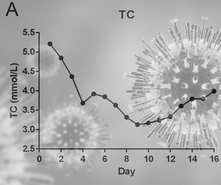
Researchers from Wenzhou, China looked at clinical laboratory features including lipid levels of patients with COVID 19. They found dramatic reductions in the cholesterol levels of patients infected with COVID 19, compared with healthy controls (1)

The study provides data to suggest that cholesterol levels decline quite rapidly during the early stages of infection and increase as the patient starts to recover. Therefore, indicating that cholesterol may have an important role to play in defending the body against such infections.

TC: TOTAL CHOLESTEROL

TC+and+virus.jpg

TC: TOTAL CHOLESTEROL

This perhaps, should not be a surprise, since we already know that cholesterol is an extremely important part of the immune system.

Back in 1997, researchers in the Netherlands published an article in the Lancet showing elderly people with the highest cholesterol levels are best protected from cancer and infections (2).

Other research, published in 2013, led by the University of California, found that derivatives of cholesterol play an important role in the immune system and could protect humans from a wide range of viruses, such as: Ebola, Rift Valley Fever, Nipah, and other deadly pathogens (3).

Two other studies in 2016 found that cholesterol-lowering statins impair the immune system and block the effectiveness of the flu vaccine (4 , 5).

Cholesterol and COVID-19 — STATIN NATION
 
EMF Mitigation - Flush Niacin - Big 5 Minerals

Similar threads

Back
Top Bottom