Richard M Fleming PhD, MD, JD - DOCUMENTATION

J.R.K

Member
Joined
Aug 4, 2020
Messages
1,837
Richard M. Fleming, PhD, MD, JD:
“From a vaccine that only carries a spike protein, it’s making antibodies to the nucleocapsid, which is another part of the virus that’s not supposedly in the vaccines. You can’t make antibodies to something not in your body, so the question is, what’s in the vaccines, that you’re not only making antibodies to the spike protein, but to the nucleocapsid? That’s the key to this paper, is that there’s antibodies to the nucleocapsid. These vaccines must have more in it than just the spike protein.
In 2017 moderna published a paper using lipid nanoparticles on the influenza vaccine, and it showed then, in the animals tested, their lipid nanoparticles vaccines spread into the brain, bone marrow, liver, spleen and the muscle site where it was injected.
If vaccine shedding doesn’t exist, Health and Human Services and the FDA, went through a lot of effort to issue a guidance to the industry in Aug. 2015, when they issued a document on shedding from viral and bacterial vaccines, to the industry. They didn’t spend all this money and time to test for something they didn’t know, and that didn’t exist. This is their document, it’s on their website.
It also doesn’t explain one other important thing. Remember those prion-like diseases we’ve talked about? Your DNA belongs inside of your nucleus or mitochondria. Your RNA belongs inside of your cells. If your body doesn’t see it, it’s not a part of your body, your immune system doesn’t see your genetic code, because it’s not outside of your cells. RNA outside of your cells is a Prion. The vaccines contain mRNA. Any leaking of that material produces a Prion-like disease.
- Richard M. Fleming, PhD, MD, JD
(Prions are misfolded proteins with the ability to transmit their misfolded shape onto normal variants of the same protein. They characterize several fatal and transmissible neurodegenerative diseases in humans and many other animals.)
Yes md_a! I just got to that part myself last night. This brings up another serious question on what else is in as well as what else do these drugs do.
You mentioned that you believe that having a strong metabolism is the best thing to deal with anything that is transmitted by these gene therapy treatments. But I am curious if you have any other thoughts on what a person could do that has to work, live, or just interact daily with people that are treated with these gene therapy treatments.
The one thing I feel that we have an advantage in is that it is not the actual lipid coated mRNA particle that we are being exposed to but rather the shed finished spike protein, and I would presume that our immune system has various defensive mechanisms in place to deal with foreign proteins that we are exposed to on a continuous basis that have evolved over millennia.
However I also have to take into account that this SN1 spike protein has been manipulated in if memory serves 12 different neucleotides and has various components added to it that are not from nature. I believe Dr Fleming mentioned that in the natural evolutionary process it would take around a hundred thousand years for the original spike protein to evolve into this form. So I would have to presume that our bodies are possibly not ready for this unnaturally evolved protein sent back from the future.
I am curious on your take on this matter.
 
OP
md_a

md_a

Member
Joined
Aug 31, 2015
Messages
468
Yes md_a! I just got to that part myself last night. This brings up another serious question on what else is in as well as what else do these drugs do.
You mentioned that you believe that having a strong metabolism is the best thing to deal with anything that is transmitted by these gene therapy treatments. But I am curious if you have any other thoughts on what a person could do that has to work, live, or just interact daily with people that are treated with these gene therapy treatments.
The one thing I feel that we have an advantage in is that it is not the actual lipid coated mRNA particle that we are being exposed to but rather the shed finished spike protein, and I would presume that our immune system has various defensive mechanisms in place to deal with foreign proteins that we are exposed to on a continuous basis that have evolved over millennia.
However I also have to take into account that this SN1 spike protein has been manipulated in if memory serves 12 different neucleotides and has various components added to it that are not from nature. I believe Dr Fleming mentioned that in the natural evolutionary process it would take around a hundred thousand years for the original spike protein to evolve into this form. So I would have to presume that our bodies are possibly not ready for this unnaturally evolved protein sent back from the future.
I am curious on your take on this matter.
From my experience I have noticed that a slightly elevated metabolism to a state of hyperthyroidism from an efficient thyroid sustained by sufficient food plus periodic exposure to the sun, is kept at a temperature of 37.2 C, pulse 85-100 at rest, with good symptoms and a slightly euphoric state with a permanent desire to smile and make pleasant jokes. On this condition, Ray Peat believes that a person can be cured of serious health problems that include cancer. This condition is different from the diagnosis given by doctors as hyper people who are actually hypo but with high stress hormones, or have been exposed to all sorts of environmental estrogenic chemicals that mimic our internal hormones and produce different signals, put the body in a state of confusion. For example, this older Times article shows the changed behavior of cats and their unhealthy reactions. I think this happens to most people through this vast exposure to all kinds of chemicals and radiation, so it's a long process started by many years of hormone mimicking and thyroid damage. In fact, I believe that the body responds differently to chemicals or spike protein depending on its metabolic status, pulse and temperature. At a low temperature the chemicals are incorporated and produce changes and at a high temperature I think they are eliminated and the body recovers quickly and maybe without symptoms. Ideally, each of us should move to a farm of a few hectares and work the land without pesticides and other chemicals, and in time eliminate all the plastic that affects us quite severely. We are bombarded by these chemicals that mimic our hormones everywhere, from clothes, furniture, air, electronics, water, food .... They are toxic substances introduced by mobsters everywhere in the environment, and we certainly do not know what hidden substances introduced in these vaccines, for this reason we will not know what will happen from now on but we will see the reactions in time, and I think a high metabolism could eliminate everything that is foreign to it.
............

PBDEs also happen to have a chemical structure that resembles thyroid hormones and may mimic or compete with these hormones in the body, binding to their receptors and interfering with their transport and metabolism. By the mid-2000s, it was clear that they could alter thyroid function in rodents, birds and fish, and the United States and the European Union have now largely phased the chemicals out. (They remain ubiquitous, however; PBDEs take years to degrade, and many people still own products manufactured before they were taken off the market.)
As the health risks of PBDEs became clear, two scientists at the Environmental Protection Agency — Linda Birnbaum, a toxicologist, and Janice Dye, a veterinarian — began to wonder whether the chemicals might also be responsible for the rise of hyperthyroidism in cats.
Several years later, a group in Illinois discovered that pet cats had higher PBDE levels than feral ones and that hyperthyroid cats tended to live in homes that were particularly saturated with the flame retardants. In 2015, a Swedish team found that hyperthyroid cats had significantly higher levels of three types of PBDEs in their blood than healthy cats did. Last year, researchers in California reported a similar result: Total PBDE levels were higher in cats with hyperthyroidism than those without.
The findings are tantalizing but not definitive. Cats’ lengthening life spans may explain some of the increased incidence of the disease, and it’s possible that high PBDE levels are a result of hyperthyroidism, rather than a cause; the compounds, which are stored in fat, may be released into the bloodstream when cats lose weight. Even if flame retardants do contribute to the disease, they may not be the sole cause. Researchers at the California Department of Toxic Substances Control recently identified more than 70 different compounds that seem to be present in especially high concentrations in hyperthyroid cats. “It’s terribly complicated to nail,” says Ake Bergman, who led the Swedish study and is the director of the Swedish Toxicology Sciences Research Center. “Because you are, and I am, and we are all, including the cats, exposed to such a mixture of chemicals.”
Could hyperthyroid cats be modern-day canaries? We know that flame retardants accumulate in our own bodies; scientists find PBDEs in nearly every person they test, including newborns. “It’s almost 100 percent detection,” says Heather Stapleton, an environmental chemist and exposure scientist at Duke University. The compounds turn up in human blood, breast milk and tissue and can persist for years in fat.

Over the course of decades, human PBDE levels skyrocketed, increasing 100-fold from the 1970s to the early 2000s. (These levels now appear to be declining, most likely as a result of the phasing out of the chemicals.) The rate of human thyroid cancer more than doubled over the same time period. These parallel trends may be more than coincidence: Multiple studies have shown that men and women with high concentrations of PBDEs in their bodies tend to have altered levels of thyroid hormones circulating in their bloodstreams. Last year, researchers reported that thyroid problems were more common among American women with elevated levels of PBDEs in their blood. And at a conference this spring, Stapleton and her colleagues presented findings suggesting that long-term exposure to PBDEs may be a risk factor for papillary thyroid cancer; according to the unpublished data, living in a home with high levels of one type of PBDE in the dust more than doubled the odds of having the disease.

Thyroid hormones also play a crucial role in brain development; a deficiency of these hormones, known as hypothyroidism, may cause neurological abnormalities. If PBDEs cause unusual fluctuations in hormone levels in early life, they may do lasting damage. Scientists have found that those who are exposed to high concentrations of PBDEs in utero or during early childhood score lower on tests of motor skills and cognition. These findings are particularly worrisome given that young children — who are not uncatlike in their behavior, ingesting up to 200 milligrams of dust a day — tend to have higher body burdens of PBDEs than adults. The data are not conclusive, and the underlying mechanisms remain unclear. But further studies of cats could help scientists clarify what’s happening. “I remain convinced that paying more attention to what the animals are trying to tell us is a really good idea,” Rabinowitz says. “There are still many disease outbreaks in animals that remain sort of unexplored or unexplained.”

..........

Urgent Announcement: COVID-19 is caused by Graphene Oxide introduced by several ways into the body
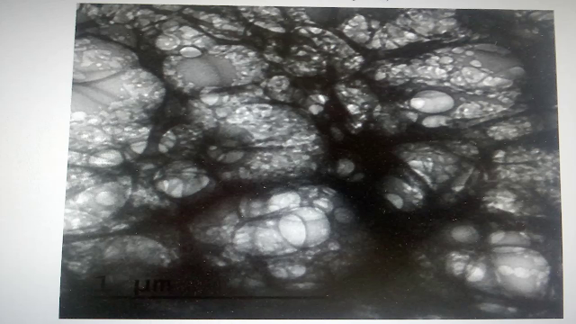
The COVID vaccines in all their variants, AstraZeca, Pfizer, Moderna, Sinovac, Janssen, Johnson & Johnson, etc., also contain a considerable dose of graphene oxide nanoparticles. This has been the result of their analysis by electron microscopy and spectroscopy, among other techniques used by various public universities in our country.

The anti-flu vaccine contained nanoparticles of graphene oxide and the new anti-flu vaccines and the new and supposedly intranasal anti-COVID vaccines they are preparing also contain enormous doses of graphene oxide nanoparticles. Graphene oxide is a toxic that generates thrombi in the organism, graphene oxide is a toxic that generates blood coagulation. Graphene oxide causes alteration of the immune system. By decompensating the oxidative balance in relation to the gulation reserves. If the dose of graphene oxide is increased by any route of administration, it causes the collapse of the immune system and subsequent cytokine storm.

Graphene oxide accumulated in the lungs generates bilateral pneumonias by uniform dissemination in the pulmonary alveolar tract. Graphene oxide causes a metallic taste. Perhaps this is starting to make sense to you now. Inhaled graphene oxide causes inflammation of the mucous membranes and thus loss of taste and partial or total loss of smell.

Graphene oxide acquires powerful magnetic properties inside the organism. This is the explanation for the magnetic phenomenon that billions of people around the world have already experienced after various routes of administration of graphene oxide. Among them the vaccine.

In short, graphene oxide is the supposed SARS-CoV-2, the supposed new coronvirus provoked before the disease called COVID-19. Therefore, we never had real isolation and purification of a new coronvirus, as recognized by most health institutions at the highest level and in different countries when they were questioned about it. COVID-19 disease is the result of introducing graphene oxide by various routes of administration.

Graphene oxide is extremely potent and strong in aerosols, as is the alleged SARS-CoV-2. Like any material, graphene oxide has what we call an 'electronic absorption band'. This means a certain frequency above which the material is excited and oxidizes very rapidly, thus breaking the equilibrium with the proliferation in the organism of the toxicant against our natural antioxidant glutathione reserves. Precisely this frequency band is emitted in the new emission bandwidths of the new 5G wireless technology. That is why the deployment of these antennas never stopped during the pandemic.

In fact, they were among the few services that were maintained, apart from a special surveillance by the State Security Forces and Corps to these antennas. We suspect in that the 2019 anti-flu campaign graphene oxide was introduced in these vials, since it was already used as an adjuvant.

With subsequent 5G technology trials in different parts of the world, COVID-19 disease developed in interaction of external electromagnetic fields and graphene oxide now in their bodies. Remember that it all started in Wuhan, and this was the first pilot sample city in the world to do the 5G technology trial in late November 2019. It's a coincidence in space and time.

Both the pangolin and bat soup versions were simply distracting elements. The purpose of the introduction of graphene oxide is even more obscure than you might imagine. Therefore, it is more than enough for you to assimilate this information and 'reset' the knowledge you had of the disease up to now from the highest governmental institutions telling the population to protect themselves and even forcing them with that which will potentially make them sick with the disease itself. Logically, now that we know that the cause or etiological agent of the disease is precisely a chemical toxicant and not a biological agent, we know how to attenuate it: by increasing glutathione levels. Glutathione is a natural antioxidant that we present in reserves in the organism.

A few details will help you to understand perfectly everything that has been reported in the media. Glutathione is extremely high in children. Therefore, the disease has hardly any impact on the child population. Glutathione drops very considerably after 65 years of age. Therefore, COVID-19 is especially prevalent in the senile population. Glutathione is at very high levels in the intensive sports population. This is why only 0.22% of athletes had the disease.

You will now understand why countless studies in practice have shown that treatment with N-acetylcysteine (which is a precursor of glutathione in the body), or glutathione administered directly, cured COVID-19 disease very quickly in patients. Plain and simple because the glutathione levels were raised to cope with the administered toxicant called graphene oxide.

The discovery made here by La Quinta Columna is a full-fledged attack of State bioterrorism, or at least with the complicity of governments to the entire world population, now constituting crimes against humanity.

It is therefore absolutely essential and vital that you make this information available to your medical community. General practitioners, nursing and health services in general, but also local and regional media and press, as well as all your environment. La Quinta Columna estimates that tens of thousands of people will die every day. In our country alone when they make the new and upcoming 5G technological switch-on.

Bearing in mind that now it is not only the elderly in nursing homes who are vaccinated in that flu vaccine with graphene oxide, but, as you know, a large part of the population has been vaccinated, or graphenated, with gradual doses of graphene oxide.

The body has a natural capacity to eliminate this toxicant, which is why we propose you up to a third dose per year for all the years to keep the graphene in your bodies.

We have each and every one of the proofs of what has been manifested here. Meanwhile justice is trying to act, people will continue to be pushed off a bottomless cliff. If you are watching this audiovisual material, you will understand that for more than a year you have been totally and naively deceived from the highest institutions. Only now will you understand all the incongruities that you observed on your television news.
 

Vileplume

Member
Joined
Jun 10, 2020
Messages
1,697
Location
California
@md_a, I find it interesting that you paired the cat PBDE article and the graphene oxide article. Your introduction helps to connect the articles, prefacing that a well-functioning organism can filter out and expel damaging material much better than a poorly functioning one. "Young children — who are not uncatlike in their behavior," (favorite excerpt) gather these PBDEs throughout childhood, and might not express troubled thyroid function until their twenties or even later. Similarly, children's high levels of the antioxidant glutathione protects them from graphene oxide AKA COVID-19. Still, over time, these harmful particles build up.

To summarize the second article, graphene oxide enters the body through spray, through vaccines, flu shots, causing the problems we know as COVID. the article claims that COVID is caused solely by graphene oxide. If this is true, then there is no COVID virus. The vaccines, the lockdowns, the claims about COVID spreading through the air, even spike proteins--all of it is untrue. I find this hard to believe. Isn't it more likely that graphene oxide harms us in conjunction with the spike protein?
 
Last edited:
OP
md_a

md_a

Member
Joined
Aug 31, 2015
Messages
468
@md_a, I find it interesting that you paired the cat PBDE article and the graphene oxide article. Your introduction helps to connect the articles, prefacing that a well-functioning organism can filter out and expel damaging material much better than a poorly functioning one. "Young children — who are not uncatlike in their behavior," (favorite excerpt) gather these PBDEs throughout childhood, and might not express troubled thyroid function until their twenties or even later. Similarly, children's high levels of the antioxidant glutathione protects them from graphene oxide AKA COVID-19. Still, over time, these harmful particles build up.

To summarize the second article, graphene oxide enters the body through spray, through vaccines, flu shots, causing the problems we know as COVID. the article claims that COVID is caused solely by graphene oxide. If this is true, then there is no COVID virus. The vaccines, the lockdowns, the claims about COVID spreading through the air, even spike proteins--all of it is untrue. I find this hard to believe. Isn't it more likely that graphene oxide harms us in conjunction with the spike protein?
It is said that the virus binds to ACE2 and thus causes the serious disease seen in patients with Covid. I think this is a trick used by mobsters, by the fact that they discovered this vital enzyme for the body that normally decreases with age, on hypothyroidism with the stimulation of AT1 and they thus hastened the death of many people. This panic induced by mobsters through the media has led to the death of patients in hospitals due to the wrongly intentional protocol implemented by blocking ACE2 and producing the vaccine that does the same. In contrast, ACE2 stimulation and AT1 blockade keep us young and protect us from degenerative diseases.

ATR-1 Receptors increase with age and are increased in cancer, diabetes, hypertension, chronic obstructive pulmonary disease.
SARS-COV2 virus attaches to the ACE2 it causes a decrease in ACE2 availability/activity. This would lead to a higher Angiotensin II and in patients with more AT-1
In patients with low ACE2 by age, sickness or virus binding to ACE2 means that it leaves the ACE1 which produces angiotensin.
The most common complication leading to the CoV-induced mortality can be justified through ACE-AngII-AT1-Spike protein overactivation caused by virus / toxin / drugs / nocebo effect induced by media.
The spikes protein have high affinity to ACE2, uses ACE2 to enter a cell.
It is suggested that this “occupation” of the virus of ACE2 might reduce the ability of Ang II to bind to ACE2.
Accumulated Ang II result in an increased activation of AT1.
Activation of AT1 results in vasoconstriction, increased pro-inflammatory response.

And so you can discover a whole series of studies that indicate the inflammatory part of the AT1 receptors.

........
The role of the angiotensin II type I receptor blocker telmisartan in the treatment of non-alcoholic fatty liver disease

Abstract

Non-alcoholic fatty liver disease (NAFLD) is currently considered an important component of metabolic syndrome (MetS). The spectrum of NAFLD includes conditions that range from simple hepatic steatosis to non-alcoholic steatohepatitis. NAFLD is correlated with liver-related death and is predicted to be the most frequent indication for liver transplantation by 2030. Insulin resistance is directly correlated to the central mechanisms of hepatic steatosis in NAFLD patients, which is strongly correlated to the imbalance of the renin–angiotensin system, that is involved in lipid and glucose metabolism. Among the emerging treatment approaches for NAFLD is the anti-hypertensive agent telmisartan, which has positive effects on liver, lipid, and glucose metabolism, especially through its action on the renin–angiotensin system, by blocking the ACE/AngII/AT1 axis and increasing ACE2/Ang(1–7)/Mas axis activation. However, treatment with this drug is only recommended for patients with an established indication for anti-hypertensive therapy. Thus, there is an increased need for large randomized controlled trials with the aim of elucidating the effects of telmisartan on liver disease, especially NAFLD. From this perspective, the present review aims to provide a brief examination of the pathogenesis of NAFLD/NASH and the role of telmisartan on preventing liver disorders and thus to improve the discussion on potential therapies.


Conclusion

In conclusion, blocking RAS to inhibit the ACE/Ang II/AT1R axis and increase ACE2/Ang-(1–7)/Mas axis activation is an important strategy in treating NAFLD. Of all the RAS blockers, telmisartan is the most promising drug, not only because of its favorable pharmacokinetic proprieties and safety, but also because it seems to exert additional AT1-independent benefits on metabolism via partial PPAR-γ agonism and/or a positive central action on the HPA axis. However, large multicenter randomized-controlled trials are needed to consolidate these findings. Currently, treatment with telmisartan and with other RAS blockers can only be formally recommended in NALFD patients with an established indication of anti-hypertensive therapy.

The role of the angiotensin II type I receptor blocker telmisartan in the treatment of non-alcoholic fatty liver disease: a brief review | Hypertension Research


Cancer, inflammation and the AT1 and AT2 receptors

The evidence relating to over-expression of AT1 with cancer progression is compelling. To this effect, AT1 blockade has been hypothesised as the mechanism to overcome cancer associated complications in organ graft recipients [45]. Additionally, a study undertaken in 1998 suggested that hypertensive patients taking ACE inhibitors were significantly less at risk of developing cancer than those taking other hypertensive treatments [46].


Tumour progression has been significantly slowed with AT1 receptor antagonists [47, 48]. The results appeared to far exceed the expectations of simple inhibition of angiogenesis. Reduction of MCP-1 was noted [48], as was the expression of many pro-inflammatory cytokines. The activity of tumour-associated macrophages was also noticed to be severely impaired [48]. The importance in reducing the action of tumour-associated macrophages in extracellular matrix decomposition is not to be underestimated, since, in this action, they further progress remodelling by releasing stored TGF-β [49]. The similarity of action of tumour associated macrophages with those in the tissue healing and repair environment has been noted [49]. The tumour suppressant action of tranilast, an AT1 antagonist, [50] has been more widely explored [51–54]. In one study on the inhibition of uterine leiomyoma cells, Tranilast also induced p21 and p53 [55]. Similarly, the AT1 blocker losartan has been shown to antagonise platelets, which are thought to modulate cell plasticity and angiogenesis via the vascular endothelial growth factor (VEGF) [56]. It has been postulated that losartan and other AT1 blockers can act as novel anti-angiogenic, anti-invasive and anti-growth agents against neoplastic tissue [56]. Furthermore, it has been shown that angiotensin II induces the phosphorylations of mitogen-activated protein kinase (MAPK) and signal transducer and activator of transcription 3 (STAT3) in prostate cancer cells. In contrast, AT1 inhibitors have been shown to inhibit the proliferation of prostate cancer cells stimulated with EGF or angiotensin II, through the suppression of MAPK or STAT3 phosphorylation [57]. Angiotensin II also induces (VEGF), which plays a pivotal role in tumour angiogenesis and has been the target of various therapeutics, including antibodies and aptamers [58]. Although the role of angiotensin II in VEGF-mediated tumour development has not yet been elucidated, an ACE inhibitor significantly attenuated VEGF-mediated tumour development, accompanying the suppression of neovascularisation in the tumour and VEGF-induced endothelial cell migration [59]. Perindopril, another ACE inhibitor has also been shown to be a potent inhibitor of tumour development and angiogenesis through suppression of the VEGF and the endothelial cell tubule formation [60].


Conclusions

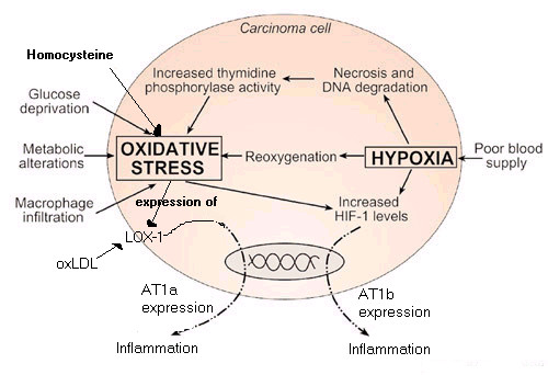
The invasiveness and immunosuppression of many cancers appears dependent on inflammation and the upregulation of AT1. Two mechanisms for upregulation of AT1 are discussed: 1) evolutionary changes to take advantage of this pro-inflammatory control mechanism, 2) AT1 expression induced by an alternating environment of hypoxia and oxidative stress. Immunosuppression as a common protection mechanism of solid tumours against immune responses has been verified from current literature and experimental procedures, as has the implication of cytokines and chemokines in tumour growth and metastasis. Given the involvement of AT1 in the immunosuppression and inflammatory processes, as well as in the expression of the pro-inflammatory cytokines and chemokines, it becomes evident that the AT1 receptor is essential for tumour protection and progression. A combination therapy consisting of AT1 receptor antagonists, NSAID for further control of the inflammation and immune therapy in the form of tumour vaccines should provide a novel and successful treatment for solid tumours.


In the renin-angiotensin system, the angiotensin II receptors AT1 and AT2 seem to have opposing functions. The actions of AT1 being principally pro-inflammatory whilst AT2 provides protection against hypoxia, draws inflammatory action to a close and promotes healing. The various direct and indirect mechanisms for feedback between the receptors, their induced products and the external hormonal system in the control of inflammation and healing are summarised in a highly simplified model which none the less can be used to explain how many key promoters and inhibitors of disease exert their effects.


From a review of the current disease literature, it has been demonstrated that the role of AT1 and AT2 in inflammation is not limited to cancer-associated inflammation, but is generally consistent and system wide. Potential therapy by manipulation of these receptors, although at an early stage, has been demonstrated for some of these diseases and it is proposed that this approach will provide an effective basis for the treatment of autoimmune, inflammatory and neurodegenerative disorders using existing drugs. AT1 receptor blockade should, in addition, provide a treatment to alleviate the damage caused by bacterial and viral infections, where their destructive action is through chronic inflammation. Given the importance of the immune suppressant effect of inflammation in cancer, it is anticipated that AT1 blockade should also serve to elicit a more effective immune response to other invaders that seek to corrupt the wound recovery process.


Manipulation of the AT1 and AT2 receptors has profound and exciting implications in the control of disease.


Related to stress

For anxiety, angiotensin II may have a role through the HPA axis and sympatho-adrenal axis. Another contemporary review argues that excessive brain AT1R activity is associated not only with hypertension and heart failure but also with brain ischaemia, abnormal stress responses, blood-brain barrier breakdown and inflammation {1}. AT1R blockers decrease brain inflammation and have numerous other effects, pointing to the potential for such treatments in reducing brain inflammatory processes. One such example is mood disorders associated with pro-inflammatory cytokines. This is a novel approach to addressing stress-induced and inflammatory brain diseases that eventually may produce new treatments.

Faculty Opinions Recommended Article: The link between angiotensin II-mediated anxiety and mood disorders with NADPH oxidase-induced oxidative stress.


Angiotensin II (AngII) plays important roles in the regulation of cardiovascular function. Both peripheral and central actions of AngII are involved in this regulation, but mechanisms of the latter actions as a neurotransmitter/neuromodulator within the brain are still unclear. Here we show that (1) intracerebroventricularly (i.c.v.) administered AngII in urethane-anesthetized male rats elevates plasma adrenaline derived from the adrenal medulla but not noradrenaline with valsartan- (AT1 receptor blocker) sensitive brain mechanisms, (2) peripheral AT1 receptors are not involved in the AngII-induced elevation of plasma adrenaline, although AngII induces both noradrenaline and adrenaline secretion from bovine adrenal medulla cells and (3) i.c.v. administered AngII elevates blood pressure but not heart rate with the valsartan-sensitive mechanisms. From these results, i.c.v. administered AngII acts on brain AT1 receptors, thereby inducing the secretion of adrenaline and pressor responses. We propose that the central angiotensinergic system can activate central adrenomedullary outflow and modulate blood pressure.

Angiotensin II acting on brain AT1 receptors induces adrenaline secretion and pressor responses in the rat


May have beneficial effects in polycystic ovary syndrome


According to our knowledge, these are the first case reports demonstrating that the angiotensin II receptor antagonist telmisartan, besides being an effective antihypertensive drug, may have beneficial effects in polycystic ovary syndrome patients. The case reports provide interesting new information about unexpected effect and possible new indication of telmisartan in patients with polycystic ovary syndrome.


Since insulin resistance plays a major role in the pathogenesis of polycystic ovary syndrome, several trials have confirmed that insulin-sensitizing drugs, metformin (7-9), and thiazolidinediones (11-13,23,24) have favorable endocrine, reproductive, and metabolic effects in polycystic ovary syndrome. Considering that telmisartan may have insulin-sensitizing effects related to its ability to activate PPAR gamma (14-18), we hypothesized that the administration of telmisartan would also improve menstrual dysfunction, hyperandrogenemia, and hyperinsulinemia of patients with polycystic ovary syndrome.


As we assumed, after 6 months of treatment with telmisartan, we observed significant reduction in free testosterone (around 35% on average), DHEAS (around 40% on average), and androstenedione concentration (around 20% on average) in all 4 patients. Three out of four women improved their menstrual cyclicity. One additional menstruation in one woman and two additional menstruations in the other two women were attained in 6 months of telmisartan treatment. The intensity of telmisartan effects appeared to be in the usual range for insulin-sensitizing drugs, metformin, or thiazolidinediones in patients with polycystic ovary syndrome.

Decreased Androgen Levels and Improved Menstrual Pattern after Angiotensin II Receptor Antagonist Telmisartan Treatment in Four Hypertensive Patients with Polycystic Ovary Syndrome: Case Series


Angiotensin II rapidly increases phosphatidate-phosphoinositide synthesis and phosphoinositide hydrolysis and mobilizes intracellular calcium in cultured arterial muscle cells.

Abstract

Smooth muscle cells were cultured from rat thoracic aorta and labeled to a stable specific activity with 45Ca2+, myo-[2-3H]inositol, or 32Pi. The efflux of 45Ca2+ was monitored over 10-sec intervals. Angiotensin II (AII) increased the amount of 45Ca2+ lost by 5-fold in the first 10-sec interval after the addition of AII and by 10-fold in the second 10-sec interval. AII-stimulated 45Ca2+ release was blocked by the angiotensin antagonist [1-sarcosine, 8-leucine]AII and by La3+. The removal of external Ca2+ had no effect on AII-stimulated 45Ca2+ release. Depolarization with high external K+ only slightly increased 45Ca2+ efflux and had no effect on AII-induced 45Ca2+ release. AII had no effect on the initial rate of 45Ca2+ influx. These results indicate that the rapid 45Ca2+ efflux evoked by AII is probably due to the release of 45Ca2+ sequestered intracellularly rather than to an increase in the Ca2+ permeability of the plasma membrane. AII provoked rapid increases in the levels of phosphatidic acid and phosphoinositides in the cells. These increases in phospholipids were associated with increases in phospholipase C-generated inositol phosphates (tri-, di-, and mono-). It appears that AII simultaneously increases phosphoinositide hydrolysis and synthesis in vascular smooth muscle, and both phospholipid effects may contribute to inositol triphosphate generation, which was sufficiently rapid to have a role in intracellular Ca2+ mobilization.


The Renin-Angiotensin-Aldosterone System and Calcium-Regulatory Hormones

..These results suggest that in the acute setting, angiotensin II may exert a stimulatory effect on PTH that can be mitigated by lowering angiotensin II with an angiotensin converting enzyme inhibitor; however, acute increases in aldosterone do not appreciably alter PTH. In contrast, in the chronic setting aldosterone may increase PTH, and this effect may be mitigated by a mineralocorticoid antagonist--observations that were similar to those reported in the aforementioned cohorts of subjects with primary aldosteronism. Thus, this study by Brown and colleagues provides evidence implicating multiple RAAS components interacting with PTH, and a differential temporal effect dictating their relationships(67). In assessments of parathyroid pathology, both normal and adenomatous parathyroid tissue have been shown to express the angiotensin type1 receptor and the mineralocorticoid receptor, and the expression of these receptors was 3-4 fold greater among adenomatous tissue, giving further support to direct actions of angiotensin II and/or aldosterone on the parathyroid(67)….

CONCLUSIONS

In summary, growing evidence suggests interactions between calcium-regulatory hormones and the renin-angiotensin-aldosterone system with potentially important clinical implications. Calcium and vitamin D have been shown to inhibit renin secretion and expression, whereas a bi-directional and stimulatory relationship between parathyroid hormone and aldosterone (and possibly angiotensin II) has been observed. With this mounting evidence, new questions are raised that may have notable implications for future clinical outcomes research.


High Calcium Diet Down-Regulates Kidney Angiotensin-Converting Enzyme in Experimental Renal Failure

Abstract

Background: Calcium salts are used as phosphate binders in renal failure, while high calcium diet also improves vasorelaxation and enhances natriuresis. The influences of calcium intake on renal renin-angiotensin system (RAS) are largely unknown.


Methods: Four weeks after NTX, rats were put on 3.0% or 0.3% calcium diet for 8 weeks (12-week study). In additional experiments, 15 weeks after NTX, rats were put on similar diets for 12 weeks (27-week study). Appropriate blood, urine, and kidney samples were taken. Renal angiotensin-converting enzyme (ACE) and angiotensin II receptors (AT1, AT2) were examined using autoradiography, ACE also using Western blotting, and connective tissue growth factor (CTGF) using immunohistochemistry.


Results: In the 12-week study, albuminuria increased 5-fold in NTX rats, but only 2-fold in calcium NTX rats on 3.0% calcium. In the 27-week study, high calcium intake decreased blood pressure, retarded progression of renal failure, reduced glomerulosclerosis, interstitial damage, and aortic calcifications, and improved survival from 50% to 92% in NTX rats. In both experiments plasma parathyroid hormone and phosphate were elevated after NTX, and suppressed by high calcium diet, while kidney ACE was down-regulated by 40% or more after increased calcium intake. In the 27-week study renal CTGF was decreased and cortical AT1 receptor density reduced after high calcium diet.


Conclusion: High calcium diet down-regulated kidney ACE, reduced albuminuria and blood pressure, and favorably influenced kidney morphology in experimental renal failure. These findings suggest a link between calcium metabolism and kidney ACE expression, which may play a role in the progression of renal damage.


J.R.K

Member
Joined
Aug 4, 2020
Messages
1,837
From my experience I have noticed that a slightly elevated metabolism to a state of hyperthyroidism from an efficient thyroid sustained by sufficient food plus periodic exposure to the sun, is kept at a temperature of 37.2 C, pulse 85-100 at rest, with good symptoms and a slightly euphoric state with a permanent desire to smile and make pleasant jokes. On this condition, Ray Peat believes that a person can be cured of serious health problems that include cancer. This condition is different from the diagnosis given by doctors as hyper people who are actually hypo but with high stress hormones, or have been exposed to all sorts of environmental estrogenic chemicals that mimic our internal hormones and produce different signals, put the body in a state of confusion. For example, this older Times article shows the changed behavior of cats and their unhealthy reactions. I think this happens to most people through this vast exposure to all kinds of chemicals and radiation, so it's a long process started by many years of hormone mimicking and thyroid damage. In fact, I believe that the body responds differently to chemicals or spike protein depending on its metabolic status, pulse and temperature. At a low temperature the chemicals are incorporated and produce changes and at a high temperature I think they are eliminated and the body recovers quickly and maybe without symptoms. Ideally, each of us should move to a farm of a few hectares and work the land without pesticides and other chemicals, and in time eliminate all the plastic that affects us quite severely. We are bombarded by these chemicals that mimic our hormones everywhere, from clothes, furniture, air, electronics, water, food .... They are toxic substances introduced by mobsters everywhere in the environment, and we certainly do not know what hidden substances introduced in these vaccines, for this reason we will not know what will happen from now on but we will see the reactions in time, and I think a high metabolism could eliminate everything that is foreign to it.
............

PBDEs also happen to have a chemical structure that resembles thyroid hormones and may mimic or compete with these hormones in the body, binding to their receptors and interfering with their transport and metabolism. By the mid-2000s, it was clear that they could alter thyroid function in rodents, birds and fish, and the United States and the European Union have now largely phased the chemicals out. (They remain ubiquitous, however; PBDEs take years to degrade, and many people still own products manufactured before they were taken off the market.)
As the health risks of PBDEs became clear, two scientists at the Environmental Protection Agency — Linda Birnbaum, a toxicologist, and Janice Dye, a veterinarian — began to wonder whether the chemicals might also be responsible for the rise of hyperthyroidism in cats.
Several years later, a group in Illinois discovered that pet cats had higher PBDE levels than feral ones and that hyperthyroid cats tended to live in homes that were particularly saturated with the flame retardants. In 2015, a Swedish team found that hyperthyroid cats had significantly higher levels of three types of PBDEs in their blood than healthy cats did. Last year, researchers in California reported a similar result: Total PBDE levels were higher in cats with hyperthyroidism than those without.
The findings are tantalizing but not definitive. Cats’ lengthening life spans may explain some of the increased incidence of the disease, and it’s possible that high PBDE levels are a result of hyperthyroidism, rather than a cause; the compounds, which are stored in fat, may be released into the bloodstream when cats lose weight. Even if flame retardants do contribute to the disease, they may not be the sole cause. Researchers at the California Department of Toxic Substances Control recently identified more than 70 different compounds that seem to be present in especially high concentrations in hyperthyroid cats. “It’s terribly complicated to nail,” says Ake Bergman, who led the Swedish study and is the director of the Swedish Toxicology Sciences Research Center. “Because you are, and I am, and we are all, including the cats, exposed to such a mixture of chemicals.”
Could hyperthyroid cats be modern-day canaries? We know that flame retardants accumulate in our own bodies; scientists find PBDEs in nearly every person they test, including newborns. “It’s almost 100 percent detection,” says Heather Stapleton, an environmental chemist and exposure scientist at Duke University. The compounds turn up in human blood, breast milk and tissue and can persist for years in fat.

Over the course of decades, human PBDE levels skyrocketed, increasing 100-fold from the 1970s to the early 2000s. (These levels now appear to be declining, most likely as a result of the phasing out of the chemicals.) The rate of human thyroid cancer more than doubled over the same time period. These parallel trends may be more than coincidence: Multiple studies have shown that men and women with high concentrations of PBDEs in their bodies tend to have altered levels of thyroid hormones circulating in their bloodstreams. Last year, researchers reported that thyroid problems were more common among American women with elevated levels of PBDEs in their blood. And at a conference this spring, Stapleton and her colleagues presented findings suggesting that long-term exposure to PBDEs may be a risk factor for papillary thyroid cancer; according to the unpublished data, living in a home with high levels of one type of PBDE in the dust more than doubled the odds of having the disease.

Thyroid hormones also play a crucial role in brain development; a deficiency of these hormones, known as hypothyroidism, may cause neurological abnormalities. If PBDEs cause unusual fluctuations in hormone levels in early life, they may do lasting damage. Scientists have found that those who are exposed to high concentrations of PBDEs in utero or during early childhood score lower on tests of motor skills and cognition. These findings are particularly worrisome given that young children — who are not uncatlike in their behavior, ingesting up to 200 milligrams of dust a day — tend to have higher body burdens of PBDEs than adults. The data are not conclusive, and the underlying mechanisms remain unclear. But further studies of cats could help scientists clarify what’s happening. “I remain convinced that paying more attention to what the animals are trying to tell us is a really good idea,” Rabinowitz says. “There are still many disease outbreaks in animals that remain sort of unexplored or unexplained.”

..........

Urgent Announcement: COVID-19 is caused by Graphene Oxide introduced by several ways into the body
The COVID vaccines in all their variants, AstraZeca, Pfizer, Moderna, Sinovac, Janssen, Johnson & Johnson, etc., also contain a considerable dose of graphene oxide nanoparticles. This has been the result of their analysis by electron microscopy and spectroscopy, among other techniques used by various public universities in our country.

The anti-flu vaccine contained nanoparticles of graphene oxide and the new anti-flu vaccines and the new and supposedly intranasal anti-COVID vaccines they are preparing also contain enormous doses of graphene oxide nanoparticles. Graphene oxide is a toxic that generates thrombi in the organism, graphene oxide is a toxic that generates blood coagulation. Graphene oxide causes alteration of the immune system. By decompensating the oxidative balance in relation to the gulation reserves. If the dose of graphene oxide is increased by any route of administration, it causes the collapse of the immune system and subsequent cytokine storm.

Graphene oxide accumulated in the lungs generates bilateral pneumonias by uniform dissemination in the pulmonary alveolar tract. Graphene oxide causes a metallic taste. Perhaps this is starting to make sense to you now. Inhaled graphene oxide causes inflammation of the mucous membranes and thus loss of taste and partial or total loss of smell.

Graphene oxide acquires powerful magnetic properties inside the organism. This is the explanation for the magnetic phenomenon that billions of people around the world have already experienced after various routes of administration of graphene oxide. Among them the vaccine.

In short, graphene oxide is the supposed SARS-CoV-2, the supposed new coronvirus provoked before the disease called COVID-19. Therefore, we never had real isolation and purification of a new coronvirus, as recognized by most health institutions at the highest level and in different countries when they were questioned about it. COVID-19 disease is the result of introducing graphene oxide by various routes of administration.

Graphene oxide is extremely potent and strong in aerosols, as is the alleged SARS-CoV-2. Like any material, graphene oxide has what we call an 'electronic absorption band'. This means a certain frequency above which the material is excited and oxidizes very rapidly, thus breaking the equilibrium with the proliferation in the organism of the toxicant against our natural antioxidant glutathione reserves. Precisely this frequency band is emitted in the new emission bandwidths of the new 5G wireless technology. That is why the deployment of these antennas never stopped during the pandemic.

In fact, they were among the few services that were maintained, apart from a special surveillance by the State Security Forces and Corps to these antennas. We suspect in that the 2019 anti-flu campaign graphene oxide was introduced in these vials, since it was already used as an adjuvant.

With subsequent 5G technology trials in different parts of the world, COVID-19 disease developed in interaction of external electromagnetic fields and graphene oxide now in their bodies. Remember that it all started in Wuhan, and this was the first pilot sample city in the world to do the 5G technology trial in late November 2019. It's a coincidence in space and time.

Both the pangolin and bat soup versions were simply distracting elements. The purpose of the introduction of graphene oxide is even more obscure than you might imagine. Therefore, it is more than enough for you to assimilate this information and 'reset' the knowledge you had of the disease up to now from the highest governmental institutions telling the population to protect themselves and even forcing them with that which will potentially make them sick with the disease itself. Logically, now that we know that the cause or etiological agent of the disease is precisely a chemical toxicant and not a biological agent, we know how to attenuate it: by increasing glutathione levels. Glutathione is a natural antioxidant that we present in reserves in the organism.

A few details will help you to understand perfectly everything that has been reported in the media. Glutathione is extremely high in children. Therefore, the disease has hardly any impact on the child population. Glutathione drops very considerably after 65 years of age. Therefore, COVID-19 is especially prevalent in the senile population. Glutathione is at very high levels in the intensive sports population. This is why only 0.22% of athletes had the disease.

You will now understand why countless studies in practice have shown that treatment with N-acetylcysteine (which is a precursor of glutathione in the body), or glutathione administered directly, cured COVID-19 disease very quickly in patients. Plain and simple because the glutathione levels were raised to cope with the administered toxicant called graphene oxide.

The discovery made here by La Quinta Columna is a full-fledged attack of State bioterrorism, or at least with the complicity of governments to the entire world population, now constituting crimes against humanity.

It is therefore absolutely essential and vital that you make this information available to your medical community. General practitioners, nursing and health services in general, but also local and regional media and press, as well as all your environment. La Quinta Columna estimates that tens of thousands of people will die every day. In our country alone when they make the new and upcoming 5G technological switch-on.

Bearing in mind that now it is not only the elderly in nursing homes who are vaccinated in that flu vaccine with graphene oxide, but, as you know, a large part of the population has been vaccinated, or graphenated, with gradual doses of graphene oxide.

The body has a natural capacity to eliminate this toxicant, which is why we propose you up to a third dose per year for all the years to keep the graphene in your bodies.

We have each and every one of the proofs of what has been manifested here. Meanwhile justice is trying to act, people will continue to be pushed off a bottomless cliff. If you are watching this audiovisual material, you will understand that for more than a year you have been totally and naively deceived from the highest institutions. Only now will you understand all the incongruities that you observed on your television news.
Once again md_a you deliver fantastic information thank you for expanding my knowledge base although it confuses my mind for now I find this is a good thing as I learn to understand that nothing makes sense until it does then new information comes and that doesn’t make sense until it makes perfect sense.
Okay enough of that, so do you believe the virus and the spike protein do not or are not produced in the vaccine or was released into the general world population but rather it was graphene instead? Or was there a virus and subsequent spike protein built by the mRNA vaccine that also has graphene in it?
Subsequently do you believe that a high metabolism that we are all trying to achieve would be enough to eliminate the graphene and as you mentioned before the spike protein.
Or do we actually need to delve into what I am understand is a dangerous supplement in the form of glutathione or it precursor N Acetyl Cystiene, which is my current understanding Dr Peats position is it is a promoter of the cancer metabolism. I hope that a high metabolism that we on this forum are trying to achieve would provide enough of this amino acid that the supplementation aspect could be avoided, especially if one heeds the concerns regarding the vaccine and stays within the control group rather than joining the experimental side. I feel that the control side has an advantage having an immune system that operates on a fully operational basis as compared with the suppression of the immune system that comes with the vaccines.
But I will concede that they may have glutathione more available due to the constant inflammatory state they are in and they demand by cancer cells for their preferred food source.
 

Forsythia

Member
Joined
Apr 13, 2014
Messages
195
I'm trying to get my head around this article by La Quinta Columna. Everything that has happened in the past 2 years has nothing to do with a virus, the virus was just the smokescreen for the poisoning of the population with graphene oxide? Graphene oxide is in the masks, it's in the nasal swabs used for PCR testing, it's in all the covid vaccines, it's in the flu shots for the past couple of flu seasons, it's toxicity is amplified by 5G frequency band. It causes all the symptoms that have been attributed to a virus. Graphene = COVID. (I know I'm basically just repeating what Vilaplume has already written).

The article/video is translated from Spanish and there are some typos. But this sentence at the end of the article, I can't figure out what is being said:

The body has a natural capacity to eliminate this toxicant, which is why we propose you up to a third dose per year for all the years to keep the graphene in your bodies.

md_a do you understand what they are trying to say in this sentence?
 

Forsythia

Member
Joined
Apr 13, 2014
Messages
195
The article goes on to say the antidote to the graphene poisoning is to increase glutathione levels:

Logically, now that we know that the cause or etiological agent of the disease is precisely a chemical toxicant and not a biological agent, we know how to attenuate it: by increasing glutathione levels. Glutathione is a natural antioxidant that we present in reserves in the organism.

A few details will help you to understand perfectly everything that has been reported in the media. Glutathione is extremely high in children. Therefore, the disease has hardly any impact on the child population. Glutathione drops very considerably after 65 years of age. Therefore, COVID-19 is especially prevalent in the senile population. Glutathione is at very high levels in the intensive sports population. This is why only 0.22% of athletes had the disease.

You will now understand why countless studies in practice have shown that treatment with N-acetylcysteine (which is a precursor of glutathione in the body), or glutathione administered directly, cured COVID-19 disease very quickly in patients. Plain and simple because the glutathione levels were raised to cope with the administered toxicant called graphene oxide.
 
OP
md_a

md_a

Member
Joined
Aug 31, 2015
Messages
468
Once again md_a you deliver fantastic information thank you for expanding my knowledge base although it confuses my mind for now I find this is a good thing as I learn to understand that nothing makes sense until it does then new information comes and that doesn’t make sense until it makes perfect sense.
Okay enough of that, so do you believe the virus and the spike protein do not or are not produced in the vaccine or was released into the general world population but rather it was graphene instead? Or was there a virus and subsequent spike protein built by the mRNA vaccine that also has graphene in it?
Subsequently do you believe that a high metabolism that we are all trying to achieve would be enough to eliminate the graphene and as you mentioned before the spike protein.
Or do we actually need to delve into what I am understand is a dangerous supplement in the form of glutathione or it precursor N Acetyl Cystiene, which is my current understanding Dr Peats position is it is a promoter of the cancer metabolism. I hope that a high metabolism that we on this forum are trying to achieve would provide enough of this amino acid that the supplementation aspect could be avoided, especially if one heeds the concerns regarding the vaccine and stays within the control group rather than joining the experimental side. I feel that the control side has an advantage having an immune system that operates on a fully operational basis as compared with the suppression of the immune system that comes with the vaccines.
But I will concede that they may have glutathione more available due to the constant inflammatory state they are in and they demand by cancer cells for their preferred food source.
Viruses modified in the laboratory exist, the mobsters deal with it, they do experiments that they could use as a biological weapon, and there is clear evidence that even in this situation everything was planned in advance and they tried with this virus to spread it to induce panic and fear among the population in order to achieve the mandatory vaccination that is part of their plan, only that the laboratory virus was not so dangerous as to kill people and lead to a rapid vaccination, and for this reason they used the media to cause panic and the medical system to produce the desired effects, because the virus does not kill but people have been killed by medical protocols that include wrong and dangerous treatments. These mobsters attack in every possible way to get the desired effect, by forcing the wearing of masks that contain dangerous chemicals, PCR tests that do the same thing. Ever since this plan started, I felt that there was something unclean in the middle, and with the help of the information from Ray Peat, I quickly made a connection with what is happening in reality. It's just that I lost my job and it was very difficult for a year to pay my installments and eat properly, and from a normal metabolism, I quickly returned to the state of hypothyroidism, my temperature dropped to 36.4 and pulse 60. At the same time I accumulated a huge internal tension that caused me a massive hair loss with his whitening and teeth problems. From the beginning of the plandemic I took methylene blue and aloe vera emodin, and maybe that was the reason I didn't have any cold symptoms, because I didn't stay away from anyone and I walked everywhere, both as much as I was allowed. For six months I found work and because I started to eat better I managed to return to normal with temperature and pulse, I stopped hair loss, and now to see if I can regenerate it as before and give it regained natural color. I think Ray Peat is the closest thing to the truth in general and I would avoid using substances like glutathione or its precursor N Acetyl Cystiene.
The interesting part is to find out what is the reason for forced vaccination and what hidden substances are in the vaccine. But I think the evil has already been produced, and I think that a high metabolism could protect the most sensitive like me, and those who have a more resistant body will probably adapt to the new chemicals.
 

J.R.K

Member
Joined
Aug 4, 2020
Messages
1,837
Viruses modified in the laboratory exist, the mobsters deal with it, they do experiments that they could use as a biological weapon, and there is clear evidence that even in this situation everything was planned in advance and they tried with this virus to spread it to induce panic and fear among the population in order to achieve the mandatory vaccination that is part of their plan, only that the laboratory virus was not so dangerous as to kill people and lead to a rapid vaccination, and for this reason they used the media to cause panic and the medical system to produce the desired effects, because the virus does not kill but people have been killed by medical protocols that include wrong and dangerous treatments. These mobsters attack in every possible way to get the desired effect, by forcing the wearing of masks that contain dangerous chemicals, PCR tests that do the same thing. Ever since this plan started, I felt that there was something unclean in the middle, and with the help of the information from Ray Peat, I quickly made a connection with what is happening in reality. It's just that I lost my job and it was very difficult for a year to pay my installments and eat properly, and from a normal metabolism, I quickly returned to the state of hypothyroidism, my temperature dropped to 36.4 and pulse 60. At the same time I accumulated a huge internal tension that caused me a massive hair loss with his whitening and teeth problems. From the beginning of the plandemic I took methylene blue and aloe vera emodin, and maybe that was the reason I didn't have any cold symptoms, because I didn't stay away from anyone and I walked everywhere, both as much as I was allowed. For six months I found work and because I started to eat better I managed to return to normal with temperature and pulse, I stopped hair loss, and now to see if I can regenerate it as before and give it regained natural color. I think Ray Peat is the closest thing to the truth in general and I would avoid using substances like glutathione or its precursor N Acetyl Cystiene.
The interesting part is to find out what is the reason for forced vaccination and what hidden substances are in the vaccine. But I think the evil has already been produced, and I think that a high metabolism could protect the most sensitive like me, and those who have a more resistant body will probably adapt to the new chemicals.
Thank you for sharing this md_a! Are you okay now? Have you returned to work? Your story is inspiring in that you were able to find a way to try to maintain a Ray Peat inspired diet, support yourself as best as possible both physically, mentally, and nutritionally, while keeping active and staving off the negative emotional traps many fall into. I for one appreciate the efforts and details you put into your posts, they are so insightful and come from a different viewpoint.
I am glad that you are able to find and use methylene blue I feel that it is an old and forgotten miracle drug much like Aspirin.
So do you think that these vaccines are designed to enhance the ability of the virus to enter the cell when the next flu season comes around, in an ADE style fashion? Since the antibodies are in the vaccinated any virus that has a spike protein that resembles the one produced by the virus there could be an autoimmune style of attack increase in the fall and winter. The healthy will stave it off but they may be weakened and unable to fully recover in time before the next exposure.
The thing with the graphene does have me wondering on its purpose, yes it is toxic on its own, but the way in which it reacts magnetically when exposed to 5G is indeed strange and opens the field to much speculation. The one thing that I am sure of is that it does have a purpose for being in there , and the fact that it is not listed on the ingredient lists has me wondering what else is in these vaccines that they are not telling us, just as Dr Fleming wants to know how a vaccine with only the mRNA of the spike protein can produce antibodies to the spike protein and the nucleioidcapsid ?
 

J.R.K

Member
Joined
Aug 4, 2020
Messages
1,837
It is said that the virus binds to ACE2 and thus causes the serious disease seen in patients with Covid. I think this is a trick used by mobsters, by the fact that they discovered this vital enzyme for the body that normally decreases with age, on hypothyroidism with the stimulation of AT1 and they thus hastened the death of many people. This panic induced by mobsters through the media has led to the death of patients in hospitals due to the wrongly intentional protocol implemented by blocking ACE2 and producing the vaccine that does the same. In contrast, ACE2 stimulation and AT1 blockade keep us young and protect us from degenerative diseases.

ATR-1 Receptors increase with age and are increased in cancer, diabetes, hypertension, chronic obstructive pulmonary disease.
SARS-COV2 virus attaches to the ACE2 it causes a decrease in ACE2 availability/activity. This would lead to a higher Angiotensin II and in patients with more AT-1
In patients with low ACE2 by age, sickness or virus binding to ACE2 means that it leaves the ACE1 which produces angiotensin.
The most common complication leading to the CoV-induced mortality can be justified through ACE-AngII-AT1-Spike protein overactivation caused by virus / toxin / drugs / nocebo effect induced by media.
The spikes protein have high affinity to ACE2, uses ACE2 to enter a cell.
It is suggested that this “occupation” of the virus of ACE2 might reduce the ability of Ang II to bind to ACE2.
Accumulated Ang II result in an increased activation of AT1.
Activation of AT1 results in vasoconstriction, increased pro-inflammatory response.

And so you can discover a whole series of studies that indicate the inflammatory part of the AT1 receptors.

........
The role of the angiotensin II type I receptor blocker telmisartan in the treatment of non-alcoholic fatty liver disease

Abstract

Non-alcoholic fatty liver disease (NAFLD) is currently considered an important component of metabolic syndrome (MetS). The spectrum of NAFLD includes conditions that range from simple hepatic steatosis to non-alcoholic steatohepatitis. NAFLD is correlated with liver-related death and is predicted to be the most frequent indication for liver transplantation by 2030. Insulin resistance is directly correlated to the central mechanisms of hepatic steatosis in NAFLD patients, which is strongly correlated to the imbalance of the renin–angiotensin system, that is involved in lipid and glucose metabolism. Among the emerging treatment approaches for NAFLD is the anti-hypertensive agent telmisartan, which has positive effects on liver, lipid, and glucose metabolism, especially through its action on the renin–angiotensin system, by blocking the ACE/AngII/AT1 axis and increasing ACE2/Ang(1–7)/Mas axis activation. However, treatment with this drug is only recommended for patients with an established indication for anti-hypertensive therapy. Thus, there is an increased need for large randomized controlled trials with the aim of elucidating the effects of telmisartan on liver disease, especially NAFLD. From this perspective, the present review aims to provide a brief examination of the pathogenesis of NAFLD/NASH and the role of telmisartan on preventing liver disorders and thus to improve the discussion on potential therapies.


Conclusion

In conclusion, blocking RAS to inhibit the ACE/Ang II/AT1R axis and increase ACE2/Ang-(1–7)/Mas axis activation is an important strategy in treating NAFLD. Of all the RAS blockers, telmisartan is the most promising drug, not only because of its favorable pharmacokinetic proprieties and safety, but also because it seems to exert additional AT1-independent benefits on metabolism via partial PPAR-γ agonism and/or a positive central action on the HPA axis. However, large multicenter randomized-controlled trials are needed to consolidate these findings. Currently, treatment with telmisartan and with other RAS blockers can only be formally recommended in NALFD patients with an established indication of anti-hypertensive therapy.

The role of the angiotensin II type I receptor blocker telmisartan in the treatment of non-alcoholic fatty liver disease: a brief review | Hypertension Research


Cancer, inflammation and the AT1 and AT2 receptors

The evidence relating to over-expression of AT1 with cancer progression is compelling. To this effect, AT1 blockade has been hypothesised as the mechanism to overcome cancer associated complications in organ graft recipients [45]. Additionally, a study undertaken in 1998 suggested that hypertensive patients taking ACE inhibitors were significantly less at risk of developing cancer than those taking other hypertensive treatments [46].


Tumour progression has been significantly slowed with AT1 receptor antagonists [47, 48]. The results appeared to far exceed the expectations of simple inhibition of angiogenesis. Reduction of MCP-1 was noted [48], as was the expression of many pro-inflammatory cytokines. The activity of tumour-associated macrophages was also noticed to be severely impaired [48]. The importance in reducing the action of tumour-associated macrophages in extracellular matrix decomposition is not to be underestimated, since, in this action, they further progress remodelling by releasing stored TGF-β [49]. The similarity of action of tumour associated macrophages with those in the tissue healing and repair environment has been noted [49]. The tumour suppressant action of tranilast, an AT1 antagonist, [50] has been more widely explored [51–54]. In one study on the inhibition of uterine leiomyoma cells, Tranilast also induced p21 and p53 [55]. Similarly, the AT1 blocker losartan has been shown to antagonise platelets, which are thought to modulate cell plasticity and angiogenesis via the vascular endothelial growth factor (VEGF) [56]. It has been postulated that losartan and other AT1 blockers can act as novel anti-angiogenic, anti-invasive and anti-growth agents against neoplastic tissue [56]. Furthermore, it has been shown that angiotensin II induces the phosphorylations of mitogen-activated protein kinase (MAPK) and signal transducer and activator of transcription 3 (STAT3) in prostate cancer cells. In contrast, AT1 inhibitors have been shown to inhibit the proliferation of prostate cancer cells stimulated with EGF or angiotensin II, through the suppression of MAPK or STAT3 phosphorylation [57]. Angiotensin II also induces (VEGF), which plays a pivotal role in tumour angiogenesis and has been the target of various therapeutics, including antibodies and aptamers [58]. Although the role of angiotensin II in VEGF-mediated tumour development has not yet been elucidated, an ACE inhibitor significantly attenuated VEGF-mediated tumour development, accompanying the suppression of neovascularisation in the tumour and VEGF-induced endothelial cell migration [59]. Perindopril, another ACE inhibitor has also been shown to be a potent inhibitor of tumour development and angiogenesis through suppression of the VEGF and the endothelial cell tubule formation [60].


Conclusions

The invasiveness and immunosuppression of many cancers appears dependent on inflammation and the upregulation of AT1. Two mechanisms for upregulation of AT1 are discussed: 1) evolutionary changes to take advantage of this pro-inflammatory control mechanism, 2) AT1 expression induced by an alternating environment of hypoxia and oxidative stress. Immunosuppression as a common protection mechanism of solid tumours against immune responses has been verified from current literature and experimental procedures, as has the implication of cytokines and chemokines in tumour growth and metastasis. Given the involvement of AT1 in the immunosuppression and inflammatory processes, as well as in the expression of the pro-inflammatory cytokines and chemokines, it becomes evident that the AT1 receptor is essential for tumour protection and progression. A combination therapy consisting of AT1 receptor antagonists, NSAID for further control of the inflammation and immune therapy in the form of tumour vaccines should provide a novel and successful treatment for solid tumours.


In the renin-angiotensin system, the angiotensin II receptors AT1 and AT2 seem to have opposing functions. The actions of AT1 being principally pro-inflammatory whilst AT2 provides protection against hypoxia, draws inflammatory action to a close and promotes healing. The various direct and indirect mechanisms for feedback between the receptors, their induced products and the external hormonal system in the control of inflammation and healing are summarised in a highly simplified model which none the less can be used to explain how many key promoters and inhibitors of disease exert their effects.


From a review of the current disease literature, it has been demonstrated that the role of AT1 and AT2 in inflammation is not limited to cancer-associated inflammation, but is generally consistent and system wide. Potential therapy by manipulation of these receptors, although at an early stage, has been demonstrated for some of these diseases and it is proposed that this approach will provide an effective basis for the treatment of autoimmune, inflammatory and neurodegenerative disorders using existing drugs. AT1 receptor blockade should, in addition, provide a treatment to alleviate the damage caused by bacterial and viral infections, where their destructive action is through chronic inflammation. Given the importance of the immune suppressant effect of inflammation in cancer, it is anticipated that AT1 blockade should also serve to elicit a more effective immune response to other invaders that seek to corrupt the wound recovery process.


Manipulation of the AT1 and AT2 receptors has profound and exciting implications in the control of disease.


Related to stress

For anxiety, angiotensin II may have a role through the HPA axis and sympatho-adrenal axis. Another contemporary review argues that excessive brain AT1R activity is associated not only with hypertension and heart failure but also with brain ischaemia, abnormal stress responses, blood-brain barrier breakdown and inflammation {1}. AT1R blockers decrease brain inflammation and have numerous other effects, pointing to the potential for such treatments in reducing brain inflammatory processes. One such example is mood disorders associated with pro-inflammatory cytokines. This is a novel approach to addressing stress-induced and inflammatory brain diseases that eventually may produce new treatments.

Faculty Opinions Recommended Article: The link between angiotensin II-mediated anxiety and mood disorders with NADPH oxidase-induced oxidative stress.


Angiotensin II (AngII) plays important roles in the regulation of cardiovascular function. Both peripheral and central actions of AngII are involved in this regulation, but mechanisms of the latter actions as a neurotransmitter/neuromodulator within the brain are still unclear. Here we show that (1) intracerebroventricularly (i.c.v.) administered AngII in urethane-anesthetized male rats elevates plasma adrenaline derived from the adrenal medulla but not noradrenaline with valsartan- (AT1 receptor blocker) sensitive brain mechanisms, (2) peripheral AT1 receptors are not involved in the AngII-induced elevation of plasma adrenaline, although AngII induces both noradrenaline and adrenaline secretion from bovine adrenal medulla cells and (3) i.c.v. administered AngII elevates blood pressure but not heart rate with the valsartan-sensitive mechanisms. From these results, i.c.v. administered AngII acts on brain AT1 receptors, thereby inducing the secretion of adrenaline and pressor responses. We propose that the central angiotensinergic system can activate central adrenomedullary outflow and modulate blood pressure.

Angiotensin II acting on brain AT1 receptors induces adrenaline secretion and pressor responses in the rat


May have beneficial effects in polycystic ovary syndrome


According to our knowledge, these are the first case reports demonstrating that the angiotensin II receptor antagonist telmisartan, besides being an effective antihypertensive drug, may have beneficial effects in polycystic ovary syndrome patients. The case reports provide interesting new information about unexpected effect and possible new indication of telmisartan in patients with polycystic ovary syndrome.


Since insulin resistance plays a major role in the pathogenesis of polycystic ovary syndrome, several trials have confirmed that insulin-sensitizing drugs, metformin (7-9), and thiazolidinediones (11-13,23,24) have favorable endocrine, reproductive, and metabolic effects in polycystic ovary syndrome. Considering that telmisartan may have insulin-sensitizing effects related to its ability to activate PPAR gamma (14-18), we hypothesized that the administration of telmisartan would also improve menstrual dysfunction, hyperandrogenemia, and hyperinsulinemia of patients with polycystic ovary syndrome.


As we assumed, after 6 months of treatment with telmisartan, we observed significant reduction in free testosterone (around 35% on average), DHEAS (around 40% on average), and androstenedione concentration (around 20% on average) in all 4 patients. Three out of four women improved their menstrual cyclicity. One additional menstruation in one woman and two additional menstruations in the other two women were attained in 6 months of telmisartan treatment. The intensity of telmisartan effects appeared to be in the usual range for insulin-sensitizing drugs, metformin, or thiazolidinediones in patients with polycystic ovary syndrome.

Decreased Androgen Levels and Improved Menstrual Pattern after Angiotensin II Receptor Antagonist Telmisartan Treatment in Four Hypertensive Patients with Polycystic Ovary Syndrome: Case Series


Angiotensin II rapidly increases phosphatidate-phosphoinositide synthesis and phosphoinositide hydrolysis and mobilizes intracellular calcium in cultured arterial muscle cells.

Abstract

Smooth muscle cells were cultured from rat thoracic aorta and labeled to a stable specific activity with 45Ca2+, myo-[2-3H]inositol, or 32Pi. The efflux of 45Ca2+ was monitored over 10-sec intervals. Angiotensin II (AII) increased the amount of 45Ca2+ lost by 5-fold in the first 10-sec interval after the addition of AII and by 10-fold in the second 10-sec interval. AII-stimulated 45Ca2+ release was blocked by the angiotensin antagonist [1-sarcosine, 8-leucine]AII and by La3+. The removal of external Ca2+ had no effect on AII-stimulated 45Ca2+ release. Depolarization with high external K+ only slightly increased 45Ca2+ efflux and had no effect on AII-induced 45Ca2+ release. AII had no effect on the initial rate of 45Ca2+ influx. These results indicate that the rapid 45Ca2+ efflux evoked by AII is probably due to the release of 45Ca2+ sequestered intracellularly rather than to an increase in the Ca2+ permeability of the plasma membrane. AII provoked rapid increases in the levels of phosphatidic acid and phosphoinositides in the cells. These increases in phospholipids were associated with increases in phospholipase C-generated inositol phosphates (tri-, di-, and mono-). It appears that AII simultaneously increases phosphoinositide hydrolysis and synthesis in vascular smooth muscle, and both phospholipid effects may contribute to inositol triphosphate generation, which was sufficiently rapid to have a role in intracellular Ca2+ mobilization.


The Renin-Angiotensin-Aldosterone System and Calcium-Regulatory Hormones

..These results suggest that in the acute setting, angiotensin II may exert a stimulatory effect on PTH that can be mitigated by lowering angiotensin II with an angiotensin converting enzyme inhibitor; however, acute increases in aldosterone do not appreciably alter PTH. In contrast, in the chronic setting aldosterone may increase PTH, and this effect may be mitigated by a mineralocorticoid antagonist--observations that were similar to those reported in the aforementioned cohorts of subjects with primary aldosteronism. Thus, this study by Brown and colleagues provides evidence implicating multiple RAAS components interacting with PTH, and a differential temporal effect dictating their relationships(67). In assessments of parathyroid pathology, both normal and adenomatous parathyroid tissue have been shown to express the angiotensin type1 receptor and the mineralocorticoid receptor, and the expression of these receptors was 3-4 fold greater among adenomatous tissue, giving further support to direct actions of angiotensin II and/or aldosterone on the parathyroid(67)….

CONCLUSIONS

In summary, growing evidence suggests interactions between calcium-regulatory hormones and the renin-angiotensin-aldosterone system with potentially important clinical implications. Calcium and vitamin D have been shown to inhibit renin secretion and expression, whereas a bi-directional and stimulatory relationship between parathyroid hormone and aldosterone (and possibly angiotensin II) has been observed. With this mounting evidence, new questions are raised that may have notable implications for future clinical outcomes research.


High Calcium Diet Down-Regulates Kidney Angiotensin-Converting Enzyme in Experimental Renal Failure

Abstract

Background: Calcium salts are used as phosphate binders in renal failure, while high calcium diet also improves vasorelaxation and enhances natriuresis. The influences of calcium intake on renal renin-angiotensin system (RAS) are largely unknown.


Methods: Four weeks after NTX, rats were put on 3.0% or 0.3% calcium diet for 8 weeks (12-week study). In additional experiments, 15 weeks after NTX, rats were put on similar diets for 12 weeks (27-week study). Appropriate blood, urine, and kidney samples were taken. Renal angiotensin-converting enzyme (ACE) and angiotensin II receptors (AT1, AT2) were examined using autoradiography, ACE also using Western blotting, and connective tissue growth factor (CTGF) using immunohistochemistry.


Results: In the 12-week study, albuminuria increased 5-fold in NTX rats, but only 2-fold in calcium NTX rats on 3.0% calcium. In the 27-week study, high calcium intake decreased blood pressure, retarded progression of renal failure, reduced glomerulosclerosis, interstitial damage, and aortic calcifications, and improved survival from 50% to 92% in NTX rats. In both experiments plasma parathyroid hormone and phosphate were elevated after NTX, and suppressed by high calcium diet, while kidney ACE was down-regulated by 40% or more after increased calcium intake. In the 27-week study renal CTGF was decreased and cortical AT1 receptor density reduced after high calcium diet.


Conclusion: High calcium diet down-regulated kidney ACE, reduced albuminuria and blood pressure, and favorably influenced kidney morphology in experimental renal failure. These findings suggest a link between calcium metabolism and kidney ACE expression, which may play a role in the progression of renal damage.

Now if I am understanding this correctly the high calcium diet actually down regulated ACE 1 which I believe would mean that it would up regulate ACE2 which I believe that Dr Peat mentioned in the last Danny Roddy podcast that would be protective in the presence of the spike protein either from the virus or the transmission from vaccinated people.
This would be a very simple and safe solution if this is indeed a correct understanding on my part.
 
OP
md_a

md_a

Member
Joined
Aug 31, 2015
Messages
468
Now if I am understanding this correctly the high calcium diet actually down regulated ACE 1 which I believe would mean that it would up regulate ACE2 which I believe that Dr Peat mentioned in the last Danny Roddy podcast that would be protective in the presence of the spike protein either from the virus or the transmission from vaccinated people.
This would be a very simple and safe solution if this is indeed a correct understanding on my part.
Any toxin / vaccine that deactivates our anti-inflammatory system will predispose us to a whole range of degenerative diseases, I think this is why those vaccinated will be sensitive and prone to other variants of the virus.
Only that ACE inhibitors are not good because they completely block ACE and accumulate bradykinin which stimulates inflammation.

The calcium-based diet reduces ACE and because it reduces AT1 I think it increases ACE2.

.......

...the angiotensin converting enzyme is one of the early parts of our immune system that sets up an inflammatory reaction that will set in action a whole chain of events, if the pathogen gets trough and is actually a treat, then you activate this proteolytic protein that create angiotensin witch is like a transmitter of a panic reaction to the organism, and it happens this coronavirus is able to bind to one of this angiotensin converting enzymes, there is one with is only pro-inflammatory and the other one that backs that up, which undoes, inhibits the inflammatory damage done by the first angiotensin producing enzyme, this is called ACE2, and ACE2 happens to be attacked by this particular virus witch binds to that enzyme, and that’s receptor its vulnerable point of organism as far this fire is concern, and binding to that ACE2 means that it leaves the ACE1 which produces angiotensin, it leaves that free to act, and ACE2 is capable in inactivating angiotensin breaking down to the first seven amino acids, they call it angiotensin 1-7, and this is a defensive anti-inflammatory peptide, so if your ACE2 is knocked out, angiotensin has a free range to cause damage, so the virus increases the inflammatory reaction by sticking to the defensive enzyme, and that enzyme combined with the virus, than acts to enter the cell by way of the angiotensin receptor which is called the AT1, that are two known receptors by which angiotensin can do damage. Angiotensin 1 is strictly an inflammation producing system, the angiotensin 2 produces somewhat defensive reactions, but it happens that the virus enzyme combination entering the cell by way of angiotensin receptor 1, AT1, and that turns on a whole range of destructive processes, nitric oxide, serotonin for example. And, so, just looking at the effects, its obvious you can defend by anything that defends you against nitric oxide and serotonin, so anti-inflammatory things are the known treatment for this kind of virus that Chinese for years have been using, cinanserin which is a serotonin blocker for other treatments, and they find that is helpful for people with the established respiratory corona infection, and losartan witch is high blood pressure drug is the most well-known blocker of angiotensin 1 receptor.. losartan is cheap and widely available. - Ray Peat
The spike protein causes inflammation by inactivating the enzyme (ACE2) that inactivates angiotensin, so the spike protein essentially turns on our inflammatory system, the angiotensin system, and the RNA allows our own cells to manufacture spike protein, so we are being prepared to manufacture the activator of our own inflammatory system which is basically the only thing that causes people to die from Covid, if they die from it, mostly none of that diagnosis or determination of the cause of death, none of that has been done in a traditional scientific manner but to the extent that virus is harmful to week people, then is causing our body to produce the agent that kills people, and they ignore the fact that we have reverse transcriptase that can turn RNA to DNA and integrated it into our genes so that we can pass on the ability to destroy our defenses against inflammation. - Ray Peat
The consequences of incorporating the spike protein of the virus into our genetic repertoire are hard to imagine. The mindless activation of our huge epigenetic system of retroelements, with no knowable benefits, should be stopped. – Ray Peat
...........

Results: In the 12-week study, albuminuria increased 5-fold in NTX rats, but only 2-fold in calcium NTX rats on 3.0% calcium. In the 27-week study, high calcium intake decreased blood pressure, retarded progression of renal failure, reduced glomerulosclerosis, interstitial damage, and aortic calcifications, and improved survival from 50% to 92% in NTX rats. In both experiments plasma parathyroid hormone and phosphate were elevated after NTX, and suppressed by high calcium diet, while kidney ACE was down-regulated by 40% or more after increased calcium intake. In the 27-week study renal CTGF was decreased and cortical AT1 receptor density reduced after high calcium diet.

Conclusion: High calcium diet down-regulated kidney ACE, reduced albuminuria and blood pressure, and favorably influenced kidney morphology in experimental renal failure. These findings suggest a link between calcium metabolism and kidney ACE expression, which may play a role in the progression of renal damage.
.........
The RAS—specifically Ang II via AT1 and AT2 receptors—has a number of effects: (1) induction of pulmonary vasoconstriction and vascular permeability in response to hypoxia resulting in pulmonary edema; (2) stimulation of the lung production of inflammatory cytokines directly and indirectly by targeting bradykinin; (3) acceleration of the Fas-induced apoptosis in alveolar epithelial cells; and (4) promotion of extracellular matrix synthesis and human lung fibroproliferation [10]. These effects of the RAS highlight the crucial role of Ang II in ACE/ACE2-regulated ARDS. Indeed, enhanced ACE activity and decreased ACE2 activity contribute to lung injury during cyclic stretch of human lung epithelial cells and to VILI in animal models [1, 6]. In models of ARDS, the use of ACE2 gene knockout mice demonstrated that ACE2 and Ang 1-7 are protective [2].

...........
Angiotensin-converting enzyme or ACE, is a central component of the renin–angiotensin system (RAS), which controls blood pressure by regulating the volume of fluids in the body. It converts the hormone angiotensin I to the active vasoconstrictor angiotensin II
ACE is also part of the kinin-kallikrein system where it degrades bradykinin, a potent vasodilator, and other vasoactive peptides.
Other less known functions of ACE are degradation of bradykinin and amyloid beta-protein.
Bradykinin is an inflammatory mediator. It is a peptide that causes blood vessels to dilate (enlarge) via the release of prostacyclin, nitric oxide, and Endothelium-Derived Hyperpolarizing Factor.
Bradykinin is a potent endothelium-dependent vasodilator and mild diuretic, which may cause a lowering of the blood pressure. It also causes contraction of non-vascular smooth muscle in the bronchus and gut, increases vascular permeability and is also involved in the mechanism of pain.
During inflammation, it is released locally from mast cells and basophils during tissue damage. Specifically, in relation to pain, bradykinin has been shown to sensitize TRPV1 receptors, thus lowering the temperature threshold at which they activate, thus presumably contributing to allodynia.
Bradykinin is also thought to be the cause of the dry cough, lack of smell and taste in some patients on widely prescribed angiotensin-converting enzyme (ACE) inhibitor drugs.
ACE inhibitors inhibit ACE competitively. That results in the decreased formation of angiotensin II and decreased metabolism of bradykinin, which leads to systematic dilation of the arteries and veins and a decrease in arterial blood pressure. In addition, inhibiting angiotensin II formation diminishes angiotensin II-mediated aldosterone secretion from the adrenal cortex, leading to a decrease in water and sodium reabsorption and a reduction in extracellular volume.
Therefore, ACE inhibitors, by blocking the breakdown of bradykinin, increase bradykinin levels, which can contribute to the vasodilator action of ACE inhibitors.
Angiotensin converting enzyme 2 (ACE2) - is a protein that sits on the lining cells within alveoli of the lung. It acts as an enzyme, being an exopeptidase that catalyses the conversion of Angiotensin II to angiotensin 1–7, which acts as a vasodilator. It also converts angiotensin I to nanopeptide angiotensin[1–9] It is a single-pass type I membrane protein expressed on the surface of epithelial cells of the pulmonary alveolus, and on small intestine enterocytes and other cell types.
ACE2 has been shown to be the entry point into human cells for some coronaviruses, including SARS-CoV, the virus that causes SARS. A number of studies have identified that the entry point is the same for SARS-CoV-2, the virus that causes COVID-19.
This might lead some to believe that decreasing the levels of ACE2, in cells, might help in fighting the infection. On the other hand, ACE2 has been shown to have a protective effect against virus-induced lung injury by increasing the production of the vasodilator angiotensin 1–7.
In fact, the interaction of the spike protein of the virus with the ACE2 induces a drop in the levels of ACE2 in cells.
Based on our examination of basic and clinical studies, we hypothesize that dysregulated bradykinin signaling is involved in COVID‐19 respiratory complications for the following reasons:
The severe acute respiratory syndrome coronavirus‐2 (SARS‐CoV‐2), which causes COVID‐19, is known to enter host cells in the respiratory system via the transmembrane protein, angiotensin converting enzyme 2 (ACE2)
SARS‐CoV infection depletes ACE2
ACE2 depletion increases levels of des‐Arg(9)‐bradykinin (DABK), which is a bioactive metabolite of bradykinin that is associated with lung injury and inflammation.
A possible role for bradykinin in COVID‐19 respiratory distress is consistent with established evidence that, bradykinin, histamine, and serotonin, have for long been known as key mediators of acute lung inflammation and respiratory distress.


Lollipop2

Member
Joined
Nov 18, 2019
Messages
5,267
Any toxin / vaccine that deactivates our anti-inflammatory system will predispose us to a whole range of degenerative diseases, I think this is why those vaccinated will be sensitive and prone to other variants of the virus.
Only that ACE inhibitors are not good because they completely block ACE and accumulate bradykinin which stimulates inflammation.

The calcium-based diet reduces ACE and because it reduces AT1 I think it increases ACE2.

.......

...the angiotensin converting enzyme is one of the early parts of our immune system that sets up an inflammatory reaction that will set in action a whole chain of events, if the pathogen gets trough and is actually a treat, then you activate this proteolytic protein that create angiotensin witch is like a transmitter of a panic reaction to the organism, and it happens this coronavirus is able to bind to one of this angiotensin converting enzymes, there is one with is only pro-inflammatory and the other one that backs that up, which undoes, inhibits the inflammatory damage done by the first angiotensin producing enzyme, this is called ACE2, and ACE2 happens to be attacked by this particular virus witch binds to that enzyme, and that’s receptor its vulnerable point of organism as far this fire is concern, and binding to that ACE2 means that it leaves the ACE1 which produces angiotensin, it leaves that free to act, and ACE2 is capable in inactivating angiotensin breaking down to the first seven amino acids, they call it angiotensin 1-7, and this is a defensive anti-inflammatory peptide, so if your ACE2 is knocked out, angiotensin has a free range to cause damage, so the virus increases the inflammatory reaction by sticking to the defensive enzyme, and that enzyme combined with the virus, than acts to enter the cell by way of the angiotensin receptor which is called the AT1, that are two known receptors by which angiotensin can do damage. Angiotensin 1 is strictly an inflammation producing system, the angiotensin 2 produces somewhat defensive reactions, but it happens that the virus enzyme combination entering the cell by way of angiotensin receptor 1, AT1, and that turns on a whole range of destructive processes, nitric oxide, serotonin for example. And, so, just looking at the effects, its obvious you can defend by anything that defends you against nitric oxide and serotonin, so anti-inflammatory things are the known treatment for this kind of virus that Chinese for years have been using, cinanserin which is a serotonin blocker for other treatments, and they find that is helpful for people with the established respiratory corona infection, and losartan witch is high blood pressure drug is the most well-known blocker of angiotensin 1 receptor.. losartan is cheap and widely available. - Ray Peat
The spike protein causes inflammation by inactivating the enzyme (ACE2) that inactivates angiotensin, so the spike protein essentially turns on our inflammatory system, the angiotensin system, and the RNA allows our own cells to manufacture spike protein, so we are being prepared to manufacture the activator of our own inflammatory system which is basically the only thing that causes people to die from Covid, if they die from it, mostly none of that diagnosis or determination of the cause of death, none of that has been done in a traditional scientific manner but to the extent that virus is harmful to week people, then is causing our body to produce the agent that kills people, and they ignore the fact that we have reverse transcriptase that can turn RNA to DNA and integrated it into our genes so that we can pass on the ability to destroy our defenses against inflammation. - Ray Peat
The consequences of incorporating the spike protein of the virus into our genetic repertoire are hard to imagine. The mindless activation of our huge epigenetic system of retroelements, with no knowable benefits, should be stopped. – Ray Peat
...........

Results: In the 12-week study, albuminuria increased 5-fold in NTX rats, but only 2-fold in calcium NTX rats on 3.0% calcium. In the 27-week study, high calcium intake decreased blood pressure, retarded progression of renal failure, reduced glomerulosclerosis, interstitial damage, and aortic calcifications, and improved survival from 50% to 92% in NTX rats. In both experiments plasma parathyroid hormone and phosphate were elevated after NTX, and suppressed by high calcium diet, while kidney ACE was down-regulated by 40% or more after increased calcium intake. In the 27-week study renal CTGF was decreased and cortical AT1 receptor density reduced after high calcium diet.

Conclusion: High calcium diet down-regulated kidney ACE, reduced albuminuria and blood pressure, and favorably influenced kidney morphology in experimental renal failure. These findings suggest a link between calcium metabolism and kidney ACE expression, which may play a role in the progression of renal damage.
.........
The RAS—specifically Ang II via AT1 and AT2 receptors—has a number of effects: (1) induction of pulmonary vasoconstriction and vascular permeability in response to hypoxia resulting in pulmonary edema; (2) stimulation of the lung production of inflammatory cytokines directly and indirectly by targeting bradykinin; (3) acceleration of the Fas-induced apoptosis in alveolar epithelial cells; and (4) promotion of extracellular matrix synthesis and human lung fibroproliferation [10]. These effects of the RAS highlight the crucial role of Ang II in ACE/ACE2-regulated ARDS. Indeed, enhanced ACE activity and decreased ACE2 activity contribute to lung injury during cyclic stretch of human lung epithelial cells and to VILI in animal models [1, 6]. In models of ARDS, the use of ACE2 gene knockout mice demonstrated that ACE2 and Ang 1-7 are protective [2].

...........
Angiotensin-converting enzyme or ACE, is a central component of the renin–angiotensin system (RAS), which controls blood pressure by regulating the volume of fluids in the body. It converts the hormone angiotensin I to the active vasoconstrictor angiotensin II
ACE is also part of the kinin-kallikrein system where it degrades bradykinin, a potent vasodilator, and other vasoactive peptides.
Other less known functions of ACE are degradation of bradykinin and amyloid beta-protein.
Bradykinin is an inflammatory mediator. It is a peptide that causes blood vessels to dilate (enlarge) via the release of prostacyclin, nitric oxide, and Endothelium-Derived Hyperpolarizing Factor.
Bradykinin is a potent endothelium-dependent vasodilator and mild diuretic, which may cause a lowering of the blood pressure. It also causes contraction of non-vascular smooth muscle in the bronchus and gut, increases vascular permeability and is also involved in the mechanism of pain.
During inflammation, it is released locally from mast cells and basophils during tissue damage. Specifically, in relation to pain, bradykinin has been shown to sensitize TRPV1 receptors, thus lowering the temperature threshold at which they activate, thus presumably contributing to allodynia.
Bradykinin is also thought to be the cause of the dry cough, lack of smell and taste in some patients on widely prescribed angiotensin-converting enzyme (ACE) inhibitor drugs.
ACE inhibitors inhibit ACE competitively. That results in the decreased formation of angiotensin II and decreased metabolism of bradykinin, which leads to systematic dilation of the arteries and veins and a decrease in arterial blood pressure. In addition, inhibiting angiotensin II formation diminishes angiotensin II-mediated aldosterone secretion from the adrenal cortex, leading to a decrease in water and sodium reabsorption and a reduction in extracellular volume.
Therefore, ACE inhibitors, by blocking the breakdown of bradykinin, increase bradykinin levels, which can contribute to the vasodilator action of ACE inhibitors.
Angiotensin converting enzyme 2 (ACE2) - is a protein that sits on the lining cells within alveoli of the lung. It acts as an enzyme, being an exopeptidase that catalyses the conversion of Angiotensin II to angiotensin 1–7, which acts as a vasodilator. It also converts angiotensin I to nanopeptide angiotensin[1–9] It is a single-pass type I membrane protein expressed on the surface of epithelial cells of the pulmonary alveolus, and on small intestine enterocytes and other cell types.
ACE2 has been shown to be the entry point into human cells for some coronaviruses, including SARS-CoV, the virus that causes SARS. A number of studies have identified that the entry point is the same for SARS-CoV-2, the virus that causes COVID-19.
This might lead some to believe that decreasing the levels of ACE2, in cells, might help in fighting the infection. On the other hand, ACE2 has been shown to have a protective effect against virus-induced lung injury by increasing the production of the vasodilator angiotensin 1–7.
In fact, the interaction of the spike protein of the virus with the ACE2 induces a drop in the levels of ACE2 in cells.
Based on our examination of basic and clinical studies, we hypothesize that dysregulated bradykinin signaling is involved in COVID‐19 respiratory complications for the following reasons:
The severe acute respiratory syndrome coronavirus‐2 (SARS‐CoV‐2), which causes COVID‐19, is known to enter host cells in the respiratory system via the transmembrane protein, angiotensin converting enzyme 2 (ACE2)
SARS‐CoV infection depletes ACE2
ACE2 depletion increases levels of des‐Arg(9)‐bradykinin (DABK), which is a bioactive metabolite of bradykinin that is associated with lung injury and inflammation.
A possible role for bradykinin in COVID‐19 respiratory distress is consistent with established evidence that, bradykinin, histamine, and serotonin, have for long been known as key mediators of acute lung inflammation and respiratory distress.

Fantastic. Thank you @md_a I completely agree with on Ray. Great news about the calcium diet. Hope things got/get better for you.
 

J.R.K

Member
Joined
Aug 4, 2020
Messages
1,837
Any toxin / vaccine that deactivates our anti-inflammatory system will predispose us to a whole range of degenerative diseases, I think this is why those vaccinated will be sensitive and prone to other variants of the virus.
Only that ACE inhibitors are not good because they completely block ACE and accumulate bradykinin which stimulates inflammation.

The calcium-based diet reduces ACE and because it reduces AT1 I think it increases ACE2.
So do you think that the graphene oxide is the main reason for the vaccines, in that it will deactivate the anti inflammatory system, and the mRNA is secondary to allow the spike protein to bind to the ACE 2 receptor and thereby allow the virus to be more effective (if you will). Do you believe in the hypothesis that the graphene oxide is the mechanism by which the primary immune system is deactivated thereby making the vaccinated totally reliant on the antibody to the spike protein as well as the new possibility of the nucleoidcapsid defence which still will take ten days to manifest?
.......

...the angiotensin converting enzyme is one of the early parts of our immune system that sets up an inflammatory reaction that will set in action a whole chain of events, if the pathogen gets trough and is actually a treat, then you activate this proteolytic protein that create angiotensin witch is like a transmitter of a panic reaction to the organism, and it happens this coronavirus is able to bind to one of this angiotensin converting enzymes, there is one with is only pro-inflammatory and the other one that backs that up, which undoes, inhibits the inflammatory damage done by the first angiotensin producing enzyme, this is called ACE2, and ACE2 happens to be attacked by this particular virus witch binds to that enzyme, and that’s receptor its vulnerable point of organism as far this fire is concern, and binding to that ACE2 means that it leaves the ACE1 which produces angiotensin, it leaves that free to act, and ACE2 is capable in inactivating angiotensin breaking down to the first seven amino acids, they call it angiotensin 1-7, and this is a defensive anti-inflammatory peptide, so if your ACE2 is knocked out, angiotensin has a free range to cause damage, so the virus increases the inflammatory reaction by sticking to the defensive enzyme, and that enzyme combined with the virus, than acts to enter the cell by way of the angiotensin receptor which is called the AT1, that are two known receptors by which angiotensin can do damage. Angiotensin 1 is strictly an inflammation producing system, the angiotensin 2 produces somewhat defensive reactions, but it happens that the virus enzyme combination entering the cell by way of angiotensin receptor 1, AT1, and that turns on a whole range of destructive processes, nitric oxide, serotonin for example. And, so, just looking at the effects, its obvious you can defend by anything that defends you against nitric oxide and serotonin, so anti-inflammatory things are the known treatment for this kind of virus that Chinese for years have been using, cinanserin which is a serotonin blocker for other treatments, and they find that is helpful for people with the established respiratory corona infection, and losartan witch is high blood pressure drug is the most well-known blocker of angiotensin 1 receptor.. losartan is cheap and widely available. - Ray Peat
The spike protein causes inflammation by inactivating the enzyme (ACE2) that inactivates angiotensin, so the spike protein essentially turns on our inflammatory system, the angiotensin system, and the RNA allows our own cells to manufacture spike protein, so we are being prepared to manufacture the activator of our own inflammatory system which is basically the only thing that causes people to die from Covid, if they die from it, mostly none of that diagnosis or determination of the cause of death, none of that has been done in a traditional scientific manner but to the extent that virus is harmful to week people, then is causing our body to produce the agent that kills people, and they ignore the fact that we have reverse transcriptase that can turn RNA to DNA and integrated it into our genes so that we can pass on the ability to destroy our defenses against inflammation. - Ray Peat
The consequences of incorporating the spike protein of the virus into our genetic repertoire are hard to imagine. The mindless activation of our huge epigenetic system of retroelements, with no knowable benefits, should be stopped. – Ray Peat
...........

Results: In the 12-week study, albuminuria increased 5-fold in NTX rats, but only 2-fold in calcium NTX rats on 3.0% calcium. In the 27-week study, high calcium intake decreased blood pressure, retarded progression of renal failure, reduced glomerulosclerosis, interstitial damage, and aortic calcifications, and improved survival from 50% to 92% in NTX rats. In both experiments plasma parathyroid hormone and phosphate were elevated after NTX, and suppressed by high calcium diet, while kidney ACE was down-regulated by 40% or more after increased calcium intake. In the 27-week study renal CTGF was decreased and cortical AT1 receptor density reduced after high calcium diet.

Conclusion: High calcium diet down-regulated kidney ACE, reduced albuminuria and blood pressure, and favorably influenced kidney morphology in experimental renal failure. These findings suggest a link between calcium metabolism and kidney ACE expression, which may play a role in the progression of renal damage.
.........
The RAS—specifically Ang II via AT1 and AT2 receptors—has a number of effects: (1) induction of pulmonary vasoconstriction and vascular permeability in response to hypoxia resulting in pulmonary edema; (2) stimulation of the lung production of inflammatory cytokines directly and indirectly by targeting bradykinin; (3) acceleration of the Fas-induced apoptosis in alveolar epithelial cells; and (4) promotion of extracellular matrix synthesis and human lung fibroproliferation [10]. These effects of the RAS highlight the crucial role of Ang II in ACE/ACE2-regulated ARDS. Indeed, enhanced ACE activity and decreased ACE2 activity contribute to lung injury during cyclic stretch of human lung epithelial cells and to VILI in animal models [1, 6]. In models of ARDS, the use of ACE2 gene knockout mice demonstrated that ACE2 and Ang 1-7 are protective [2].

...........
Angiotensin-converting enzyme or ACE, is a central component of the renin–angiotensin system (RAS), which controls blood pressure by regulating the volume of fluids in the body. It converts the hormone angiotensin I to the active vasoconstrictor angiotensin II
ACE is also part of the kinin-kallikrein system where it degrades bradykinin, a potent vasodilator, and other vasoactive peptides.
Other less known functions of ACE are degradation of bradykinin and amyloid beta-protein.
Bradykinin is an inflammatory mediator. It is a peptide that causes blood vessels to dilate (enlarge) via the release of prostacyclin, nitric oxide, and Endothelium-Derived Hyperpolarizing Factor.
Bradykinin is a potent endothelium-dependent vasodilator and mild diuretic, which may cause a lowering of the blood pressure. It also causes contraction of non-vascular smooth muscle in the bronchus and gut, increases vascular permeability and is also involved in the mechanism of pain.
During inflammation, it is released locally from mast cells and basophils during tissue damage. Specifically, in relation to pain, bradykinin has been shown to sensitize TRPV1 receptors, thus lowering the temperature threshold at which they activate, thus presumably contributing to allodynia.
Bradykinin is also thought to be the cause of the dry cough, lack of smell and taste in some patients on widely prescribed angiotensin-converting enzyme (ACE) inhibitor drugs.
ACE inhibitors inhibit ACE competitively. That results in the decreased formation of angiotensin II and decreased metabolism of bradykinin, which leads to systematic dilation of the arteries and veins and a decrease in arterial blood pressure. In addition, inhibiting angiotensin II formation diminishes angiotensin II-mediated aldosterone secretion from the adrenal cortex, leading to a decrease in water and sodium reabsorption and a reduction in extracellular volume.
Therefore, ACE inhibitors, by blocking the breakdown of bradykinin, increase bradykinin levels, which can contribute to the vasodilator action of ACE inhibitors.
Angiotensin converting enzyme 2 (ACE2) - is a protein that sits on the lining cells within alveoli of the lung. It acts as an enzyme, being an exopeptidase that catalyses the conversion of Angiotensin II to angiotensin 1–7, which acts as a vasodilator. It also converts angiotensin I to nanopeptide angiotensin[1–9] It is a single-pass type I membrane protein expressed on the surface of epithelial cells of the pulmonary alveolus, and on small intestine enterocytes and other cell types.
ACE2 has been shown to be the entry point into human cells for some coronaviruses, including SARS-CoV, the virus that causes SARS. A number of studies have identified that the entry point is the same for SARS-CoV-2, the virus that causes COVID-19.
This might lead some to believe that decreasing the levels of ACE2, in cells, might help in fighting the infection. On the other hand, ACE2 has been shown to have a protective effect against virus-induced lung injury by increasing the production of the vasodilator angiotensin 1–7.
In fact, the interaction of the spike protein of the virus with the ACE2 induces a drop in the levels of ACE2 in cells.
Based on our examination of basic and clinical studies, we hypothesize that dysregulated bradykinin signaling is involved in COVID‐19 respiratory complications for the following reasons:
The severe acute respiratory syndrome coronavirus‐2 (SARS‐CoV‐2), which causes COVID‐19, is known to enter host cells in the respiratory system via the transmembrane protein, angiotensin converting enzyme 2 (ACE2)
SARS‐CoV infection depletes ACE2
ACE2 depletion increases levels of des‐Arg(9)‐bradykinin (DABK), which is a bioactive metabolite of bradykinin that is associated with lung injury and inflammation.
A possible role for bradykinin in COVID‐19 respiratory distress is consistent with established evidence that, bradykinin, histamine, and serotonin, have for long been known as key mediators of acute lung inflammation and respiratory distress.

than doubled the odds of having the disease.

Thyroid hormones also play a crucial role in brain development; a deficiency of these hormones

Any toxin / vaccine that deactivates our anti-inflammatory system will predispose us to a whole range of degenerative diseases, I think this is why those vaccinated will be sensitive and prone to other variants of the virus.
Only that ACE inhibitors are not good because they completely block ACE and accumulate bradykinin which stimulates inflammation.
 

J.R.K

Member
Joined
Aug 4, 2020
Messages
1,837
So md_a do you think that the graphene oxide is the main reason for the vaccines, in that it will deactivate the anti inflammatory system, and the mRNA is secondary to allow the spike protein to bind to the ACE 2 receptor and thereby allow the virus to be more effective (if you will). Do you believe in the hypothesis that the graphene oxide is the mechanism by which the primary immune system is deactivated thereby making the vaccinated totally reliant on the antibody to the spike protein as well as the new possibility of the nucleoidcapsid defence which still will take ten days to manifest?
 

Blue Water

Member
Joined
Apr 26, 2020
Messages
268
I'm extremely confused about the difference between the vaccine and the wild-virus. Because I got a long case of Covid, I am concerned that there was retro transcription of the RNA into my DNA, and thus an expression of spike protein. IF that is the case then people with Covid should worry in the same way that the vaccinated individuals should be worrying. Fleming seems to think it possible. I read another blog, MIT & Harvard Study Suggests mRNA Vaccine Might Permanently Alter DNA After All, that states that it is most likely to cause the reverse integration of the nucleocapsid, the "N" protein, not the Spike protein. Even so, since we develop antibodies to the N protein as well, wouldn't this still cause autoimmune issues, just not as severe potentially as if one produces spike protein? Do people have any idea whether it is really possible that those of us who were infected with Covid are now producing spike proteins and expressing it within our own genome? Very concerned about this.
 

Lollipop2

Member
Joined
Nov 18, 2019
Messages
5,267
I'm extremely confused about the difference between the vaccine and the wild-virus. Because I got a long case of Covid, I am concerned that there was retro transcription of the RNA into my DNA, and thus an expression of spike protein. IF that is the case then people with Covid should worry in the same way that the vaccinated individuals should be worrying. Fleming seems to think it possible. I read another blog, MIT & Harvard Study Suggests mRNA Vaccine Might Permanently Alter DNA After All, that states that it is most likely to cause the reverse integration of the nucleocapsid, the "N" protein, not the Spike protein. Even so, since we develop antibodies to the N protein as well, wouldn't this still cause autoimmune issues, just not as severe potentially as if one produces spike protein? Do people have any idea whether it is really possible that those of us who were infected with Covid are now producing spike proteins and expressing it within our own genome? Very concerned about this.
From my understanding this is NOT the case for wild virus - you won’t be producing the spike. I think you are ultimately safe from the concern you are raising. The vaxxes are the real concern. There are many treatments now for the wild virus but the vaxxes and their effects are another story.
 

J.R.K

Member
Joined
Aug 4, 2020
Messages
1,837
From my understanding this is NOT the case for wild virus - you won’t be producing the spike. I think you are ultimately safe from the concern you are raising. The vaxxes are the real concern. There are many treatments now for the wild virus but the vaxxes and their effects are another story.
I would concur with @Lollypop2. I think if you got the wild version of the virus you are probably in much better shape to ward off the variants as they do not seem to be that different than the wild SARS COV2, even Michael Yeadon indicated that SARS COV1 was only 20-30% different than SARS COV2 and people from 2003 that contracted it have antibodies in place that react to SARS COV2 17 years after the fact.
As for the jabs many questions need to be answered before I would even consider being corralled into the guinea pig pen.
Not the least is how long does the spike protein really stay in the body? As well once it crossed the blood brain barrier will it leave on its own over time? If so how much time and what residuals of it presence will remain. Or how long is thrombosis and thrombocytopenia going to be a concern especially when flying or if the crew are all vaccinated?
 

Nighteyes

Member
Joined
Sep 5, 2015
Messages
411
Location
Europe
I would concur with @Lollypop2. I think if you got the wild version of the virus you are probably in much better shape to ward off the variants as they do not seem to be that different than the wild SARS COV2, even Michael Yeadon indicated that SARS COV1 was only 20-30% different than SARS COV2 and people from 2003 that contracted it have antibodies in place that react to SARS COV2 17 years after the fact.
As for the jabs many questions need to be answered before I would even consider being corralled into the guinea pig pen.
Not the least is how long does the spike protein really stay in the body? As well once it crossed the blood brain barrier will it leave on its own over time? If so how much time and what residuals of it presence will remain. Or how long is thrombosis and thrombocytopenia going to be a concern especially when flying or if the crew are all vaccinated?
These questions are super relevant but wont they be enormously difficult to answer even if vaccine manufacturers and governments actually wanted to do the necessary research? :bored:
 

Lollipop2

Member
Joined
Nov 18, 2019
Messages
5,267
Not the least is how long does the spike protein really stay in the body? As well once it crossed the blood brain barrier will it leave on its own over time?
This is why Dr. Bhakti in his early videos was horrified and so upset - he said once it reaches the blood it would never go away just keep circulating and circulating. Who knows? @tankasnowgod has said in several places about the study @Giraffe posted showing tests that very little of the spikes remained after two weeks.
 
EMF Mitigation - Flush Niacin - Big 5 Minerals

Similar threads

Back
Top Bottom