NAD/NADH Ratio - The One Metabolic Cause To Rule Them All

Texon

Member
Joined
Nov 28, 2016
Messages
671
Yeah, unfortunately they are still pursuing the same old and discredited theory that activating sirtuins is the key to reverse aging and disease. Pterostilbene is just a more bioavailable form of resveratrol and Peat wrote a whole article on the resveratrol scam. I posted about it a few times too. If you search the forum for resveratrol you will find the threads. The infamous drug Vioxx, which killed quite a few people is also a stilbene and most/all stilbenes (including resveratrol) are estrogenic.
Aside from the nicotinamide riboside being crazy expensive for no good reason, a much better supplement to raise NAD/NADH ratio and achieve a host of other benefits would be a combination of plan niacinamide with methylene blue.
@noordinary Haidut, FWIW I took one pill of this stuff and felt horrible. That was that as they say for me. And, I'm positive it wasn't the NR that made me feel that way. I am very interested to learn more about Cytoflavin though. Does anyone know of a reliable source for it?
 

LeeLemonoil

Member
Joined
Sep 24, 2016
Messages
4,265
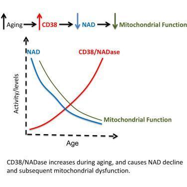
NAD is undergoes catabloic reduction by a n enzyme (CD38) that is 2,5-fold active in people over 60ys as in 35 year olds. CD38 might therefore be according to the reserchers, a promising candidate for mito-improving agents....
1-s2.0-S1550413116302248-fx1.jpg

CD38 Dictates Age-Related NAD Decline and Mitochondrial Dysfunction through an SIRT3-Dependent Mechanism
http://www.sciencedirect.com/science/art...550413116302248


Guess what: Apigenin might be one such inhibitor of CD38 activity

Flavonoid Apigenin Is an Inhibitor of the NAD+ase CD38
Flavonoid Apigenin Is an Inhibitor of the NAD+ase CD38: Implications for Cellular NAD+ Metabolism, Protein Acetylation, and Treatment of Metabolic Syndrome
 
OP
haidut

haidut

Member
Forum Supporter
Joined
Mar 18, 2013
Messages
19,799
Location
USA / Europe
NAD is undergoes catabloic reduction by a n enzyme (CD38) that is 2,5-fold active in people over 60ys as in 35 year olds. CD38 might therefore be according to the reserchers, a promising candidate for mito-improving agents....
1-s2.0-S1550413116302248-fx1.jpg

CD38 Dictates Age-Related NAD Decline and Mitochondrial Dysfunction through an SIRT3-Dependent Mechanism
http://www.sciencedirect.com/science/art...550413116302248


Guess what: Apigenin might be one such inhibitor of CD38 activity

Flavonoid Apigenin Is an Inhibitor of the NAD+ase CD38
Flavonoid Apigenin Is an Inhibitor of the NAD+ase CD38: Implications for Cellular NAD+ Metabolism, Protein Acetylation, and Treatment of Metabolic Syndrome

Yep, and there is more good news. Niacinamide itself inhibits CD38 :): Hard to get better than that - increases NAD and decreases its degradation and consumption by all sort of enzymes like PARP for example.
Evidence for a causal role of CD38 expression in granulocytic differentiation of human HL-60 cells. - PubMed - NCBI
"... Consistently, treatment of HL-60 cells with a permeant inhibitor of CD38, nicotinamide, inhibited both the CD38 activity and differentiation. "
 
Joined
Feb 26, 2018
Messages
988
@haidut You mention at the start of this thread that methelyne blue can be made more effective by assuring that pyruvate dehydrogenase is upregulated and cofactors such as niacinamide, magnesium, biotin and thiamine can be helpful. I have experimented off and on with various doses of MB, and never noticed much of anything, from microgram to 100+mg doses, however I may have been low in one of these cofactors, or perhaps I was in too poor health at the time to notice much. I am in better health now, with 98+ temps upon waking. Despite having good temps, good sleep, and generally more energy, I still feel like I’m only at 80% of where I’d like to be. I get home from work and usually feel zapped, despite a high 99 f temperature in the evening. I would like to know what a good MB plus co-factors stack would be, e.g. 1mg MB + 100mg niacinamide + 200mg magnesium... What time of day would be optimal? Also is taking with food a good idea, if so what macros are important to take along? Many thanks!
 
Last edited:
OP
haidut

haidut

Member
Forum Supporter
Joined
Mar 18, 2013
Messages
19,799
Location
USA / Europe
@haidut You mention at the start of this thread that methelyne blue can be made more effective by assuring that pyruvate dehydrogenase is upregulated and cofactors such as niacinamide, magnesium, biotin and thiamine can be helpful. I have experimented off and on with various doses of MB, and never noticed much of anything, from microgram to 100+mg doses, however I may have been low in one of these cofactors, or perhaps I was in too poor health at the time to notice much. I am in better health now, with 98+ temps upon waking. Despite having good temps, good sleep, and generally more energy, I still feel like I’m only at 80% of where I’d like to be. I get home from work and usually feel zapped, despite a high 99 f temperature in the evening. I would like to know what a good MB plus co-factors stack would be, e.g. 1mg MB + 100mg niacinamide + 200mg magnesium... What time of day woukd be optimal? Also is taking with food a good idea, if so what macros are important to take along? Many thanks!

I think 0.5mg-1mg MB combined with 100mg niacinamide, taken 2-3 times daily would be a great stack for raising NAD/NADH.
 
Joined
Feb 26, 2018
Messages
988
I think 0.5mg-1mg MB combined with 100mg niacinamide, taken 2-3 times daily would be a great stack for raising NAD/NADH.

Thank you. Is there a diurnal rhythm to NAD/NADH? Would what food was recently consumed or being in a fasted state have any significant effect on the efficacy of the stack?
 
Last edited:
OP
haidut

haidut

Member
Forum Supporter
Joined
Mar 18, 2013
Messages
19,799
Location
USA / Europe
Thank you. Is there a diurnal rhythm to NAD/NADH? Would what food was recently consumed or being in a fasted state have any significant effect on the efficacy of the stack?

I don't thin there is a rhythm unique to it but its levels may very with time of day due to other factors like cortisol, T, progesterone, DHEA, thyroid, etc. And yes, chronic fasting will probably lower NAD/NADH due to increased fat oxidation.
 

LeeLemonoil

Member
Joined
Sep 24, 2016
Messages
4,265
@haidut
from the stuy you quoted (with the excellent hint that Niacinamide inhibits CD38) --- does it not also hint at Doxycycline enhancing expression? At leat in the absence of RA?

Evidence for a causal role of CD38 expression in granulocytic differentiation of human HL-60 cells.

Granulocytic differentiation of human HL-60 cells can be induced by retinoic acid and is accompanied by a massive expression of CD38, a multi-functional enzyme responsible for metabolizing cyclic ADP-ribose (cADPR), a Ca(2+) messenger. Immunofluorescence staining showed that CD38 was expressed not only on the surface of intact HL-60 cells but also intracellularly, which was revealed after permeabilization with Triton. Concomitant with CD38 expression was the accumulation of cADPR, and both time courses preceded the onset of differentiation, suggesting a causal role for CD38. Consistently, treatment of HL-60 cells with a permeant inhibitor of CD38, nicotinamide, inhibited both the CD38 activity and differentiation. More specific blockage of CD38 expression was achieved by using morpholino antisense oligonucleotides targeting its mRNA, which produced a corresponding inhibition of differentiation as well. Similar inhibitory effects were observed when CD38 expression was reduced by the RNA interference technique targeting two separate regions of the coding sequence of CD38. Further support came from transfecting HL-60 cells with a Tet-On expression vector containing a full-length CD38. Subsequent treatments with doxycycline induced both CD38 expression and differentiation in the absence of retinoic acid. These results provide the first evidence that CD38 expression and the consequential accumulation of cADPR play a causal role in mediating cellular differentiation.
 
OP
haidut

haidut

Member
Forum Supporter
Joined
Mar 18, 2013
Messages
19,799
Location
USA / Europe
@haidut
from the stuy you quoted (with the excellent hint that Niacinamide inhibits CD38) --- does it not also hint at Doxycycline enhancing expression? At leat in the absence of RA?

Evidence for a causal role of CD38 expression in granulocytic differentiation of human HL-60 cells.

Granulocytic differentiation of human HL-60 cells can be induced by retinoic acid and is accompanied by a massive expression of CD38, a multi-functional enzyme responsible for metabolizing cyclic ADP-ribose (cADPR), a Ca(2+) messenger. Immunofluorescence staining showed that CD38 was expressed not only on the surface of intact HL-60 cells but also intracellularly, which was revealed after permeabilization with Triton. Concomitant with CD38 expression was the accumulation of cADPR, and both time courses preceded the onset of differentiation, suggesting a causal role for CD38. Consistently, treatment of HL-60 cells with a permeant inhibitor of CD38, nicotinamide, inhibited both the CD38 activity and differentiation. More specific blockage of CD38 expression was achieved by using morpholino antisense oligonucleotides targeting its mRNA, which produced a corresponding inhibition of differentiation as well. Similar inhibitory effects were observed when CD38 expression was reduced by the RNA interference technique targeting two separate regions of the coding sequence of CD38. Further support came from transfecting HL-60 cells with a Tet-On expression vector containing a full-length CD38. Subsequent treatments with doxycycline induced both CD38 expression and differentiation in the absence of retinoic acid. These results provide the first evidence that CD38 expression and the consequential accumulation of cADPR play a causal role in mediating cellular differentiation.

Yes, it does suggest that. But doxocycline is a quinone and has been shown to be a powerful oxidizing agent that increases NAD/NADH ratio. So, I guess its net effects have been shown to favor NAD (by oxidizing NADH) more so than it increases NAD consumption by CD38.
 

Amazoniac

Member
Joined
Sep 10, 2014
Messages
8,583
Location
Not Uganda
Just a collection of some publications on Raj's 'reductive stress' and related subjects..

- Reductive Stress in Inflammation-Associated Diseases and the Pro-Oxidant Effect of Antioxidant Agents

"Reductive stress (RS) is the counterpart oxidative stress (OS), and can occur in response to conditions that shift the redox balance of important biological redox couples, such as the NAD+/NADH, NADP+/NADPH, and GSH/GSSG, to a more reducing state. Overexpression of antioxidant enzymatic systems leads to excess reducing equivalents that can deplete reactive oxidative species, driving the cells to RS. A feedback regulation is established in which chronic RS induces OS, which in turn, stimulates again RS. Excess reducing equivalents may regulate cellular signaling pathways, modify transcriptional activity, induce alterations in the formation of disulfide bonds in proteins, reduce mitochondrial function, decrease cellular metabolism, and thus, contribute to the development of some diseases in which NF-κB, a redox-sensitive transcription factor, participates. Here, we described the diseases in which an inflammatory condition is associated to RS, and where delayed folding, disordered transport, failed oxidation, and aggregation are found. Some of these diseases are aggregation protein cardiomyopathy, hypertrophic cardiomyopathy, muscular dystrophy, pulmonary hypertension, rheumatoid arthritis, Alzheimer’s disease, and metabolic syndrome, among others. Moreover, chronic consumption of antioxidant supplements, such as vitamins and/or flavonoids, may have pro-oxidant effects that may alter the redox cellular equilibrium and contribute to RS, even diminishing life expectancy."

- Evidence in support of a concept of reductive stress

Too brief.

- Glutathione-dependent reductive stress triggers mitochondrial oxidation and cytotoxicity

"To investigate the effects of the predominant nonprotein thiol, glutathione (GSH), on redox homeostasis, we employed complementary pharmacological and genetic strategies to determine the consequences of both loss- and gain-of-function GSH content in vitro. We monitored the redox events in the cytosol and mitochondria using reduction-oxidation sensitive green fluorescent protein (roGFP) probes and the level of reduced/oxidized thioredoxins (Trxs). Either H2O2 or the Trx reductase inhibitor 1-chloro-2,4-dinitrobenzene (DNCB), in embryonic rat heart (H9c2) cells, evoked 8 or 50 mV more oxidizing glutathione redox potential, Ehc (GSSG/2GSH), respectively. In contrast, N-acetyl-l-cysteine (NAC) treatment in H9c2 cells, or overexpression of either the glutamate cysteine ligase (GCL) catalytic subunit (GCLC) or GCL modifier subunit (GCLM) in human embryonic kidney 293 T (HEK293T) cells, led to 3- to 4-fold increase of GSH and caused 7 or 12 mV more reducing Ehc, respectively. This condition paradoxically increased the level of mitochondrial oxidation, as demonstrated by redox shifts in mitochondrial roGFP and Trx2. Lastly, either NAC treatment (EC50 4 mM) or either GCLC or GCLM overexpression exhibited increased cytotoxicity and the susceptibility to the more reducing milieu was achieved at decreased levels of ROS. Taken together, our findings reveal a novel mechanism by which GSH-dependent reductive stress triggers mitochondrial oxidation and cytotoxicity."

- Increased reactive oxygen species production during reductive stress: The roles of mitochondrial glutathione and thioredoxin reductases

"Both extremes of redox balance are known to cause cardiac injury, with mounting evidence revealing that the injury induced by both oxidative and reductive stress is oxidative in nature. During reductive stress, when electron acceptors are expected to be mostly reduced, some redox proteins can donate electrons to O2 instead, which increases reactive oxygen species (ROS) production. However, the high level of reducing equivalents also concomitantly enhances ROS scavenging systems involving redox couples such as NADPH/NADP+ and GSH/GSSG. Here our objective was to explore how reductive stress paradoxically increases net mitochondrial ROS production despite the concomitant enhancement of ROS scavenging systems. Using recombinant enzymes and isolated permeabilized cardiac mitochondria, we show that two normally antioxidant matrix NADPH reductases, glutathione reductase and thioredoxin reductase, generate H2O2 by leaking electrons from their reduced flavoprotein to O2 when electron flow is impaired by inhibitors or because of limited availability of their natural electron acceptors, GSSG and oxidized thioredoxin. The spillover of H2O2 under these conditions depends on H2O2 reduction by peroxiredoxin activity, which may regulate redox signaling in response to endogenous or exogenous factors. These findings may explain how ROS production during reductive stress overwhelms ROS scavenging capability, generating the net mitochondrial ROS spillover causing oxidative injury. These enzymes could potentially be targeted to increase cancer cell death or modulate H2O2-induced redox signaling to protect the heart against ischemia/reperfusion damage."

- Glutathione Redox State Regulates Mitochondrial Reactive Oxygen Production

"Oxidative stress induced by 2,3,7,8-tetrachlorodibenzo-p-dioxin (TCDD; dioxin) is poorly understood. Following one dose of TCDD (5 μg/kg body weight), mitochondrial succinate-dependent production of superoxide and H2O2 in mouse liver doubled at 7–28 days, then subsided by day 56; concomitantly, levels of GSH and GSSG increased in both cytosol and mitochondria. Cytosol displayed a typical oxidative stress response, consisting of diminished GSH relative to GSSG, decreased potential to reduce protein-SSG mixed disulfide bonds (type 1 thiol redox switch) or protein-SS-protein disulfide bonds (type 2 thiol redox switch), and a +10 mV change in GSSG/2GSH reduction potential. In contrast, mitochondria showed a rise in reduction state, consisting of increased GSH relative to GSSG, increases in type 1 and type 2 thiol redox switches, and a –25 mV change in GSSG/2GSH reduction potential. Comparing Ahr(–/–) knock-out and wild-type mice, we found that TCDD-induced thiol changes in both cytosol and mitochondria were dependent on the aromatic hydrocarbon receptor (AHR). GSH was rapidly taken up by mitochondria and stimulated succinate-dependent H2O2 production. A linear dependence of H2O2 productionon thereduction potential for GSSG/2GSH exists between –150 and –300 mV. The TCDD-stimulated increase in succinate-dependent and thiol-stimulated production of reactive oxygen paralleled a four-fold increase in formamidopyrimidine DNA N-glycosylase (FPG)-sensitive cleavage sites in mitochondrial DNA, compared with a two-fold increase in nuclear DNA. These results suggest that TCDD produces an AHR-dependent oxidative stress in mitochondria, with concomitant mitochondrial DNA damage mediated, at least in part, by an increase in the mitochondrial thiol reduction state."

- Redox environment of the cell as viewed through the redox state of the glutathione disulfide/glutathione couple

"Redox state is a term used widely in the research field of free radicals and oxidative stress. Unfortunately, it is used as a general term referring to relative changes that are not well defined or quantitated. In this review we provide a definition for the redox environment of biological fluids, cell organelles, cells, or tissue. We illustrate how the reduction potential of various redox couples can be estimated with the Nernst equation and show how pH and the concentrations of the species comprising different redox couples influence the reduction potential. We discuss how the redox state of the glutathione disulfide-glutathione couple (GSSG/2GSH) can serve as an important indicator of redox environment. There are many redox couples in a cell that work together to maintain the redox environment; the GSSG/2GSH couple is the most abundant redox couple in a cell. Changes of the half-cell reduction potential (Ehc) of the GSSG/2GSH couple appear to correlate with the biological status of the cell: proliferation Ehc ≈ −240 mV; differentiation Ehc ≈ −200 mV; or apoptosis Ehc ≈ −170 mV. These estimates can be used to more fully understand the redox biochemistry that results from oxidative stress. These are the first steps toward a new quantitative biology, which hopefully will provide a rationale and understanding of the cellular mechanisms associated with cell growth and development, signaling, and reductive or oxidative stress."

- Redox compartmentalization and cellular stress

"Mammalian cells are highly organized to optimize function. For instance, oxidative energy‐producing processes in mitochondria are sequestered away from plasma membrane redox signalling complexes and also from nuclear DNA, which is subject to oxidant‐induced mutation. Proteins are unique among macromolecules in having reversible oxidizable elements, ‘sulphur switches', which support dynamic regulation of structure and function. Accumulating evidence shows that redox signalling and control systems are maintained under kinetically limited steady states, which are highly displaced from redox equilibrium and distinct among organelles. Mitochondria are most reducing and susceptible to oxidation under stressed conditions, while nuclei are also reducing but relatively resistant to oxidation. Within compartments, the glutathione and thioredoxin systems serve parallel and non‐redundant functions to maintain the dynamic redox balance of subsets of protein cysteines, which function in redox signalling and control. This organization allows cells to be poised to respond to cell stress but also creates sites of vulnerability. Importantly, disruption of redox organization is a common basis for disease. Research tools are becoming available to elucidate details of subcellular redox organization, and this development highlights an opportunity for a new generation of targeted antioxidants to enhance and restore redox signalling and control in disease prevention."

- Oxidative Shielding or Oxidative Stress?

"In this review I report evidence that the mainstream field of oxidative damage biology has been running fast in the wrong direction for more than 50 years. Reactive oxygen species (ROS) and chronic oxidative changes in membrane lipids and proteins found in many chronic diseases are not the result of accidental damage. Instead, these changes are the result of a highly evolved, stereotyped, and protein-catalyzed “oxidative shielding” response that all eukaryotes adopt when placed in a chemically or microbially hostile environment. The machinery of oxidative shielding evolved from pathways of innate immunity designed to protect the cell from attack and limit the spread of infection. Both oxidative and reductive stress trigger oxidative shielding. In the cases in which it has been studied explicitly, functional and metabolic defects occur in the cell before the increase in ROS and oxidative changes. ROS are the response to disease, not the cause. Therefore, it is not the oxidative changes that should be targeted for therapy, but rather the metabolic conditions that create them. This fresh perspective is relevant to diseases that range from autism, type 1 diabetes, type 2 diabetes, cancer, heart disease, schizophrenia, Parkinson's disease, and Alzheimer disease. Research efforts need to be redirected. Oxidative shielding is protective and is a misguided target for therapy. Identification of the causal chemistry and environmental factors that trigger innate immunity and metabolic memory that initiate and sustain oxidative shielding is paramount for human health."

- Pathogenesis of Chronic Hyperglycemia: From Reductive Stress to Oxidative Stress

"Chronic overnutrition creates chronic hyperglycemia that can gradually induce insulin resistance and insulin secretion impairment. These disorders, if not intervened, will eventually be followed by appearance of frank diabetes. The mechanisms of this chronic pathogenic process are complex but have been suggested to involve production of reactive oxygen species (ROS) and oxidative stress. In this review, I highlight evidence that reductive stress imposed by overflux of NADH through the mitochondrial electron transport chain is the source of oxidative stress, which is based on establishments that more NADH recycling by mitochondrial complex I leads to more electron leakage and thus more ROS production. The elevated levels of both NADH and ROS can inhibit and inactivate glyceraldehyde 3-phosphate dehydrogenase (GAPDH), respectively, resulting in blockage of the glycolytic pathway and accumulation of glycerol 3-phospate and its prior metabolites along the pathway. This accumulation then initiates all those alternative glucose metabolic pathways such as the polyol pathway and the advanced glycation pathways that otherwise are minor and insignificant under euglycemic conditions. Importantly, all these alternative pathways lead to ROS production, thus aggravating cellular oxidative stress. Therefore, reductive stress followed by oxidative stress comprises a major mechanism of hyperglycemia-induced metabolic syndrome."

- The role of cytosolic reductive stress in oxidant formation and diabetic complications.

Not awailable.

- A “Reductionist” View of Cardiomyopathy

"Oxidative stress due to the generation of reactive oxygen species has been implicated in many diseases. Rajasekaran et al. (2007) now make the surprising discovery that its counterpart “reductive stress,” caused by an increase in reduced glutathione, contributes to cardiomyopathy triggered by protein aggregation."

- Reductive Stress - Linking Heat Shock Protein 27, Glutathione, and Cardiomyopathy?

It's a brief commentary.

- Reductive potential — A savior turns stressor in protein aggregation cardiomyopathy

"Redox homeostasis is essential for basal signaling of several physiological processes, but a unilateral shift towards an ‘oxidative’ or ‘reductive’ trait will alter intracellular redox milieu. Typically, such an event influences the structure and the native function of a cell or an organelle. Numerous experimental research and clinical trials over the last 6 decades have demonstrated that enhanced oxygen-derived free radicals constitute a major stimulus to trigger damage in several human diseases, including cardiovascular complications supporting the theory of oxidative stress (OS). However, until our key discovery, the dynamic interrelationship between “Reductive Stress (RS)” and cardiac health has been obscured by overwhelming OS studies (Rajasekaran et al., 2007). Notably, this seminal finding spurred considerable interest in investigations of other mechanistic insights, and thus far the results indicate a similar or stronger role for RS, as that of OS. In addition, from our own findings we strongly believe that constitutive activation of pathways that enable sustained generation of reducing equivalents of glutathione (GSH), reduced nicotinamide adenine dinucleotide phosphate (NADPH) will cause RS and impair the basal cellular signaling mechanisms operating through harmless pro-oxidative events, in turn, disrupting single and/or a combination of key cellular processes such as growth, maturation, differentiation, survival, death etc., that govern healthy cell physiology. Here, we have discussed the role of RS as a causal or contributing factor in relevant pathophysiology of a major cardiac disease of human origin."

- Responses to Reductive Stress in the Cardiovascular System

"There is a growing appreciation that reductive stress represents a disturbance in the redox state that is harmful to biological systems. On a cellular level, the presence of increased reducing equivalents and the lack of beneficial fluxes of reactive oxygen species can prevent growth factor-mediated signalling, promote mitochondrial dysfunction, increase apoptosis, and decrease cell survival. In this review, we highlight the importance of redox balance in maintaining cardiovascular homeostasis and consider the tenuous balance between oxidative and reductive stress. We explain the role of reductive stress in models of protein aggregation-induced cardiomyopathies, such as those caused by mutations in αB-crystallin. In addition, we discuss the role of NADPH oxidases in models of heart failure and ischemia-reperfusion to illustrate how oxidants may mediate the adaptive responses to injury. NADPH oxidase 4, a hydrogen peroxide generator, also has a major role in promoting vascular homeostasis through its regulation of vascular tone, angiogenic responses, and effects on atherogenesis. In contrast, the lack of antioxidant enzymes that reduce hydrogen peroxide, such as glutathione peroxidase 1, promotes vascular remodeling and is deleterious to endothelial function. Thus, we consider the role of oxidants as necessary signals to promote adaptive responses, such as the activation of Nrf2 and eNOS, and the stabilization of Hif1. In addition, we discuss the adaptive metabolic reprogramming in hypoxia that lead to a reductive state, and the subsequent cellular redistribution of reducing equivalents from NADH to other metabolites. Finally, we discuss the paradoxical ability of excess reducing equivalents to stimulate oxidative stress and promote injury."

- Antioxidant-induced reductive stress has untoward consequences on the brain microvasculature

:neener

- Reductive Stress: A New Concept in Alzheimer’s Disease

"Reactive oxygen species play a physiological role in cell signaling and also a pathological role in diseases, when antioxidant defenses are overwhelmed causing oxidative stress. However, in this review we will focus on reductive stress that may be defined as a pathophysiological situation in which the cell becomes more reduced than in the normal, resting state. This may occur in hypoxia and also in several diseases in which a small but persistent generation of oxidants results in a hormetic overexpression of antioxidant enzymes that leads to a reduction in cell compartments. This is the case of Alzheimer’s disease. Individuals at high risk of Alzheimer’s (because they carry the ApoE4 allele) suffer reductive stress long before the onset of the disease and even before the occurrence of mild cognitive impairment. Reductive stress can also be found in animal models of Alzheimer’s disease (APP/PS1 transgenic mice), when their redox state is determined at a young age, i.e. before the onset of the disease. Later in their lives they develop oxidative stress. The importance of understanding the occurrence of reductive stress before any signs or symptoms of Alzheimer’s has theoretical and also practical importance as it may be a very early marker of the disease."

- Reductive stress in young healthy individuals at risk of Alzheimer disease

"Oxidative stress is a hallmark of Alzheimer disease (AD) but this has not been studied in young healthy persons at risk of the disease. Carrying an Apo ε4 allele is the major genetic risk factor for AD. We have observed that lymphocytes from young, healthy persons carrying at least one Apo ε4 allele suffer from reductive rather than oxidative stress, i.e., lower oxidized glutathione and P-p38 levels and higher expression of enzymes involved in antioxidant defense, such as glutamylcysteinyl ligase and glutathione peroxidase. In contrast, in the full-blown disease, the situation is reversed and oxidative stress occurs, probably because of the exhaustion of the antioxidant mechanisms just mentioned. These results provide insights into the early events of the progression of the disease that may allow us to find biomarkers of AD at its very early stages."

- Reductive stress after exercise: The issue of redox individuality

"Exercise has been consistently used as an oxidant stimulus in redox biology studies. However, previous studies have focused on group differences and did not examine individual differences. As a result, it remains untested whether all individuals experience oxidative stress after acute exercise. Therefore, the main aim of the present study was to investigate whether some individuals exhibit unexpected responses after an acute eccentric (i.e., muscle-damaging) exercise session. Ninety eight (N = 98) young men performed an isokinetic eccentric exercise bout with the knee extensors. Plasma, erythrocytes and urine samples were collected immediately before and 2 days post-exercise. Three commonly used redox biomarkers (F2-isoprostanes, protein carbonyls and glutathione) were assayed. As expected, the two oxidant biomarkers (F2-isoprostanes and protein carbonyls) significantly increased 2 days after exercise (46% and 61%, respectively); whereas a significant decrease in glutathione levels (by −21%) was observed after exercise. A considerable number of the participants exhibited changes in the levels of biomarkers in the opposite, unexpected direction than the group average. More specifically, 13% of the participants exhibited a decrease in F2-isoprostanes and protein carbonyls and 10% of the participants exhibited an increase in glutathione levels. Furthermore, more than 1 out of 3 individuals exhibited either unexpected or negligible (from 0% to ± 5%) responses to exercise in at least one redox biomarker. It was also observed that the initial values of redox biomarkers are important predictors of the responses to exercise. In conclusion, although exercise induces oxidative stress in the majority of individuals, it can induce reductive stress or negligible stress in a considerable number of people. The data presented herein emphasize that the mean response to a redox stimulus can be very misleading. We believe that the wide variability (including the cases of reductive stress) described is not limited to the oxidant stimulus used and the biomarkers selected."

- Redox regulation of antioxidants, autophagy, and the response to stress: Implications for electrophile therapeutics

"Redox networks in the cell integrate signaling pathways that control metabolism, energetics, cell survival, and death. The physiological second messengers that modulate these pathways include nitric oxide, hydrogen peroxide, and electrophiles. Electrophiles are produced in the cell via both enzymatic and nonenzymatic lipid peroxidation and are also relatively abundant constituents of the diet. These compounds bind covalently to families of cysteine-containing, redox-sensing proteins that constitute the electrophile-responsive proteome, the subproteomes of which are found in localized intracellular domains. These include those proteins controlling responses to oxidative stress in the cytosol—notably the Keap1-Nrf2 pathway, the autophagy-lysosomal pathway, and proteins in other compartments including mitochondria and endoplasmic reticulum. The signaling pathways through which electrophiles function have unique characteristics that could be exploited for novel therapeutic interventions; however, development of such therapeutic strategies has been challenging due to a lack of basic understanding of the mechanisms controlling this form of redox signaling. In this review, we discuss current knowledge of the basic mechanisms of thiol-electrophile signaling and its potential impact on the translation of this important field of redox biology to the clinic. Emerging understanding of thiol-electrophile interactions and redox signaling suggests replacement of the oxidative stress hypothesis with a new redox biology paradigm, which provides an exciting and influential framework for guiding translational research."

- Simultaneous generation of methane, carbon dioxide, and carbon monoxide from choline and ascorbic acid: a defensive mechanism against reductive stress?

"Recent evidences suggest that reductive stress rather than oxidative stress is a common cause of abnormal biological oxygen radical activity. We hypothesized that under such conditions, electrophilic methyl groups (EMGs) bound to positively charged nitrogen or sulfur moieties may act as protective electron acceptors and that this poising mechanism may entail the generation of methane gas."

- Electrophilic methyl groups present in the diet ameliorate pathological states induced by reductive and oxidative stress: a hypothesis

"Reductive stress, characterised by an increased NADH:NAD+ ratio, may be as common and as important a consequence of redox imbalance as oxidative stress. It may also be an important predisposing cause of the generation of reactive oxygen species. Considerable experimental and indirect clinical evidence suggests that protection against reductive stress depends on biomolecules with electrophilic methyl groups (EMG) such as S-adenosylmethionine, betaine, carnitine and phosphatidylcholine. Pathological processes leading to reductive stress and their relief by such protective agents is reviewed and the proposed molecular mechanism is outlined. These and other EMG-containing biomolecules are part of the daily diet and may represent an important control system for redox balance."

- Hypoxia-Induced Generation of Methane in Mitochondria and Eukaryotic Cells - An Alternative Approach to Methanogenesis

"Background/Aims: Electrophilic methyl groups bound to positively charged nitrogen moieties may act as electron acceptors, and this mechanism could lead to the generation of methane from choline. The aims were to characterize the methanogenic potential of phosphatidylcholine metabolites, and to define the in vivo relevance of this pathway in hypoxia-induced cellular responses. Methods: The postulated reaction was investigated (1) in model chemical experiments, (2) in rat mitochondrial subfractions and (3) in bovine endothelial cell cultures under hypoxic conditions and in the presence of hydroxyl radical generation. The rate of methane formation was determined by gas chromatography with flame-ionisation detectors. The lucigenin-enhanced chemiluminescence assay was used to determine the reactive oxygen species-scavenging capacity of the choline metabolites. Results: Significant methane generation was demonstrated in all three series of experiments. Phosphatidylcholine metabolites with alcoholic moiety in the molecule (i.e. choline, N,N-dimethylethanolamine and N-methylethanolamine), inhibited oxygen radical production both in vitro and in vivo, and displayed an effectiveness proportional to the amount of methane generated and the number of methyl groups in the compounds. Conclusion: Methane generation occurs in aerobic systems. Phosphatidylcholine metabolites containing both electron donor and acceptor groups may have a function to counteract intracellular oxygen radical production."

- Molecular Mechanisms of Hydrogen Sulfide Toxicity

"Rationale. The toxicity of H2S has been attributed to its ability to inhibit cytochrome c oxidase in a similar manner to HCN. However, the successful use of methemoglobin for the treatment of HCN poisoning was not successful for H2S poisonings even though the ferric heme group of methemoglobin scavenges H2S. Thus, we speculated that other mechanisms contribute to H2S induced cytotoxicity. Experimental procedure. Hepatocyte isolation and viability and enzyme activities were measured as described by , and . Results. Incubation of isolated hepatocytes with NaHS solutions (a H2S source) resulted in glutathione (GSH) depletion. Moreover, GSH depletion was also observed in TRIS-HCl buffer (pH 6.0) treated with NaHS. Several ferric chelators (desferoxamime and DETAPAC) and antioxidant enzymes (superoxide dismutase [SOD] and catalase) prevented cell-free and hepatocyte GSH depletion. GSH-depleted hepatocytes were very susceptible to NaHS cytotoxicity, indicating that GSH detoxified NaHS or H2S in cells. Cytotoxicity was also partly prevented by desferoxamine and DETAPC, but it was increased by ferric EDTA or EDTA. Cell-free oxygen consumption experiments in TRIS-HCl buffer showed that NaHS autoxidation formed hydrogen peroxide and was prevented by DETAPC but increased by EDTA. We hypothesize that H2S can reduce intracellular bound ferric iron to form unbound ferrous iron, which activates iron. Additionally, H2S can increase the hepatocyte formation of reactive oxygen species (ROS) (known to occur with electron transport chain). H2S cytotoxicity therefore also involves a reactive sulfur species, which depletes GSH and activates oxygen to form ROS."

- The NAD ratio redox paradox: why does too much reductive power cause oxidative stress?

"The reductive power provided by nicotinamide adenine dinucleotides is invaluable for several cellular processes. It drives metabolic reactions, enzymatic activity, regulates genetic expression and allows for the maintenance of a normal cell redox status. Therefore, the balance between the oxidized (NAD+) and the reduced (NADH) forms is critical for the cell’s proper function and ultimately, for its survival. Being intimately associated with the cells’ metabolism, it is expected that alterations to the NAD+/NADH ratio are to be found in situations of metabolic diseases, as is the case of diabetes. NAD+ is a necessary cofactor for several enzymes’ activity, many of which are related to metabolism. Therefore, a decrease in the NAD+/NADH ratio causes these enzymes to decrease in activity (reductive stress), resulting in an altered metabolic situation that might be the first insult toward several pathologies, such as diabetes. Here, we review the importance of nicotinamide adenine dinucleotides in the liver cell and its fluctuations in a state of type 2 diabetes mellitus."

- Complementation of mitochondrial electron transport chain by manipulation of the NAD+/NADH ratio

"A decline in electron transport chain (ETC) activity is associated with many human diseases. Although diminished mitochondrial ATP production is recognized as a source of pathology, the contribution of the associated reduction in the ratio of the amount of oxidized nicotinamide adenine dinucleotide (NAD+) to that of its reduced form (NADH) is less clear. We used a water-forming NADH oxidase from L. brevis (LbNOX) as a genetic tool for inducing a compartment-specific increase of the NAD+/NADH ratio in human cells. We used LbNOX to demonstrate the dependence of key metabolic fluxes, gluconeogenesis, and signaling on the cytosolic or mitochondrial NAD+/NADH ratios. Expression of LbNOX in the cytosol or mitochondria ameliorated proliferative and metabolic defects caused by an impaired ETC. The results underscore the role of reductive stress in mitochondrial pathogenesis and demonstrate the utility of targeted LbNOX for direct, compartment-specific manipulation of redox state."

- NAD+ and NADH in cellular functions and cell death

"Increasing evidence has indicated that NAD+ and NADH play critical roles not only in energy metabolism, but also in cell death and various cellular functions including regulation of calcium homeostasis and gene expression. It has also been indicated that NAD+ and NADH are mediators of multiple major biological processes including aging. NAD+ and NADH produce the biological effects by regulating numerous NAD+/NADH-dependent enzymes, including dehydrogenases, poly(ADP-ribose) polymerases, Sir2 family proteins (sirtuins), mono(ADP-ribosyl)transferases, and ADP-ribosyl cyclases. Of particular interest, [NAD+]-dependent generation of ADP-ribose, cyclic ADP-ribose and O-acetyl-ADP-ribose can mediate calcium homeostasis by affecting TRPM2 receptors and ryanodine receptors; and sirtuins and PARPs appear to play key roles in aging, cell death and a variety of cellular functions. It has also been indicated that NADH and NAD+ can be transported across plasma membranes of cells, and that extracellular NAD+ may be a new signaling molecule. Our latest studies have shown that intranasal NAD+ administration can profoundly decrease ischemic brain damage. These new pieces of information have fundamentally changed our understanding about NAD+ and NADH, suggesting novel paradigms about the metabolism and biological activities of NAD+ and NADH. Based on this information, it is tempted to hypothesize that NAD+ and NADH, together with ATP and Ca2+, may be four most fundamental components in life, which can significantly affect nearly all major biological processes. Future studies on NAD+ and NADH may not only elucidate some fundamental mysteries in biology, but also provide novel insights for interfering aging and many disease processes."

- Coenzyme Q Cytoprotective Mechanisms for Mitochondrial Complex I Cytopathies Involves NAD(P)H: Quinone Oxidoreductase 1(NQO1)

"The commonest mitochondrial diseases are probably those impairing the function of complex I of the respiratory electron transport chain. Such complex I impairment may contribute to various neurodegenerative disorders e.g. Parkinson's disease. In the following, using hepatocytes as a model cell, we have shown for the first time that the cytotoxicity caused by complex I inhibition by rotenone but not that caused by complex III inhibition by antimycin can be prevented by coenzyme Q (CoQ1) or menadione. Furthermore, complex I inhibitor cytotoxicity was associated with the collapse of the mitochondrial membrane potential and reactive oxygen species (ROS) formation. ROS scavengers or inhibitors of the mitochondrial permeability transition prevented cytotoxicity. The CoQ1 cytoprotective mechanism required CoQ1 reduction by DT-diaphorase (NQO1). Furthermore, the mitochondrial membrane potential and ATP levels were restored at low CoQ1 concentrations (5 uM). This suggests that the CoQ1H2 formed by NQO1 reduced complex III and acted as an electron bypass of the rotenone block. However cytoprotection still occurred at higher CoQ1 concentrations (>10 uM), which were less effective at restoring ATP levels but readily restored the cellular cytosolic redox potential (i.e. lactate: pyruvate ratio) and prevented ROS formation. This suggests that CoQ1 or menadione cytoprotection also involves the NQO1 catalysed reoxidation of NADH that accumulates as a result of complex I inhibition. The CoQ1H2 formed would then also act as a ROS scavenger."
 
Last edited:

Amazoniac

Member
Joined
Sep 10, 2014
Messages
8,583
Location
Not Uganda
"[..]chronic RS can induce OS [operational system], which stimulates again RS by a feedback regulation. For example, during RS, when electron acceptors are expected to be mostly reduced, some redox proteins can donate electrons to O2 instead, thus increasing ROS production [18]. However, a high level of reducing equivalents also enhances ROS scavenging systems, involving redox couples such as the NAD/NADH+, NADPH/NADP+, and glutathione reduce (GSH)/glutathione oxidized (GSSG) ratio [18,19], resulting in a net H2O2 spillover from mitochondria that favors RS [19].

On the other hand, the term mitochondrial homeostasis refers to how low doses of mitochondrial ROS produced by the respiratory electron transport chain (RETC) can activate the biogenesis and the antioxidant capacity, in order to counteract OS and to re-establish homeostasis [1]."

"Mitochondrial ROS and their depletion by RS play an essential and necessary role in the correct folding of proteins and in the formation of disulfide bonds, which determine the normal structure and function of many proteins [19]. When the mitochondrial oxidant production is inhibited, there is an important decrease in the levels of cellular disulfide bonds in many cells [20]. RS leads to the loss of disulfide bond formation and induces the unfolded protein response of the ER endoplasmic reticulum (UPR[ER]). The recuperation of the correct folding of proteins is necessary to regain proteostasis in this compartment [21]. It has been reported that H2O2 accumulation during RS attenuated the UPR[ER] amplitude by altering translation, without any discernible effect on transcription in Saccharomyces cerevisiae [22]. In yeast with RS, some proteins showed delayed folding, disordered transport and failed oxidation, and were finally aggregated [15]."

"[..]overproduction of NADH or lack of NAD+ can induce the accumulation of NADH [24]. Overproduction of NADH induces an electron pressure upon mitochondrial complex I, which responds within its capacity, to oxidize more NADH to NAD+. This leads to an increase in electron leakage that decreases oxygen to yield O2−. These free radicals, in turn, enhance OS. Due to a high level of reducing equivalents, such as NADH, an oxidative condition appears [25], and it achieves the transition to RS by the polyol pathway. This pathway converts NADPH to NADH, leading to a redox imbalance between NADH and NAD+ [26]. This condition could be linked to metabolic syndrome (MS) and diabetes."

"GSH excess could decrease the basal ROS and contribute to RS [34,35]."

"Several biological functions in the human body depend on the balance of Se levels, and decreased or elevated levels can cause damaging effects." "The synthesis of Se proteins such as GPx isoforms is affected by levels of Se supplementation; however, exceeding and inadequate Se intake can produce damaging health effects and contribute to RS by upregulated Se-protein W (SelW) mRNA expression [54]."

"[..]under hyperglycemic conditions, the polyol pathway utilizes more than 30% of the body glucose, which significantly contributes to RS [105]. Moreover, iNOS also uses NADPH as a cofactor, contributing to hypertension in MS. Therefore, RS followed by OS could act as an important process of glucotoxicity when chronic hyperglycemic conditions are present. It would induce RS, which is linked to the inhibition of insulin release by pancreatic β-cells [24]."

Kidney section for @yerrag, to be conservative with N and A and C supplementation. I suspect low doses are fine if you really need it, and NAC might be preferable to straight reduced glutathione for requiring its synthesis.

"The products of the prototypical glucose regulated (grp) genes: grp94 and grp78 play important roles as chaperones during protein folding and processing in the ER [76], and are also linked to inflammatory conditions, such renal disease. These genes are members of the gene battery that is responsive to RS, while the hsp genes respond to OS [121,122]. Thiol reductions are also cytotoxic and increase expression of grp genes. Agents that interfere with ER protein folding include thiols that activate grp78 transcription [75]. In LLC-PK1 renal epithelial cells, DTT treatment induces grp78 gene expression and gadd153 gene transcription. In addition, in human embryonic kidney cells N-acetyl-l-cysteine treatment led to 3- to 4-fold increase of GSH. This increased the level of mitochondrial oxidation, and drove to RS that could later on lead to oxidative stress [14]. RS associated to hypoxia causes the L2HG enantiomer accumulation in renal cell carcinoma of children. Cell lines with RETC defects and D2HG have been identified as the product of cancer-associated mutant enzyme cytosolic isocitrate dehydrogenase-1 [65]."

"[..]the use of antioxidants is not completely effective for treating neurodegenerative diseases, chronic inflammation, cardiovascular diseases, and cancer, and can even increase the production of free radicals. High doses of antioxidants can also lead to cellular dysfunction, by altering the redox balance after interacting with physiological concentrations of ROS [113]. Thereby, antioxidants may increase the damage to the body by interfering with the metabolism of some nutrients, increase the risk of cancer, or reduce the effectiveness of cancer treatments (e.g., radiation therapy, chemotherapy), thus decreasing the health-promoting effects of exercise, and even decreasing life expectancy [129]."
 
Joined
Feb 26, 2018
Messages
988
"[..]chronic RS can induce OS [operational system], which stimulates again RS by a feedback regulation. For example, during RS, when electron acceptors are expected to be mostly reduced, some redox proteins can donate electrons to O2 instead, thus increasing ROS production [18]. However, a high level of reducing equivalents also enhances ROS scavenging systems, involving redox couples such as the NAD/NADH+, NADPH/NADP+, and glutathione reduce (GSH)/glutathione oxidized (GSSG) ratio [18,19], resulting in a net H2O2 spillover from mitochondria that favors RS [19].

On the other hand, the term mitochondrial homeostasis refers to how low doses of mitochondrial ROS produced by the respiratory electron transport chain (RETC) can activate the biogenesis and the antioxidant capacity, in order to counteract OS and to re-establish homeostasis [1]."

"Mitochondrial ROS and their depletion by RS play an essential and necessary role in the correct folding of proteins and in the formation of disulfide bonds, which determine the normal structure and function of many proteins [19]. When the mitochondrial oxidant production is inhibited, there is an important decrease in the levels of cellular disulfide bonds in many cells [20]. RS leads to the loss of disulfide bond formation and induces the unfolded protein response of the ER endoplasmic reticulum (UPR[ER]). The recuperation of the correct folding of proteins is necessary to regain proteostasis in this compartment [21]. It has been reported that H2O2 accumulation during RS attenuated the UPR[ER] amplitude by altering translation, without any discernible effect on transcription in Saccharomyces cerevisiae [22]. In yeast with RS, some proteins showed delayed folding, disordered transport and failed oxidation, and were finally aggregated [15]."

"[..]overproduction of NADH or lack of NAD+ can induce the accumulation of NADH [24]. Overproduction of NADH induces an electron pressure upon mitochondrial complex I, which responds within its capacity, to oxidize more NADH to NAD+. This leads to an increase in electron leakage that decreases oxygen to yield O2−. These free radicals, in turn, enhance OS. Due to a high level of reducing equivalents, such as NADH, an oxidative condition appears [25], and it achieves the transition to RS by the polyol pathway. This pathway converts NADPH to NADH, leading to a redox imbalance between NADH and NAD+ [26]. This condition could be linked to metabolic syndrome (MS) and diabetes."

"GSH excess could decrease the basal ROS and contribute to RS [34,35]."

"Several biological functions in the human body depend on the balance of Se levels, and decreased or elevated levels can cause damaging effects." "The synthesis of Se proteins such as GPx isoforms is affected by levels of Se supplementation; however, exceeding and inadequate Se intake can produce damaging health effects and contribute to RS by upregulated Se-protein W (SelW) mRNA expression [54]."

"[..]under hyperglycemic conditions, the polyol pathway utilizes more than 30% of the body glucose, which significantly contributes to RS [105]. Moreover, iNOS also uses NADPH as a cofactor, contributing to hypertension in MS. Therefore, RS followed by OS could act as an important process of glucotoxicity when chronic hyperglycemic conditions are present. It would induce RS, which is linked to the inhibition of insulin release by pancreatic β-cells [24]."

Kidney section for @yerrag, to be conservative with N and A and C supplementation. I suspect low doses are fine if you really need it, and NAC might be preferable to straight reduced glutathione for requiring its synthesis.

"The products of the prototypical glucose regulated (grp) genes: grp94 and grp78 play important roles as chaperones during protein folding and processing in the ER [76], and are also linked to inflammatory conditions, such renal disease. These genes are members of the gene battery that is responsive to RS, while the hsp genes respond to OS [121,122]. Thiol reductions are also cytotoxic and increase expression of grp genes. Agents that interfere with ER protein folding include thiols that activate grp78 transcription [75]. In LLC-PK1 renal epithelial cells, DTT treatment induces grp78 gene expression and gadd153 gene transcription. In addition, in human embryonic kidney cells N-acetyl-l-cysteine treatment led to 3- to 4-fold increase of GSH. This increased the level of mitochondrial oxidation, and drove to RS that could later on lead to oxidative stress [14]. RS associated to hypoxia causes the L2HG enantiomer accumulation in renal cell carcinoma of children. Cell lines with RETC defects and D2HG have been identified as the product of cancer-associated mutant enzyme cytosolic isocitrate dehydrogenase-1 [65]."

"[..]the use of antioxidants is not completely effective for treating neurodegenerative diseases, chronic inflammation, cardiovascular diseases, and cancer, and can even increase the production of free radicals. High doses of antioxidants can also lead to cellular dysfunction, by altering the redox balance after interacting with physiological concentrations of ROS [113]. Thereby, antioxidants may increase the damage to the body by interfering with the metabolism of some nutrients, increase the risk of cancer, or reduce the effectiveness of cancer treatments (e.g., radiation therapy, chemotherapy), thus decreasing the health-promoting effects of exercise, and even decreasing life expectancy [129]."

Wouldn’t this all corroborate with the fundamental theory behind ozone and hydrogen peroxide therapy? Essentially the theory is that providing small doses of pro oxidants is health promoting, in that the immune system is strengthened and the cells become more geared towards aerobic metabolism and handling oxidative stress...
 

Amazoniac

Member
Joined
Sep 10, 2014
Messages
8,583
Location
Not Uganda
Wouldn’t this all corroborate with the fundamental theory behind ozone and hydrogen peroxide therapy? Essentially the theory is that providing small doses of pro oxidants is health promoting, in that the immune system is strengthened and the cells become more geared towards aerobic metabolism and handling oxidative stress...
Reminded me of this idea:
- Antioxidants prevent health-promoting effects of physical exercise in humans
- Are Bigger Muscles Better? Antioxidants and the Response to Exercise - Perfect Health Diet
 

magnesiumania

Member
Joined
Jul 17, 2018
Messages
607
Greetings Haidut @Travis
Haidut, this is a super interesting thread, but problematic in some ways for me, as I I tend to have counterintuitive responses to things discussed like ALA, NDT, supplemental vitamin D (sunshine...no problemo though), etc. And, lower doses of almost anything are safer in my case. Anything over 100 mgs niacinamide leaves me groggy and with a dull headache. Glycine I have found to be detrimental too. IOW, as a hospital worker once told me, I am one of the aliens among us. I was one of the 1,500 or so tryptophan poisoning victims in 1989, and I believe elevated histamine and serotonin have been pretty much lifelong issues for me. Lately. I believe I have uncovered an extreme problem with glutamate exitotoxicity...seeing disturbing neurological sides from milk, fermented foods, dark chocolate, etc. I was giving serious thought to ordering some oxaloacetate to try to alleviate some of this. I just recently started 0.5 to 1 mg of Periactin at night, and believe it or not, I think it is helping in different ways, particularly with sleep, possible liver detox (based mainly on darker fecal color and better consistency), easier breathing, calmer perspective, etc. Then better sleep is enough to make me ignore the grogginess and stick it out with cyproheptadine in hopes it will be a game changer. I've tried lots of supplements and herbs and am currently on T Cypionate 50 mgs E3D with approximately 1 mg arimidex as needed as well as methyl B12 as needed to lower homocysteine. Gut should be very clean due to recent antibiotics, but can't say for sure, and increasing food sensitivities are definitely an issue along with unfriendly EM frequencies. An MD once told me to get out of town every weekend. Anyway, based on all this, I would very much appreciate any ideas you might have about some of the items mentioned in this thread you feel might impart long term benefits in my case. I really appreciate your involvement on this forum, and if you would rather correspond via PM, I fully understand.

You're not the only one that react to glycine. Ive experiemented some on and off and eventually it seem to have induced irreversible brain damage. I guess this a NMDA problem, im also glutamate dominant.
 

Texon

Member
Joined
Nov 28, 2016
Messages
671
You're not the only one that react to glycine. Ive experiemented some on and off and eventually it seem to have induced irreversible brain damage. I guess this a NMDA problem, im also glutamate dominant.
B6 and taurine help to process glutamate afaik. Also you may want to try methyl or hydroxo forms of b12 just in case, regardless of what blood tests may show, as this vitamin is notoriously difficult to absorb sometimes.
 
EMF Mitigation - Flush Niacin - Big 5 Minerals

Similar threads

Back
Top Bottom