rockarolla
Member
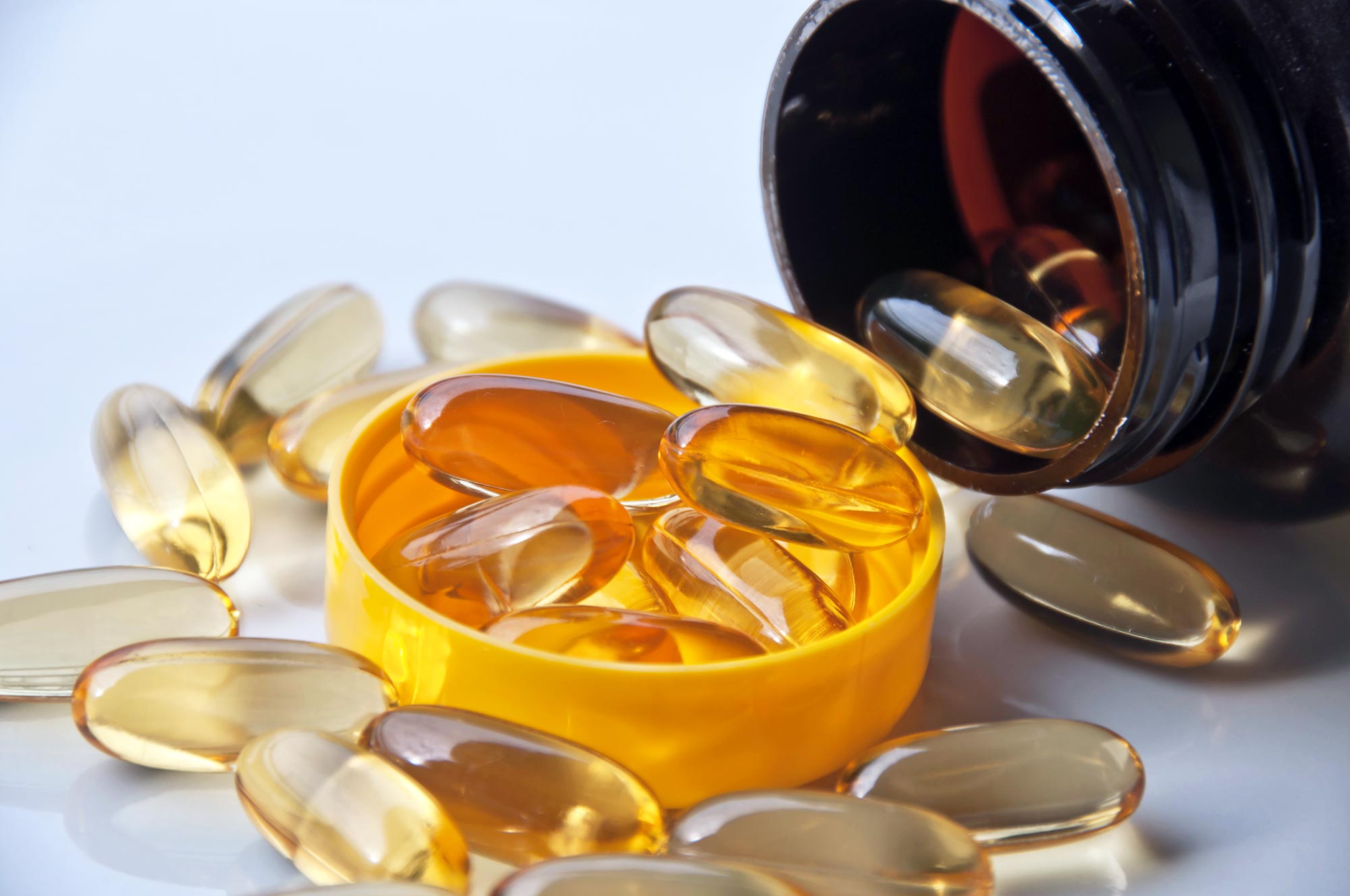
I've tried PUFA in isolation as a supplement and it brought a lot of stress response(fatigue, insomnia, etc) like I was eating some sort of toxin. Does PUFA raises endotoxemia as an antibiotic?
Anyway I've overcame the effect next time eating an extra 500 carb-calories, i.e. it seems that negative effects from PUFA could be mitigated via increased glucose calories intake, as if extra toxicity spikes requirements for glucose drastically.
Anyone noticed the same thing?
Anyway I've overcame the effect next time eating an extra 500 carb-calories, i.e. it seems that negative effects from PUFA could be mitigated via increased glucose calories intake, as if extra toxicity spikes requirements for glucose drastically.
Anyone noticed the same thing?
New Defense Against Bacterial Superbugs: Taking Fish Oil May Reduce Antibiotic Resistance
For the first time, Australian scientists have confirmed a link between the role of regular fish oil to break down the ability of 'superbugs' to become resistant to antibiotics. The discovery, led by Flinders University and just published in international journal mBio, found that the antimicrobia
scitechdaily.com