Linoleic Acid: Is This The Key That Unlocks The Quantum Brain?

OP
Travis

Travis

Member
Joined
Jul 14, 2016
Messages
3,189
It must affect the reflective properties of zinc oxide and at the same time interact with radiation and skin unsaturated oils.
To an uncertain, and perhaps insignificant, extent. This sunscreen here has beeswax and stearic acid listed. These heavy and unsaturated carbons could form a barrier on the skin, as well as insulate the iron oxide preventing a lipid peroxidation chain reaction. Even olive oil makes a terrible painting oil with 71.6% oleic, 9.0% linoleic, and 1.0% linolenic acids; the reaction is too slow to be practical. The linseed oils shift more towards the unsaturated with its respective concentrations of 17.7%, 15.7%, and 57.8%. And the difference in iodine value is illuminating, with olive oil at ~88 and linseed (flaxseed) over 200.
 
Last edited:

schultz

Member
Joined
Jul 29, 2014
Messages
2,653
Either it's the iron doing this, or you're forced to believe that the body creates a special enzyme only for the lysosomes of old people which only works at the unphysiological pH of 6.

I love the theory you're proposing. (Re: scientists discovering things + ego, it reminds me of how Duesberg portrays virologists in his book). I think I will need to go back and read about these systems again and try to think about it from this perspective. Things may "click" that didn't click before.

I went read a few lipofuscin articles a few months ago. The best ones, in my opinion, are from Terman and Brunk. Here is a full-text link to one of the review articles.

Thanks!

From what I remember Brunk and Terman saying: Lipofuscin accumulates mainly in non-renewing cells. The first time that I had become aware of such non-mitotic cells was from them. Some cells always divide and dilute the lipofuscin, but some cells do not. Some cells of the central nervous system accumulate the most lipofuscin since they don't have the benefit of dividing at a high rate.

This is what I remember reading, but I am wondering then what causes the cells to become post-mitotic? Ray doesn't seem to subscribe to the Hayflick limit idea (and I think this is the gentleman he called stupid when talking to John Barhausen?) (okay I found the quote where he says this...)

JOHN BARKHAUSEN: I’d say that's true. So when you're talking about regeneration, you mentioned Hayflick last week as being one of the – the person who did the experiment that ended up convincing most of science that you can regenerate, only a certain limit…

RAY PEAT: Yeah. The fact that he worked at a very influential laboratory that sold material, biological material, cell cultures and such and rats to laboratories all over the country, that helped to give him a reputation. People just assumed that an idiot wouldn’t be working for a big company like that.

JOHN BARKHAUSEN: I see.

RAY PEAT: But I don't know whether – I suspect – in one of my newsletters, I give reasons for suspecting that he wasn’t as stupid as he seems. For 30 years, I thought he was just a very remarkably stupid person. And I saw he had financial motives that might save his mental reputation.


Perhaps of interest is the action of prolactin on the cell. This hormone powerfully increase the intracellular calcium concentration. It can do this in single-digit nanomolar concentrations. It's stunning, really. I just got done reading about this yesterday. Here is a series of photographs taken every 400 milliseconds (their shutter speed was actually quicker than this, but they only posted every ten frames.)

Wow that's really impressive. Now I have to go read about prolactin... you've given me a lot of homework and it's going to take me a while to get through it. :writer:
 
OP
Travis

Travis

Member
Joined
Jul 14, 2016
Messages
3,189
This is what I remember reading, but I am wondering then what causes the cells to become post-mitotic?
I think they must be actively restrained by the organism. In the case of prolactin, this hormone is actively restrained by dopamine impinging on the pineal gland. Growth hormone—another protein hormone released by the pituitary which has high homology, and is the evolutionary fore-runner, to prolactin—is not restrained by dopamine. Growth hormone responds to serotonin. So selective inhibition of certain cells appears to be a normal function.

Perhaps looking at what does the opposite, what creates growth, could be illuminating. There are hundred of things which do this, and it would be nice the narrow-it down to irreducible factors—small molecules and ions. I had first heard, incidentally from @Amazoniac, that calcium could cause cancer. It didn't take me long to find confirmations of this: first from that prolactin study, and then from one on 11β-hydroxysteroid dehyrogenase Type I* where I became familiar with the term: calcium-induced proliferation. They used it without explanation, an aside, so I would assume that it is a common phrase in biochemistry. This does appear to be the case, as a quick Google search—with verbatim quotes—indicates as much.

Polyamines are another small molecule which stimulate growth. They have a peculiar affinity for DNA, where they are thought to stabilize it for division. This is thought to be a direct chemical interaction and has even been modeled. Perhaps they function to separate the two DNA coils from eachother? I certainly plan on reading more about polyamines in the future.

Methylglyoxal is a big one. This is a small molecule which inhibits cells division, in all cases. You could bet the farm on this. It is known to interact with basically two things: amines and sulfhydryls. It could work to inhibit growth by binding polyamines. It is also known to react with arginine to form an imidizole ring, which both prevents it from becoming nitrix oxide and could be an important post-translational modification in itself. It was shown by Thornally† that this methyl-hydroxy-imidazole, the methylglyoxsal–arginine adduct, was actually created at an important location on a transcription factor. This caused the transcription factor to be released, a new one take its place, and a downregulation of a vascular growth factor confirmed both through Western blot and PCR. Arginine is also found more often than any other amino acid in the catalytic domain of enzymes. In this way, methylglyoxal could turn off or on certain enzymes. The enzyme that turns methylgloxal into lactic acid has an arginine in its active site. It is tempting to think that this could function to control the rate of methylglyoxal transformation through a post-translational modification. This is not a modification of the substrate itself, as methylgloxal is turned into lactic acid first by spontaneously-reacting with glutathione at the sulfhydryl group. The enzymes glyoxylase I and glyoxylase II then turn this adduct into glutathione and lactic acid—the molecule formerly-known as Prince methylglyoxal. The affinity of methylgloxal for sulfhydryl appears to be higher than it's affinity for amines, and the addition of cysteine with methylglyoxal in a 1:1 ratio completely nullifies it's effect.‡

These are the most irreducible controllers of growth that I know of. They are small molecules and they always work. They don't need a receptor and work on a physical level. It could be that many growth factors, vascular endothelial growth factor and growth hormone, simply raise calcium in the cell like prolactin—with selectivity conferred by whether or not the cell membrane has a receptor for that particular hormone.

You might then expect homocysteine to be doubleplusbad; first by having the capability of becoming a stable free radical, traversing the blood–brain barrier like a Trojan Horse resulting in a free-radical lipid peroxidation cascade; and second, by nullifying methylglyoxal with its sulfhydryl group. Also, homocysteine derives from methionine. This amino acid is one of the three, besides lysine and arginine, which can form a polyamine. This is probably why only small changes of less than a fraction of one percent reduces the lifespan of rats about 35%. This result has been replicated: With one showing a reduction of 44%, one of 35%, and one of 21%.

*This is actually a misnomer at times. This enzyme has been found in all places where they have looked, and it is actually bidirectional: So not only does dehyrogenate hyroxysteroids, it actually reduces them as well—in the exact same position. It turns cortisone into cortisol, and vice versa. It depends on where the cell is located what direction the arrow lies, and this depends on the NADH/NAD⁺ ratio. This is the cofactor, and when it has extra hydrogens it donates them to cortisone: When it has no hyrogens, it takes them from cortisol. This enzyme, when in certain tissues, would more appropriately be called 11β-oxysteroid reductase. The skin is one of these tissues, and here the bidirectional rate ratio is 10:1 in favor of the production of cortisol from cortisone. The enzyme 11β-hydroxysteroid dehyrogenase Type I should not to be confused with 11β-hydroxysteroid dehyrogenase Type II, which is found in the kidneys. These two enzymes are structurally different.

†Yao, Dachun, et al. "High glucose increases angiopoietin-2 transcription in microvascular endothelial cells through methylglyoxal modification of mSin3A." Journal of Biological Chemistry 282.42 (2007): 31038-31045.

‡Együd, Laszlo G., and A. Szent-Györgyi. "On the regulation of cell division." Proceedings of the National Academy of Sciences 56.1 (1966): 203-207.
 
Last edited:

noordinary

Member
Joined
Jun 1, 2016
Messages
209
@Travis could you please elaborate on vision from conductivity point of view?
I mean if something as common as nearsightedness is not a mechanical structural problem (elongated eyeball, muscle tone etc) may it be a conductivity loss to some degree?
i'm trying to catch up in betwen work...
 
OP
Travis

Travis

Member
Joined
Jul 14, 2016
Messages
3,189
I'm not sure how small reductions in microtubules would affect vision, but there are perhaps ways to find out...[searching: "colchicine vision loss"]...Wow. This is interesting and proves what w... reinforces what has already been proven about the physical nature of vision. Apparently the disruption of microtubules can cause physical changes as a result of altered nerve flow.

Colchicine is the classic microtubule disruptor. It destabilizes them and inhibits their growth. Microtubules are made-up of two nearly-identical proteins called α- and β-tubulin, which always alternate and form a cylindrical tube. Guanosine triphosphate (GTP) is essential for their formation. But colchicine is also a drug, used to treat gout, so there is a lot of data on the side-effects. The side-effects that I'd previously read about were such things as muscle weakness, loss of sensation, and nerve tingling—which reinforced my conviction that microtubules inside of nerves are the physical structures which facilitate nerve conduction.

Here are just a few titles demonstrating the colchicine has long-been-known as the go-to microtubule-disrupting molecule, both in vitro and in several species of small furry animals.

I'm pretty sure that colchicine is the most powerful one known, but there are others such as taxol and 2-methoxyestradiol. Here are some side-effects taken from the article called "Colchicine poisoning: the dark side of an ancient drug" from the journal Clinical toxicology:
Myopathy, neuropathy, and combined myoneuropathy have all been reported after acute poisoning. Proximal limb weakness, distal sensory abnormalities, distal areflexia, and nerve conduction impairment compatible with axonal neuropathy are characteristics. ―Yaron Finkelstein
Perhaps what some would predict from colchicine poisoning.
Mechanisms of toxicity: Colchicine binds to the intracellular protein tubulin, preventing its alpha and beta forms from polymerizing to form microtubules. This disruption of the microtubular network results in impaired protein assembly in the Golgi apparatus, decreased endocytosis and exocytosis, altered cell shape, depressed cellular motility, and arrest of mitosis. [...] Disruption of the microtubular network also leads to decreased expression of adhesion molecules on neutrophil membranes and modulates cytokine production. Colchicine may also have a direct toxic action on myocardial cells. This effect may be related to binding of the drug to microtubules in myocytes, which interferes with cardiac conduction and contractility. The inhibition of other important microtubular functions such as cytoplasmic motility and extracellular secretion of hormones and neurotransmitters may be involved in the devastating processes leading to multi-organ failure in cases of drug overdose. Colchicine also inhibits the release of histamine from mast cells . . . ―Yaron Finkelstein
He gives a litany of reported side-effects from the literature. I think it could be described more mechanistically: Colchicine inhibits the tubulin assembly which forms the physical basis for nerve transmission. All biological systems dependent upon nervous stimulation could be affected. Secondary effects arising from the attenuation or amplification of hormones and neurotransmitters normally released as a result of nerve stimulation (such as in pituitary and adrenals) are also possible. In this way, systemic effects could result from hormonal secondary effects far removed from the actual pharmacodynamic dispersion of the drug.

Since we all know how colchicine works, we can now we can see how it effects the eye directly:

Colchicine greatly increased axial length, equatorial diameter, eye weight, and myopic refractive error, while reducing corneal curvature. ―Dr. Andy J Fischera
He starts-off describing near-sightedness as a loss of focal control and introduces the word emmetropia, which he defines a pathological change in the distance between the retinal and the lens and changing normal focal length. He used only 0.2 μg of colchicine into a chicken's eye and spared the other. After bird-death, he uses flourophore-conjugated antibodies to view specific proteins with the microscope. The list of proteins stained-for was a let-down, as he uses no less than 14 separate fluoro-antibodies but not the most important ones: ones for α- or β-tubulin. But the small 0.2 μg dose of colchicine did cause complete unilateral blindness in the injected eye, as 'confirmed by an approaching hand.' Noteworthy is that he had noticed a 48.3% reduction of optic nerve thickness:
Colchicine caused significant losses of thickness from several retinal layers, particularly the optic fibre layer (OFL) (28.7±6.2% of the control; mean±S.D.; n=8) and IPL (55.5±8.3% of the control). ―Dr. Andy J Fischera
With eight chickens; so this wasn't a random event.
The photoreceptor layer and retinal pigmented epithelium (RPE) appeared dysplastic, thin or discontinuous in some parts, while appearing thicker in other regions (Fig. 1). ―Dr. Andy J Fischera
The entire hisological data shows nothing too exciting. As mentioned, he hadn't even stained for tubulin. About half of the things he stained for showed no change. The chicken's were blind, so there is little reason to worry about slight reductions in tyrosine hydroxylase.
On average, colchicine-treated eyes were 10.2±2.9% longer, 5.0±1.9% wider, and weighed 20.6±4.9% more than contralateral control eyes.
He did notice shape changes. He seems to get on track towards the end of the article where he says:
Colchicine also resulted in the activation of growth-promoting pathways or the destruction of growth-suppressing pathways, thereby allowing ocular growth to run unchecked or by default. These changes in ocular growth may have been caused by colchicine-induced damage to the RPE, photoreceptors, ganglion cells, and/or subsets of amacrine cells, or by undetected damage to ocular tissues. [...] Colchicine causes the disassembly of microtubules, and thereby interferes with many cellular functions. One function disrupted by colchicine is axonal transport [...] ―Dr. Andy J Fischera
It's seems like he's getting with the idea that these changes are happening as a result of disturbed nerve function. He hypthesizes that some nerves have a suppressive effect on growth. This is not too unusual, as cortisol inhibits growth of the skin and dopamine inhibits prolactin release in the pituitary. Perhaps the changes in tyrosine hydroxylase he noted have importance here, as this is the dopamine-synthesizing enzyme.
Colchicine destroyed most ganglion cells, as well as subtypes of amacrine cells including those thought to participate in visual guidance of ocular growth. Colchicine also damaged the RPE and photoreceptors. As a result of the damage caused by colchicine, eyes were unable to emmetropize, grew excessively, became myopic, and had flattened corneas. We propose that colchicine-induced elongation of the vitreous chamber results from the destruction of one or more subsets of amacrine cells that normally suppress growth in response to visual stimuli, and that colchicine-induced corneal flattening results from the destruction of one or more subsets of ganglion cells that normally promote proper corneal shape. ―Dr. Andy J Fischera
I think we all know what happened here. The tubulin disassembled the microtubules of the optic nerve leading to blindness. All of other changes were the result of microtubule disruption elsewhere. These things run through the mitochondria and could very well function as a source of intracellular signalling. How else can you explain a signal running from the brain to the fingers at typing speed by anything other than microtubules coupled-directly to mitochondria?


I found another study:
It seems that more is known about changes in neurofilaments than microtubules after RGC injury, but the work of Knighton and colleagues suggests that microtubules are the primary source of birefringence in the retinal nerve fiber layer [RNFL]. Although it is possible that other thin cylindrical parallel structures, such as neurofilaments or the axonal membranes themselves, could also contribute to form birefringence of the RNFL, Knighton et al. found that RNFL birefringence declines to essentially zero after colchicine treatment of rat retinal explant preparations. The latter results are consistent with their earlier theoretical work and a model in which MT are the primary contributor to RNFL birefringence, with other mechanisms contributing less than 15%. ―Dr. Brad Fortune
Colchicine causes total blindness, destabilizes microtubules, and can be measured by birefringence—a brilliant technique that I never would have thought to apply to measure microtubule density in the retina. I should have a look at that Knighton Study sometime in the future, as he reported greater changes with an even smaller dose—and he could have more interesting images . . .
Because the conversion is linear, the decline in RNFL “thickness” measured by SLP represents a 20% decline in RNFL birefringence within approximately 100 minutes of intravitreal colchicine injection. This effect is smaller than that obtained by Huang and Knighton, which might be attributed to one or several differences in the experimental conditions. [...] Moreover, using intravitreal colchicine doses 10 to 100 times lower than what was used here, Davidson et al. found near total loss of neurotubules within the intraretinal axons of the monkey after only 1 hour. ―Dr. Brad Fortune
I think light is more of a particle than a wave, and polarization would then represent all light photons having parallel spins. I think @Meatbag would agree with this.
Newton initially tried to explain it with spin, but he was shouted down by Hooke and Huygens, and his spin model is still being repressed as an embarrassment. Without spin, the modern explanations are not really mechanical explanations at all. They are just descriptions. A substance is given a refractive index, and this index causes the bend. But of course that is heuristic, not mechanical. It explains nothing. It is to say that violet is bent more because the substance bends it more. The refractive index causes the bend and the bend determines the refractive index: mechanics=zero. ―Miles Mathis

[1]Finkelstein, Yaron, et al. "Colchicine poisoning: the dark side of an ancient drug."
Clinical toxicology 48.5 (2010): 407-414.
[2]Fischer, Andy J., Ian G. Morgan, and William K. Stell. "Colchicine causes excessive ocular growth and myopia in chicks." Vision research 39.4 (1999): 685-697.
[3]Fortune, Brad, et al. "Intravitreal colchicine causes decreased RNFL birefringence without altering RNFL thickness." Investigative ophthalmology & visual science 49.1 (2008): 255-261.
 
Last edited:
OP
Travis

Travis

Member
Joined
Jul 14, 2016
Messages
3,189
Here's the Knighton Article alluded to above. This one is really worth checking-out.
This study shows how quickly colchicine can disassemble microtubules:
knighton2.png (click to embiggen)

(A) Image showing microtubules 20 minutes before the addition of colchicine (B) Image of the same retinal region as in (A) after 30 minutes of colchicine treatment.

knighton1.png (click to embiggen)

Temporal change of bundle contrast in colchicine experiments. (A) Time course of B̄ for 15 bundles in 5 retinas. B̄ was high during baseline measurements, declined after the solution was switched to colchicine, and was close to 0 after approximately 30 minutes of treatment; (B) normalized and averaged B̄ displayed as in Figure 3B . Curves stop at 70 minutes because only a few bundles were observed longer than that.
 
Last edited:

noordinary

Member
Joined
Jun 1, 2016
Messages
209
@Travis thank you!
Reading on tubulin inhibitors. Which are plenty, one of them 2-Methoxyestradiol (metabolite of estradiol) surprise surprise...
most of info on them in context of cancer treatment.
I wonder in the chick eyes in Dr Andy J. Fichera study got bigger and heavier because cell mutiplied or because the eye accululated water or smth else?
On a good side:
What do you think would be protective (promoting?) for microtubular stabilization, density increase?
 
OP
Travis

Travis

Member
Joined
Jul 14, 2016
Messages
3,189
What do you think would be protective (promoting?) for microtubular stabilization, density increase?
I think maybe progesterone, but I'm not for certain yet.

Stabilization can be seen as good, but hyperstabillization can be pathological. Taxol works by freezing microtubules; and since the cell cannot divide without breaking-down its microtubule network, it is stuck in the G₂-phase of the cell cycle. It cannot enter mitosis.
tubulin1.png

The three images on the right (F–H) are cells with taxol-frozen microtubule networks; the image on the left (E) is a cell entering mitosis. The microtubles are visualized by using an antibody for β-tubulin with a fluorescent molecule attached. They create specific antibodies to certain proteins by injecting goats with the target protein.

Some drugs can stabilize microtubules so well that they can even be elongated without GTP—the cell's normal molecule for this. Here are some images which demonstrate this effect:
tubulin2.png

But this over-rides the body's control system, and division cannot take place. Not only that, short microtubules can form spontaneously everywhere in the cell leading to disorder. Perhaps very small concentrations might be helpful, I'm not sure. This is an extreme drug, but it demonstrates why we need some plasticity involved in the microtubule network.

Taxol sits on the other end of the extreme—opposite to colchicine, which can rapidly dissolve them.

The first hint of 2-methoxyestradiol having an effect came in 1994, although effects were noted with the synthetic estrogen DES before that . . .

. ..something which the author speaks about. The entire first paragraph is packed-full of interesting information, and D'Amato is a good writer with good insight.

"There is growing evidence that estrogenic compounds affect cell division and act directly on microtubules by interacting with tubulin. The most extensively studied of these compounds is the synthetic analog diethylstilbestrol and related agents. These drugs cause disturbances in mitosis, inhibit microtubule assembly at relatively high concentrations, and inhibit the binding of colchicine to tubulin. Estradiol, the major estrogenic hormone of human beings, also causes disturbances of mitosis in cultured cells (1, 4, 5). These perturbations include aneuploidy, multinucleation, and mitotic arrest, and estradiol has been reported to inhibit the polymerization of rat brain tubulin (6). Seegers et al. (5) found that 2-methoxyestradiol, a metabolite of both estradiol and the oral contraceptive agent 17-ethynylestradiol, was more potent than estradiol in producing mitotic perturbations and proposed that it was 2ME rather than estradiol that caused the observed disturbances. Although 2-methoxyestrogens are extremely weak in binding to cytosol estrogen receptors, 2ME is found in blood and urine after sequential hepatic hydroxylation and methylation of estradiol." ―D'Amato

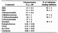
He came into this study knowing that synthetic estrogen drug diethylstilbestrol interacts with microtubules, and that estradiol caused disturbances in mitosis. He then tested a large group of compounds on microtubule formation: twelve estrogens, diethylstilbestrol, and colchicine. The estrogens were mainly synthetic but can reveal molecular clues on what binds and what doesn't. I don't even think that there are twelve natural estrogens; I can only think of a few (not counting the sulfated metabolites of course, which immediately doubles the number). The ones that were the most disruptive are listed in Table 1:

tubulin3.png click to embiggen


Methoxyextradiol was about 40% as strong as colchicine in inhibiting polymerization, and methoxyestrone was close. Estradiol was the only other natural one tried, and it was not nearly as effective. I think methoxyestradiol can represent the extreme for natural, endogenous microtubule disruptors; I haven't heard of any other steroid hormone with such an effect.

"Our observations with 2ME suggest that other, perhaps unknown, steroid compounds could play important roles in microtubule assembly, possibly functioning as negative regulators." ―D'Amato

I had this thought earlier, and was glad to see it in print. It's glad to know that one other person is taking this idea seriously. Perhaps the original function of steroid hormones was in microtubule stabilization and the hormonal—transcriptional effects only came later in evolution? All steroids, from cortisol to estradiol, are all very similar.

steroids1.png

The interaction of 2ME with the colchicine site may also provide structure-activity insights. ―D'Amato
But estrogen is the only one with a resonant ring. This is planar, and has a different geometry. Two-dimensional representations of molecules generally leave-out important 3-D geometries. A short article on that can be found here: http://www.chem.ucla.edu/~harding...

steroid2.png

This would be consistent with spectroscopic evidence suggesting the C ring of colchicine interacts by base stacking with a tryptophan residue of tubulin (29). ―D'Amato
So the resonant ring, is thought to intercolate within the microtubule interior and base-stack with the tryptophan residues. I would expect that to perhaps inhibit the photoelectrical transmission inside of the microtuble. The effect of this light could be to direct growth at the terminal end. Melatonin also has a methoxy group, and I've read studies that indicate all small molecules which act to quench fluorescence are anesthetic. I couldn't find which article I had read that in, but this one below shows that some photochemists are aware of the relationship:

  • Koblin, Donald D., Wilson D. Pace, and Howard H. Wang. "The penetration of local anesthetics into the red blood cell membrane as studied by fluorescence quenching." Archives of biochemistry and biophysics 171.1 (1975): 176-182
Studies from the '70's correlate a drug's hallucinogenic potential to it's highest occupied molecular orbital.
And they are all planar, meaning that they all might be expected to intercolate into the microtubule and stack between the flourescent tryptophan indole rings. The methoxy group is a quencher of flourescence.

"Although the effects of the steroid derivative are weaker than those of many antimitotic agents, 2ME is the most potent endogenous inhibitor of tubulin polymerization produced by mammalian tissues yet described. This compound or related steroid derivatives may play a significant role in modulating microtubule assembly and function in mammals." ―D'Amato

I get the impression that microtubules run from the centriole to the cell membrane, based on the images with stained tubulin. This could perhaps be the source of the "ultraweak emission" that was historically-seen emanating from cells.

Nerve cells have longer microtubule structures that run from cell-to-cell, and also through the mitochondria. This is part of the central nervous system. Other cells have short-range intracellular networks that function locally

The reason why I think progesterone could be involved in microtubule formation is that it was recently found to protect mice from blindness. By recently I mean . . . it was posted just a few days ago. It's on Researchgate: https://www.researchgate.net...

"Retinitis pigmentosa (RP) refers to a group of inherited retinal diseases, characterized by genetic mutations in rod photoreceptors . These mutations lead to rod cell death, subsequent cone cell loss and eventual blindness. The rd10 mouse is a model of RP, with a mutation in
phosphodiesterase-6b." ―Roche

This is interesting because phosphodiestere-6b is involved in GMP production. This is only two phosphates away from GTP, the microtubule forming molecule. Anything that would interfere with GTP formation would be expected to interfere with microtubule polymerization. The Waldian Theory of Photoconduction, of course, has a different explanation:
Photon absorption triggers a signaling cascade in rod photoreceptors that activates cGMP phosphodiesterase (PDE), resulting in the rapid hydrolysis of cGMP, closure of cGMP-gated cation channels, and hyperpolarization of the cell.
How does hyperpolarization of cells explain vision? Is this some special quantum long-range hyperpolarization? (lol) You need microtubules to explain vision because diffusing chemicals cannot possible work that quickly. You also need microtubules to explain long-range nerve conduction.

Table 1 from the 2-methoxyestradiol study shows that estradiol actually binds with tubulin better that diethylstilbesterol, yet interferes with polymerization way less. Taxol is proof that some drugs bind and then stabilize, while others bind a depolymerize. The difference must be the planar electrically-resonant ring with a methoxy substituent. Colchicine has four methoxy groups attached to planar rings. Perhaps these intercolate and quench flourescence? I just looked-up podophyllotoxin, the only drug which inhibits polymerization more than colchicine (Table 1), and it has three methoxy groups. I'm going to keep an eye-out not for molecules with methoxy groups and perhaps read that article cited by D'Amato (29), in which describes how the methoxylated estradiol ring intercolates between the stacked fluorescent tryptophans in the microtubule core.

Melatonin has sedative effects, and also has a planar methoxy group.

Microglial-induced Müller cell gliosis is attenuated by progesterone in a mouse model of retinitis pigmentosa. Available from: https://www.researchgate.net/public...rone_in_a_mouse_model_of_retinitis_pigmentosa [accessed Oct 19 2017].
 
Last edited:
OP
Travis

Travis

Member
Joined
Jul 14, 2016
Messages
3,189
It looks they've must have read the Massimo article. I think we should tell them in the YouTube comment section that it's not going to work, their metabolism will slow, lipofuscin will accumulate, and prostate cancer will become more than a remote possibility.
 

noordinary

Member
Joined
Jun 1, 2016
Messages
209
@Travis nevermind.
They didn't have control group and basically compared children from mothers, who supplemented cold liver oil, and mothers, who were supplementing corn oil,
for the period from 18 week of pregnancy to 3 months after delivery (all of the subjects breastfed at 3 month age).
And showed that cod liver oil babies scored tiny bit more than corn oil babies on Kaufman Assessment Battery for Children test: 106.4 vs 102.3
Which is laughable difference really, considering that K-ABC test has a standard deviation of 16 and average score of 100.
Maternal Supplementation With Very-Long-Chain n-3 Fatty Acids During Pregnancy and Lactation Augments Children’s IQ at 4 Years of Age
"Augments Children's IQ" are just big words, considering this table for K-ABC: average 85 - 115 and above average starting at 116.
Pediatric versus adult assessment
pediatric-versus-adult-assessment-58-638.jpg

But Dr. Rhonda Patrick announces to all her subscribers (under the saint salmon picture of course) about DHA advantages:
Instagram post by Dr. Rhonda Patrick • Jan 4, 2018 at 8:14pm UTC
"On the baby front...a randomized, controlled trial found that maternal supplementation with omega-3 fatty acids during pregnancy and lactation improves children's IQ at four years old.
Study link:
Maternal Supplementation With Very-Long-Chain n-3 Fatty Acids During Pregnancy and Lactation Augments Children’s IQ at 4 Years of Age"

Improves comparing to what? Comparing to supplementing 10mL of corn oil daily?!
@haidut check this out.
 
J

jb116

Guest
In that silly little study they compare n-6 to n-3 without considering what the mothers diets consist of. With a typical diet and not paying attention to the already overt amounts of pufa (especially n-6) from their diets, the corn oil group is merely compounding the hazard, where as the n-3 is at least balancing those n-6 abundant groups. There is probably some benefit from the Vit A/D of the cod oil as well. So there seems to be a "lesser of two evils." If you recall Dr. Peat talking about a particular French group who was set out to prove any and all pufas are beneficial to baby brains, instead inadvertently found that the group with the lowest n-6 and n-3 and highest glucose intake were the smartest babies.
 

noordinary

Member
Joined
Jun 1, 2016
Messages
209
In that silly little study they compare n-6 to n-3 without considering what the mothers diets consist of. With a typical diet and not paying attention to the already overt amounts of pufa (especially n-6) from their diets, the corn oil group is merely compounding the hazard, where as the n-3 is at least balancing those n-6 abundant groups. There is probably some benefit from the Vit A/D of the cod oil as well. So there seems to be a "lesser of two evils." If you recall Dr. Peat talking about a particular French group who was set out to prove any and all pufas are beneficial to baby brains, instead inadvertently found that the group with the lowest n-6 and n-3 and highest glucose intake were the smartest babies.
Thanks, I figured that out: they could supplement lead and mercury and conclude that lead "Augments Children's IQ" just as well.
By any case you have a link to that study Dr. Ray Peat mentioned. I also heard him saying that several times. Did someone locate the study?
 

haidut

Member
Forum Supporter
Joined
Mar 18, 2013
Messages
19,799
Location
USA / Europe
@Travis nevermind.
They didn't have control group and basically compared children from mothers, who supplemented cold liver oil, and mothers, who were supplementing corn oil,
for the period from 18 week of pregnancy to 3 months after delivery (all of the subjects breathed at 3 month age).
And showed that cod liver oil babies scored tiny bit more than corn oil babies on Kaufman Assessment Battery for Children test: 106.4 vs 102.3
Which is laughable difference really, considering that K-ABC test has a standard deviation of 16 and average score of 100.
Maternal Supplementation With Very-Long-Chain n-3 Fatty Acids During Pregnancy and Lactation Augments Children’s IQ at 4 Years of Age
"Augments Children's IQ" are just big words, considering this table for K-ABC: average 85 - 115 and above average starting at 116.
Pediatric versus adult assessment
View attachment 7826
But Dr. Rhonda Patrick announces to all her subscribers (under the saint salmon picture of course) about DHA advantages:
Instagram post by Dr. Rhonda Patrick • Jan 4, 2018 at 8:14pm UTC
"On the baby front...a randomized, controlled trial found that maternal supplementation with omega-3 fatty acids during pregnancy and lactation improves children's IQ at four years old.
Study link:
Maternal Supplementation With Very-Long-Chain n-3 Fatty Acids During Pregnancy and Lactation Augments Children’s IQ at 4 Years of Age"

Improves comparing to what? Comparing to supplementing 10mL of corn oil daily?!
@haidut check this out.

Yeah, I am not surprised. It is a common trick Peat spoke about. Comparing a poison to a more potent poison and then concluding the poison is protective. If you can find data on what is the average IQ of children with EFA deficiency we may have some blockbuster counter-conclusions that we can send to Dr. Patrick.
 

noordinary

Member
Joined
Jun 1, 2016
Messages
209
Yeah, I am not surprised. It is a common trick Peat spoke about. Comparing a poison to a more potent poison and then concluding the poison is protective. If you can find data on what is the average IQ of children with EFA deficiency we may have some blockbuster counter-conclusions that we can send to Dr. Patrick.
Unfortunately, when comfronted with older studies, Dr. Patrick says something along the lines: Science did not exist before 2000s.
 

haidut

Member
Forum Supporter
Joined
Mar 18, 2013
Messages
19,799
Location
USA / Europe
Unfortunately, when comfronted with older studies, Dr. Patrick says something along the lines: Science did not exist before 2000s.

I see. So, what did she learn in medical school then, given she went there before the 2000s? If what she learned back then was not science then should she have her MD revoked?
I would actually ask her that question directly if I ever meet her.
 
J

jb116

Guest
I see. So, what did she learn in medical school then, given she went there before the 2000s? If what she learned back then was not science then should she have her MD revoked?
I would actually ask her that question directly if I ever meet her.
:joyful: great point!
 
OP
Travis

Travis

Member
Joined
Jul 14, 2016
Messages
3,189
@Travis nevermind.
They didn't have control group and basically compared children from mothers, who supplemented cold liver oil, and mothers, who were supplementing corn oil,
for the period from 18 week of pregnancy to 3 months after delivery (all of the subjects breastfed at 3 month age).
And showed that cod liver oil babies scored tiny bit more than corn oil babies on Kaufman Assessment Battery for Children test: 106.4 vs 102.3
Which is laughable difference really, considering that K-ABC test has a standard deviation of 16 and average score of 100.
Maternal Supplementation With Very-Long-Chain n-3 Fatty Acids During Pregnancy and Lactation Augments Children’s IQ at 4 Years of Age
"Augments Children's IQ" are just big words, considering this table for K-ABC: average 85 - 115 and above average starting at 116.
Pediatric versus adult assessment
View attachment 7826
But Dr. Rhonda Patrick announces to all her subscribers (under the saint salmon picture of course) about DHA advantages:
Instagram post by Dr. Rhonda Patrick • Jan 4, 2018 at 8:14pm UTC
"On the baby front...a randomized, controlled trial found that maternal supplementation with omega-3 fatty acids during pregnancy and lactation improves children's IQ at four years old.
Study link:
Maternal Supplementation With Very-Long-Chain n-3 Fatty Acids During Pregnancy and Lactation Augments Children’s IQ at 4 Years of Age"

Improves comparing to what? Comparing to supplementing 10mL of corn oil daily?!
@haidut check this out.
Corn oil is actually worse because it has much more linoleic acid, the only precursor to the prostaglandins.

The old studies on high‐fat diets in rats, dating back to the '40s, had shown a high fat diet to promote cancer. Later on, in the '70s, it had been shown that some fats were far more carcinogenic than others. The most carcinogenic fatty acid was always corn oil.

And then later, in the '80s, review articles from all these fat‐feeding rat studies had teased‐out one important finding: linoleic acid content! While it's common to separate fatty acids into two groups, saturated and unsaturated, this is a simplification which hides important details. Only arachidonic acid can become prostaglandins, and arachidonic acid is exclusively made from linoleic acid. When arachidonic acid is concentrated on the cell membrane, you have potential for 'inflammation' and cancer.

The enzyme phospholipase A₂ cleaves arachidonic acid from a phospholipid on the cell membrane. This is a cool enzyme, and it can do this completely isolated in vitro. This enzyme can be shown to work using simple micelles—or artificial lipid droplets in water—constructed of phosphotidylcholine, with arachidonic acid at the sn‐2 position. This enzyme has an affinity for the lipid membrane, and can catalyze the cleavage of the arachidonic acid–phospholipid bond.

Released arachidonic acid then diffuses towards cyclooxygenase, which adds O₂ to the lipid forming the endoperoxide ring characteristic of prostaglandin H. This is then isomerized to either prostaglandin D₂ or prostaglandin E₂. These have opposing functions, to a degree, as prostaglandin D₂ usually constricts the blood vessels while prostaglandin E₂ always relaxes them.

But they have hormonal functions to, besides those mediated through their G protein‐coupled receptors on microtubules. The PPAR series of nuclear receptors can bind prostaglandins, and then induce transcription of enzymes and proteins.

But there is another twist, since the entire process can be mitigated by competing fatty acids. Oleic and eicosapentaenoic acids have been shown to inhibit prostaglandin production by: (1) Preventing its incorporation into the cell membrane, and (2) Competing with it for the cyclooxygenase enzymes. This is why ω−3 fatty acids are considered 'protective.'

But omega−3 fatty acids cannot be said to be 'protective' against spontaneous oxidation or lipofuscin. In fact, some of them are even more prone to oxidation since they can have upwards of six double‐bonds. So between corn oil and fish oil you are presented with a dilemma: Either you can have the growth promoting, proliferative, and carcinogenic corn oil . . . or you can have the less‐proliferative, yet more unstable fish oil. If a person had cancer, they'd probably have to choose the fish oil; but if a person was underweight and taking iron supplements for some reason, the more stable corn oil could be a better choice.

Choosing between corn oil and fish oil is tantamount to choosing between cancer and lipofuscin.

But I would think the IQ point difference could have been influenced more by the vitamins in the fish oil, since lipofuscin does not form in rapidly growing cells (it is diluted through mitosis). This oil is naturally rich in vitamin A, but it also has significant amounts of vitamin D. However, it is often deodorized—a process which largely removes these vitamins. Sometimes they are added back in, but the concentrations are always somewhat unnatural (vitamin A‐heavy). Vitamin A is powerful lipid hormone which can affect nearly everything. Active vitamin A, or retinoic acid, has two nuclear receptors which double as transcription factors. Upon binding retinoic acid, these receptors cumulatively transcribe DNA for thousands of genes.

It would be nice to see a study controlling for vitamin A—also having a coconut oil group!

There's been over one hundred such fat‐feeding rat studies, and about one dozen articles compiling the results from these. Stearic acid has consistently been found protective against cancer. As a saturated fatty acid, it can displace arachidonic acid from the cell membrane—reducing prostaglandin potential. But also, it cannot form lipofuscin since it will not spontaneously oxidize. So although much attention is given to oleic acid and ω−3 fatty acids in inhibiting cancer, the fully‐saturated stearic acid is better for the brain and skin long‐term since it cannot form lipofuscin. This could be why so many centenarians report using chocolate, a food rich in the relatively‐rare stearic acid.
 
Last edited:
EMF Mitigation - Flush Niacin - Big 5 Minerals

Similar threads

Back
Top Bottom