FoodForeal

Member
Joined
Feb 9, 2022
Messages
333
Location
Midwest
Got arround that
Awesome will look tomorrow I think.

If anyone wants to buy this book it probably has a lot of good info for $150 lol. The Apex Apicomplexan.
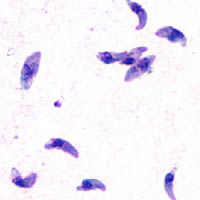
I found the book from a wikipedia citation link on a sentence about the parasitophorous vacuole forming the bradyzoite cyst because I want to know more about how it works but it's behind the paywall: Toxoplasma gondii - Wikipedia
"The cyst wall is formed by the parasitophorous vacuole membrane.[30]:"

Really it's unbelievable how sophisticated it is and how cats are such a great host allowing it to spread to so many different animals all over the world.
 
Last edited:
OP
K

Karmeleon

Member
Joined
Jan 30, 2020
Messages
84
"Tissue cysts can be maintained in host tissue for the lifetime of the animal.[30]: 580  However, the perpetual presence of cysts appears to be due to a periodic process of cyst rupturing and re-encysting, rather than a perpetual lifespan of individual cysts or bradyzoites.[30]: 580  At any given time in a chronically infected host, a very small percentage of cysts are rupturing,[30]: 45  although the exact cause of this tissue cysts rupture is, as of 2010, not yet known.[4]: 47 "

Toxoplasma eats tryptophan
"Initially, a T. gondii infection stimulates production of IL-2 and IFN-γ by the innate immune system.[38] Continuous IFN-γ production is necessary for control of both acute and chronic T. gondii infection.[38] These two cytokines elicit a CD4+ and CD8+ T-cell mediated immune response.[38] Thus, T-cells play a central role in immunity against Toxoplasma infection. T-cells recognize Toxoplasma antigens that are presented to them by the body's own Major Histocompatibility Complex (MHC) molecules. The specific genetic sequence of a given MHC molecule differs dramatically between individuals, which is why these molecules are involved in transplant rejection. Individuals carrying certain genetic sequences of MHC molecules are much more likely to be infected with Toxoplasma. One study of >1600 individuals found that Toxoplasma infection was especially common among people who expressed certain MHC alleles (HLA-B*08:01, HLA-C*04:01, HLA-DRB 03:01, HLA-DQA*05:01 and HLA-DQB*02:01).[48]

IL-12 is produced during T. gondii infection to activate natural killer (NK) cells.[38] Tryptophan is an essential amino acid for T. gondii, which it scavenges from host cells. IFN-γ induces the activation of indole-amine-2,3-dioxygenase (IDO) and tryptophan-2,3-dioxygenase (TDO), two enzymes that are responsible for the degradation of tryptophan.[49] Immune pressure eventually leads the parasite to form cysts that normally are deposited in the muscles and in the brain of the hosts.[38]

Immune response and behavior alterations[edit]​

The IFN-γ-mediated activation of IDO and TDO is an evolutionary mechanism that serves to starve the parasite, but it can result in depletion of tryptophan in the brain of the host. IDO and TDO degrade tryptophan to N-formylkynurenine. Administration of L-kynurenine is capable of inducing depressive-like behavior in mice.[49] T. gondii infection has been demonstrated to increase the levels of kynurenic acid (KYNA) in the brains of infected mice and KYNA has also been demonstrated to be increased in the brain of schizophrenic persons.[49] Low levels of tryptophan and serotonin in the brain were already associated with depression.[50]"
Hu nice find about the role of tryptophan as an essential nutrient.
About the phosphate earlier mentioned, toxoplasma induces a calcium ca2+ influx into host cells. So compounds with calcium channel inhibition are a growth suppressor for toxoplasma, artemisinin as an example. Also Histone Deacylase Inhibitors are tested against toxoplasma, some do very well, but interestingly niacinamide has no action on it. I have to search and find the study about the HDAC Inhibitors again.

found it: SCRIPTAID AND SUBEROYLANILIDE HYDROXAMIC ACID ARE HISTONE DEACETYLASE INHIBITORS WITH POTENT ANTI–TOXOPLASMA GONDII ACTIVITY IN VITRO
RESEARCH ARTICLE| JUNE 01 2007

SCRIPTAID AND SUBEROYLANILIDE HYDROXAMIC ACID ARE HISTONE DEACETYLASE INHIBITORS WITH POTENT ANTI–TOXOPLASMA GONDII ACTIVITY IN VITRO​

Toxoplasma gondii is a well-recognized cause of disease in congenitally infected and immunocompromised individuals. Histone deacetylases (HDAC) comprise a family of enzymes that participate in the regulation of chromatin structure, gene expression, and cell signaling in eukaryotes. Toxoplasma gondii expresses a HDAC Class I enzyme homologous to human hdac3. Previous work showed that the histone deacetylase inhibitors (HDI) apicidin and valproic acid inhibit T. gondii infections in vitro. The present study compares the activity of hydroxamic-acid histone deacetylase inhibitors against the RH strain of T. gondii growing in HS68 human foreskin fibroblast cells. Nanomolar concentrations of suberoylanilide hydroxamic acid (SAHA), suberic bishydroxamic acid (SBHA), scriptaid, and trichostatin A (TSA) inhibited T. gondii tachyzoite proliferation. Scriptaid was the most potent hydroxamic acid inhibitor (IC50 = 39 nM). In comparison, the carboxylate histone deacetylase inhibitors sodium valproate, sodium butyrate, and 4-phenylbutyrate were less potent (IC50 range 1–5 mM). All of the inhibitors tested, except SBHA, completely protected the HS68 monolayers from T. gondii at concentrations 3–6 times greater than their respective IC50. In contrast, nicotinamide, an inhibitor of NAD+-dependent Class III HDAC, had minimal activity against T. gondii in our in vitro assays. We conclude that the hydroxamic acid class of histone deacetylase inhibitors exhibit potent anti–T. gondii activity in vitro.
 

FoodForeal

Member
Joined
Feb 9, 2022
Messages
333
Location
Midwest
T. gondii infection has been demonstrated to increase the levels of kynurenic acid (KYNA) in the brains of infected mice and KYNA has also been demonstrated to be increased in the brain of schizophrenic persons.[49] Low levels of tryptophan and serotonin in the brain were already associated with depression.[50]"
Adding to this: Jaroslav Flegr - Wikipedia
"His work on how toxoplasmosis—an infection caused by the protozoan parasite T. gondii—influences personality,[5] sex ratios,[6] and rates of traffic accidents,[7][8] has received coverage in The Atlantic,[9] Salon,[10] and The Guardian.[11] Flegr maintains that toxoplasmosis might increase the rate of traffic accidents by as much as one million collisions per year.[12][13][14] He also believes that T. gondii contributes to suicides and mental disorders such as schizophrenia.[9]"
 
Last edited:
OP
K

Karmeleon

Member
Joined
Jan 30, 2020
Messages
84

Possible Link Between Toxoplasma Gondii and the Anosmia Associated With Neurodegenerative Diseases​


Toxoplasma gondii is an intracellular protozoan infecting 30% to 50% of global human population. Recently, it was suggested that chronic latent neuroinflammation caused by the parasite may be responsible for the development of several neurodegenerative diseases manifesting with the loss of smell. Studies in animals inoculated with the parasite revealed cysts in various regions of the brain, including olfactory bulb. Development of behavioral changes was paralleled by the preferential persistence of cysts in defined anatomic structures of the brain, depending on the host, strain of the parasite, its virulence, and route of inoculation. Olfactory dysfunction reported in Alzheimer’s disease, multiple sclerosis, and schizophrenia was frequently associated with the significantly increased serum anti–T gondii immunoglobulin G antibody levels. Damage of the olfactory system may be also at least in part responsible for the development of depression because T gondii infection worsened mood in such patients, and the olfactory bulbectomized rat serves as a model of depression.


The olfactory route for various infectious and/or toxic agents may initiate or exacerbate classical neurodegenerative and autoimmune diseases, especially in persons with genetic predisposition.1-9 Several authors showed that many neurologic and neurodegenerative abnormalities are first demonstrable in the olfactory system with loss of smell (anosmia) up to 10 years before the onset of cognitive or motor dysfunction.7 Neuroinflammation is a common feature of these diseases mostly emerging in the elderly individuals9 and marked by activated glial cells that secrete numerous pro- and anti-inflammatory cytokines and other neurobiomediators.10 For example, an exacerbation of Alzheimer’s symptoms lasting for few months following a systemic infection was also capable of elevating serum interleukin (IL) 1b. 11 Recently, it was suggested that chronic T gondii infection may be the key infectious agent responsible for triggering and development of several neurodegenerative diseases associated with an increased generation of several pro- and anti-inflammatory cytokines, including IL-1b. 12-14 Toxoplasmosis is one of the most frequent infections affecting both healthy and immunocompromised humans with approximately 6 billion people infected.15,16 During its life cycle, the pathogen interacts with approximately 3000 host genes or proteins, and many of them represent an extensive Toxoplasma gondii host–pathogen interactome enrichment in several psychiatric and neurological diseases.17 At present, in immunocompetent individuals T gondii infection is believed to be asymptomatic,18,19 but an increasing body of literature strongly suggests that the parasite is slowly emerging as a global health threat,16,19-22 especially in neurodegenerative diseases. Seroprevalence of the parasite measured by specific serum anti–toxo immunoglobulin G (IgG) antibodies varies widely in different countries depending on diagnostic tests used, environmental and socioeconomic conditions, including eating habits, health-related practices, and host susceptibility (Table 1). All these factors considerably hinder attempts to establish clear-cut connections between the highly prevalent infection of T gondii and the development of neurological diseases that are heralded by anosmia. In 1994, the National Health Interview Survey data obtained from 42 000 US households showed a 1.4% prevalence of self-reported olfactory dysfunction exponentially increasing with age.24 Pregnancy is one of potential risk factors for olfactory disorders,25 and the relationship between development of these abnormalities and chronic latent T gondii infection may be supported by the fact that at that time hormonal storm markedly affecting cellular and humoral immunity of pregnant woman may also exacerbate latent toxoplasmosis and increase the risk of congenital infection in the fetus. At present, one cannot exclude that isolated/syndromic congenital anosmia26 is due to perinatal infection with the parasite, especially that structural differences in the brains of individuals with congenital anosmia are extending well beyond olfactory bulb and tract, including the piriform and orbitofrontal cortices.27,28 Thus, from the first days/weeks of life, these neuroinflammatory processes may play an important role in the progress of pathophysiological abnormalities developing in the brain that finally lead to the olfactory system dysfunction also in neurodegenerative diseases. Toxoplasma gondii tachyzoites may invade different types of brain cells including neurons, astrocytes, microglial cells, and Purkinje cells in the cerebellum. Intracellular tachyzoites manipulate signaling pathways and several signs for transduction mechanisms involved in apoptosis, immune cell maturation, and antimicrobial effectors functions.29 It was demonstrated that in neurons infected by T gondii not only parasitic cysts but also the host cell cytoplasm and some axons were stained positive for the parasite antigens, thus supporting the notion that it may interfere with neuronal function.20,30 It must be noted that in mice (at day 60 postinoculation with the parasite type II ME49 strain), a calculation of total cyst number per brain volume of various regions of the brain revealed that although cyst number decreased in cortex, thalamus, hippocampus, and striatum, their number slightly increased in olfactory bulb, hypothalamus, cerebellum, and brain stem.30,31 The development of behavioral changes was paralleled by the preferential persistence of cysts in defined anatomic structures of the brain,30-32 depending on the host, strain of the parasite, its virulence, and the route of inoculation.32-34 Localization of T gondii cysts in different brain regions and cell types in both embryonal and adult animal brain tissues are presented in Tables 2 to 7. Immunochemistry study revealed that all major parts of neurons including the soma, dendrites, and axons harbored cysts, whereas intraneuronal T gondii antigen was present in the cytoplasm of cyst harboring neurons, and the parasite antigen–positive axons could be followed over long distances.31 Astrocyte interactions with neuronal cysts were frequently observed.33 Exposure of lipopolysaccharide (LPS) to neurons in the central nervous system (CNS) induced strong neurodegeneration in vivo and in vitro in substantia nigra and midbrain dopaminergic neurons49-51 as well as in hippocampal and cortical neurons.52,53 Similar neuronal cell death was also reported in the enteric nervous system (ENS).54,55 It was found that the increased production of nitric oxide (NO) by inducible nitric oxide synthase was a major cause of cell death in LPStreated cell cultures.49,52 Toxoplasma gondii infection of different host brain cells was associated with an enhanced generation of various cytokines, including interferon (IFN) g, tumor necrosis factor (TNF) a, IL-1b, NO, and reactive oxygen/nitrogen species56,57as well as with an increased production of many neurotic biomolecules (Table 8). These molecular disturbances could affect the sense of smell also in children with autism,13,64 Asperger’s syndrome,65 and migraine patients66,67 and result in olfactory impairment along with age68-70(Table 9). This reasoning may be supported by the progressive decline in the levels of serum heat shock protein (HSP) 60 and HSP70 with age, whereas HSP70 antibody levels tend to increase (Table 10). On the other hand, it is known that host-derived HSPs play an important role in the development of innate immune defense against T gondii infection.72 It must be noted that different strains of T gondii induced several constellations of cytokine responses73 important for the development of various clinical signs and symptoms in the infected host. Virulence of the parasite has been linked with strain-dependent distinct dendritic cell responses and reduced number of activated CD8þ T cells.74 In animals, oral/peritoneal inoculation with T gondii genotypes I to III resulted in atrophy or hypoplasia of some segments of the gastrointestinal tract and death/hypertrophy of part of myenteric neurons.75-77 Similar morphometric abnormalities of the ENS may be responsible for the development of gastrointestinal tract dysfunction reported in patients with autism, inflammatory bowel and/or autoimmune diseases, and in many other gastrointestinal tract disturbances.55 Glial cells in the ENS appear to be very similar in origin, gross morphology, and ultrastructure to astrocytes of the CNS and bear similar relationships with neuronal cell bodies and processes to peripheral Schwann cells.78 All these abnormalities in the brain and other organs associated with chronic T gondii infection strongly suggest that similar neuroinflammatory processes are also taking place in the olfactory system, leading to its progressing damage. Xiao et al79 showed that in male mice, infection with the parasite led mainly to modulation of genes associated with olfactory function, such as downregulation of the number of olfactory receptors and dopamine receptor D4. However, general olfactory tests and sensorimotor gating were normal in both male and female infection.79,80 The discrepancy between the findings in rodents and impaired sense of smell reported in the patients with Alzheimer’s disease, as well as in the individuals with various autoimmune diseases having chronic T gondii infection, may be at least in part explained by the markedly greater morphometric parameters of rhinencephalon in animals (lobus olfactorius) than in humans (bulbus olfactorius),81 which must be clearly associated with a considerably smaller extent of the olfactory tissue subjected to neuroinflammatory destruction.82 It must be emphasized that low olfactory bulb volumes have been found in patients with schizophrenia (left and right bulb) and their first-degree relatives (right bulb) as compared with healthy individuals (Table 11).83 In 1 study, the significant atrophy was also reported in 43.9% of 150 patients with systemic lupus erythematous (SLE), with progression of reduction in right and left hippocampal volumes related to disease duration (P < .001).84 Moreover, patients with neuropsychiatric SLE had amygdala damage.85 In patients with Parkinson’s disease, olfactory loss was considered as a marked early symptom that correlated with the progression of the disease,86 and parkinsonian symptoms have been observed as an initial manifestation in a Japanese patient with acquired immunodeficiency syndrome and T gondii infection.87 Olfactory dysfunction has also been reported in HIVinfected and AIDS individuals,88,89 in patients with Alzheimer’s disease,90 in patients with Down’s syndrome,91 in patients with multiple sclerosis,92 in patients with SLE,93 in patients with schizophrenia94 and their relatives,95 and during severalpregnancies,96 that is, the clinical entities with significantly increased serum anti–T gondiiIgG antibody levels compared with healthy controls.23,97-104 Furthermore, the above-mentioned disturbed brain regions were consistently more infected than other sites in animals with toxoplasmosis.31-33 Depression is highly prevalent in various medical conditions, including infectious, autoimmune, and neurodegenerative diseases. It seems that damage of the olfactory system is at least in part responsible also for development of depression because it was found that T gondii infection worsened mood in pregnant women,105 female veterans,106 older persons,107 and patients with multiple sclerosis.108,109 Higher incidence of depression also preceded the onset of Parkinson’s disease,110 and the olfactory bulbectomized rat is usually serving as a model of depression.111 Moreover, depression was reported in a sample of patients with obsessive–compulsive disorder,112 and an important role of the parasite was suggested in the pathogenesis of this clinical entity.113 Also, patients with recurrent mood disorders with history of suicide attempt had higher T gondii antibody titers than nonsuicide attempters (P ¼ .004).114 It must be added that antidepressants act on the host immune system causing neuroendocrine alterations associated with an increased generation of several bioneurotic molecules (Table 12).115 Thus, the relationship between chronic latent T gondii infection and brain damage resulting in the development of depression should be seriously taken into consideration. In such patients, treatment of the infestation together with estimation of clinical course of depression would be helpful in more beneficial modification of actual therapeutic regimens. This suggestion is in line with the finding that the antipsychotic haloperidol and the mood stabilizer valproic acid most effectively inhibited the parasite growth in vitro with synergistic activity.29 Interestingly, T gondii infection can convert the rodents’ natural aversion to cat odors into attraction,82,116,117 probably because of altered neuronal activity in limbic brain regions that is necessary for innate defensive behavior associated with the activation of adjacent sexual arousal pathways79,80 and generation of various neurotransmitters.115 It should be noted that a surface SAG1 antigen of T gondii combined with nontoxic mutants of cholera toxin and enterotoxin (powerful mucosal adjuvants) administered intranasally in mice provided a beneficial high-level protection after virulent challenge infection with the parasite cysts.118 In addition, treatment with monoclonal antibody against IL-6 resulted in a remarkable decrease in inflammation and numbers of cysts in the brain of animals with toxoplasmic encephalitis.119 This beneficial effect may be partly explained by the fact that IL-6 enhances intracellular replication of the parasite acting through interactions with IFN-g and TNF-a molecular activities.120 Unfortunately, despite development of many serological and molecular methods in recent years, diagnosis of toxoplasmosis still faces difficulties because most of the commercially available tests are not fully specific and sensitive, representing wide variations in accuracy due to the fact that the parasite exhibits several protein and LPS antigens depending on its virulence, strain type, infection stage (tachyzoites, bradyzoites, oocysts), innate and/or acquired host immunity, and so on.121-123 In summary, damage of the olfactory system caused by chronic latent T gondii infection may affect olfactory bulb volume and various olfactory functions, therefore being responsible for the smell impairment in patients with several neuropsychiatric and/or autoimmune diseases. In addition, it seems that damage of the olfactory system may also be at least in part responsible for development of depression, which is frequently observed in those individuals.

1648463111438.png

1648463179681.png

1648463286425.png

1648463341420.png

1648463370791.png

1648463398852.png

1648463426158.png


FoodForeal

Member
Joined
Feb 9, 2022
Messages
333
Location
Midwest

No, the Case of Paul Karason Wasn’t ‘Disinformation’ To Scare People Away from Using Colloidal Silver​

Well thanks for setting things straight snopes, guess I'll just take you at your word.

"His death was believed to be unrelated to argyria, as the condition isn’t known to be life threatening."
"In mid-January 2022, a Reddit user wondered whether the story of Karason was “disinformation” intended to discourage people from using the substance"


View: https://youtu.be/T58YRgdrljM?t=181

"Doctor OZ" says that colloidal silver prevents bacteria from making energy and it does the same for our cells which is blatantly false. Also, he's holding a murky cup of water. Colloidal silver looks clear if made correctly.
completebatterysetuplight.jpg
finalgenerator1.jpg
dark_tyndal_with_h2o2.jpg
bobsgeneratorimage.jpg
tyndal4good.jpg


View: https://youtu.be/RHN406rUv04

CNN.

View: https://www.youtube.com/watch?v=GDz75iaSW2s


Besides the fact that he didn't prepare the solution correctly and that's why he got a much higher dose and different silver form than what is supposed to be silver ions and similar sized particles, the dedication of multiple media organizations that followed the life of this guy seems to at least be an effort to obscure what is really going on with colloidal silver if properly used. They even mention a lot of times how silver has been used since ancient times. Paul Karason didn't seem to have too much difficulty thinking or remembering in the interviews I've seen. He certainly didn't have schizophrenia. If I knew I had a toxoplasmosis infection I would consider taking some colloidal silver, because it seems that the neurological effects of colloidal silver are negligible compared to the neurological effects of toxoplasmosis. Still long term, the effects of accumulation may be different, but toxoplasmosis also worsens over time.

I looked up a bunch of studies on colloidal silver in the brain and they seem to say that there are inflammatory and neurodegenerative effects from colloidal silver particles. Silver ions generated by low voltage electrolysis may be different. I didn't look too closely but in general it seems that they were talking about larger silver particles. The study replication crisis is also something to consider. It's just as easy to buy a scientist as a politician. If they have such a big media campaign against colloidal silver, why not for studies too. Also, some AgNP studies seem to indicate that the particles were coated? I'm not sure, if anybody could help me in interpreting them that would be great. Let me know what a citrate buffer is too. I wish they would just do low voltage electrolysis to make mostly silver ions and test silver ions then.
"Since most colloidal silver produced via LVDC ( Low Voltage Direct Current ) is between 80 - 85% ionic"
You can make it with a 5v usb charger and two strips of .9999 silver and pure distilled water.

Here's the study that was pro colloidal silver:

Silver nanoparticles reduce brain inflammation and related neurotoxicity through induction of H2S-synthesizing enzymes​


Here's the anti colloidal silver studies:

Again, look at my earlier post linking to the posts by @Travis about colloidal silver.
 
Last edited:

FoodForeal

Member
Joined
Feb 9, 2022
Messages
333
Location
Midwest
Got arround that. I bet it's also around retinol self-self identification like you mentioned, cows somehow do a way better job to identify and exterminate foreign visitors. I was also able to find the connection of IFN-y to allow macrophages to sterilize the terrain so to speak and NK Cells do the job of injecting IFN-y.
View attachment 35108
I got to reading the study and it doesn't go into the Cell Mediated Immunity response of cattle but it does for sheep! That's the whole reason I was interested in the study in the first place... *sigh*

"Studies of CMI in cattle in response to T. gondii infection have not been published, however, in view of the perceived importance of CD4 + and CD8 + cells and IFN-7 in Toxoplasma gondii infection in sheep and cattle 195 ovine immunity to the parasite, it would be of value to make comparative studies in cattle and combine those results with investigations of the humoral responses in both species."
"in cattle the parasite is eliminated quickly from the tissues and clinical abortion has not been reported. "
"The factors that confer this natural resistance or susceptibility to toxoplasmosis are not yet known. Comparative studies on T. gondii infection in sheep and cattle may offer a valuable insight into the development of protective immunity to the parasite. "

On the "vaccine" idea I mentioned earlier, apparently the S48 strain can't form cysts so it will not persist and can be completely eliminated. Then when infected naturally, the body already has an immune response ready before the parasite can spread to the lymph nodes and then to the rest of the body. From my understanding, the cells that recognize toxoplasma as a pathogen have to travel to the lymph nodes and then coordinate the immune system there to attack t. gondii, but if the parasite is riding to the lymph nodes it will be one step ahead and able to spread before the immune system updates.
"the incomplete S48 strain of T. gondii, which lacks the ability to produce tissue cysts (and therefore to establish persistent infection)"
"The oocysts, after excysting in the small intestine, release sporozoites, which reach the mesenteric lymph node within 4 days of infection"
"Experiments suggest that the tachyzoites become widely disseminated during the parasitaemia that occurs between the fifth and the twelfth days of infection [9, 10]. This is followed by the onset of a protective immune response, although the organism then persists as bradyzoites within tissue cysts in a variety of organs [11]"
"Interferon gamma (IFN-7) was detected in cell-free lymph supernatant from the third to the tenth day after primary infection but appeared in lymph as early as 24 h after secondary challenge [24]"

So yeah, probably if everybody was innoculated with nonpersistent S48 strain t. gondii then the immune response would happen within 24 hours and toxoplasmosis wouldn't have a chance to spread and persist since the immune system would eliminate it immediately.


Cattle seem to eliminate toxoplasma very well so it is undetectable:
"The clinical features and demonstration of T. gondii-like structures were reported in four separate herds, in which some of the adult cattle died but greater losses occurred in young animals. Parasites were demonstrated in tissue sections from calves and cows. The surviving animals had a positive reaction to the intradermal toxoplasmin test and respiratory and central nervous system symptoms were described [14]. These findings have been challenged since further examinations of the tissues failed to reveal T. gondii or related protozoan parasites [15]."

Maybe they didn't use this method to detect toxoplasma:
"The most sensitive method of isolating T. gondii from infected cattle is by feeding cats with tissues and subsequently demonstrating oocysts in the faeces of the latter. The tissues to be examined in this way should include the heart, tongue, intestines, liver [29] and brain (unpublished data). "
Interesting that you simply have to feed tissue to cats and use microscope to find oocysts in poop to confirm infection.
 
Last edited:

FoodForeal

Member
Joined
Feb 9, 2022
Messages
333
Location
Midwest
What if instead of "vaccinating" people with the S48 strain of t. gondii people could drink colostrum from toxoplasmosis infected women who have developed immunity and thereby gain the same immunity?


FoodForeal

Member
Joined
Feb 9, 2022
Messages
333
Location
Midwest

Serum Derived Transfer Factor Stimulates the Innate Immune System to Improve Survival Traits in High Risk Pathogen Scenarios​

"In the present study, a serum derived TF improved the ability of the recipient animal to survive high-risk infectious challenges (salmonellosis and canine parvoviral enteritis (CPV)) by altering the host's cytokine response profile."
"Twenty-four hours post-administration, the treated murine population expressed a rapid temporal increase in serum IL-6 (26-fold) and INF-γ (77-fold) concentrations."

Even if the colostrum immunity doesn't last forever it could change the immune system to respond faster to toxoplasmosis maybe preventing it from gaining a foothold. Probably this isn't a practical treatment though.
 

FoodForeal

Member
Joined
Feb 9, 2022
Messages
333
Location
Midwest

Silver nanoparticles reduce brain inflammation and related neurotoxicity through induction of H2S-synthesizing enzymes​

"
Summary of the physicochemical characteristics of AgNPs in DI water.
Average Size (TEM)​
49.7 ± 10.5 nm in diameter​
Capping Agent
Citrate​
"
"this study employs the murine microglial N9 cell line and thoroughly characterized citrate-capped AgNPs to test the hypothesis that Ag+ ions released from AgNPs following endocytosis by microglial cells induce expression of H2S-synthesizing enzymes, leading to reprecipitation of silver ions as insoluble Ag2S, reducing the toxicity of the AgNPs."
"Silver nanoparticles were synthesised via the chemical bath reduction method in which trisodium citrate (Na3C6H5O7) (Fisher Scientific, UK) serves a dual role as both reductant and stabilizer."
"In order to remove impurities and excess citrate, the AgNPs suspension was washed with DI water and centrifuged at a relative centrifugal force maximum value (RCFmax) of 10,000 g. This washing process was repeated three times to ensure no citrate remained in the solutions. Finally, citrate-coated AgNPs were suspended in DI water at the desired concentration, sealed and stored in the dark at 4 °C."


Here's the anti colloidal silver studies:

"The aim of this study was to investigate in CD-1 (ICR) male mice the tissue distribution and in vivo effects of 4-week oral exposure to 0.25 and 1 mg Ag/kg bw 10 nm citrate coated silver nanoparticles (AgNPs) and 1 mg Ag/kg bw silver acetate (AgAc) at the end of treatment (EoT) and after 4 weeks of recovery."

"The present paper aims at elucidating the impact of in vivo treatment with AgNPs on learning, memory and social interaction. AgNPs were prepared by using the well-known chemical reduction method starting from an aqueous solution of AgNO3 and using tri-sodium citrate (Na3C6H5O7) as reducing and capping agent."

This one is the same as the last one

"Animals were exposed for 2 weeks to 0.2 mg/kg b.w. of small (10 nm) AgNPs stabilized in citrate buffer or silver citrate established as a control to compare the effects of particulate and ionic forms of silver."
"Three groups of 12 individual rats were formed: (1) a saline-treated group (negative control), (2) a AgNP-treated group, and (3) a silver citrate-treated group. AgNPs and silver citrate "

"AgNPs Versus Ag+ Effects​

The release of ions from the surface of AgNPs is considered as one of the mechanisms related to the toxicity of AgNPs (Singh and Ramarao 2012). Silver ions are liberated inside the cell, particularly in lysosomes, where the relevant conditions are met for oxidation of particulate matter within acidic environment (Setyawati et al. 2014). We therefore used Ag citrate-exposed group of animals as “a positive control” to distinguish between determinants of action of both forms—particulate and ionic.

In cerebral myelin, both forms of silver interacted with –SH groups in a similar manner. Likewise, they both were found to induce peroxidation of lipids, although AgNPs seem to be more effective than Ag+ (#p < 0.05 AgNPs vs. Ag citrate). Simultaneously, Sod1 mRNA did not differ from control in AgNP-treated rats and protein expression of SOD2 increased less than in Ag citrate-treated rats (#p < 0.05 AgNPs vs. Ag citrate).

This indicates that toxicological effects observed in cerebral myelin of AgNP- and Ag citrate-exposed rats are slightly different, particularly in the context of antioxidant defense, suggesting that some of AgNP-mediated effects may be characteristic for nano-formulation. The results of our study may be helpful in the ongoing discussion whether the cellular response to AgNPs is driven by ions, specific features of nanosized material, or a combination of both (Hadrup and Lam 2014; Garcia-Reyero et al. 2014; Skalska et al. 2015, 2016). However, these effects are difficult to distinguish since, in light of recent reports, de novo formation of small secondary AgNPs after injection of ionic silver is also possible inside the cell (Juling et al. 2016).

In conclusion, changes in examined parameters such as enhanced lipid peroxidation and decreased protein and non-protein –SH groups as well as diminished effectiveness of the glutathionylation process provide evidence of oxidative stress which is not counterbalanced by overexpressed SOD enzymes in case of AgNPs. The results of the current study confirm that oxidative stress is a significant mechanism of AgNP/Ag+-induced neurotoxicity, highlighting the impact of a low dose of AgNPs on protein and lipid components of myelin membranes which in turn may influence the proper structure of myelin sheaths. Pathological implications of myelin disintegration may include dysmyelination/demyelination of axons, degeneration of demyelinated nerve fibers, and disturbed neuronal transmission. Hence, neurotoxic potency of AgNPs raises substantial question about their safety usage in a wide range of medical and commercial products.
"

Citrate-silver nanoparticles and their impact on some environmental beneficial fungi​

"Colloidal suspensions of silver nanoparticles (AgNPs) with surface modified by capping with citrate ions were synthesized by chemical reduction method."
"The reagents we used were silver nitrate (AgNO3 from Merck Chemical Company), as precursor salt, and dihydrated trisodium citrate (Na3C6H5O7 × 2H2O from Sigma Aldrich), as reducing and capping agent. "
"The reduced silver, Ag0, forms AgNPs with citrate attached to the surface"
"This could be caused by the complex composition of the citrate-AgNP suspension, involving dynamical electric phenomena at the AgNP surface, for example due to silver and sodium ions available for electron acceptance"
"We worked with citrate-silver nanoparticles"

"In vitro studies have found low toxic potential of AgNPs towards mammalian cells (Bouwmeester et al. 2009; Tolaymat et al. 2010; Rai, Yadav, and Gade 2009; Ahamed, Alsalhi, and Siddiqui 2010; Chen and Schluesener 2008). However, despite their widespread usage, there are many concerns regarding the effects of long-term exposure to AgNPs (Christensen et al. 2010; Sharma 2009). Although the applications and benefits of these engineered nanomaterials" (???)
"Suspensions of nanoparticles (both bare and precoated with protein corona) at 0.02 mg/mL in Milli Q water were sonicated for 5 min and then transferred into the plastic disposable cuvettes."
"the sizes of spherical PVP- stabilized AgNPs. The diameters of these AgNPs were 55.7 ± 2.3 nm (Figure 1(A)). For protein corona formation, the PVP-coated AgNPs were mixed with 10% human plasma in 50 ml falcon tubes and incubated for 2 h on rotation at 4 °C."
"naked PVP coated AgNPs with no protein corona was (ζ = −34 mV) and for the PVP coated AgNPs"

"a colloidal solution of nanoparticles (10 ± 4 nm in diameter) suspended in citrate buffer in a concentration of 0.02 mg AgNPs/mL in order to provide proper dispersion. According to the manufacturer, each batch of AgNPs is characterized by dispersity and lack of agglomerations in coating agent"
"a stock solution of citrate-coated AgNPs was vortexed for 15 s and deposited immediately on TEM formvar-coated grids."

2.3. Experimental design​

"
72 pups were randomly allocated into 3 groups (n = 24 pups per group): a negative control group (treated with saline); a group treated with colloidal solution of citrate-stabilized AgNPs (Sigma-Aldrich, St. Louis, MO, USA), and a group treated with Ag citrate as an ionic form of silver (Ag+). Solutions of AgNPs stabilized in citrate buffer or Ag citrate were administered once daily via a gastric tube at a dose of 0.2 mg/kg body weight (b.w.)/day for 21 consecutive days, starting from postnatal day 14 (PND 14). Control animals received a corresponding volume of saline"
"

4.1. Significant retention of silver in the brain of rats exposed developmentally to a low dose of AgNPs​

"
As highlighted above, the effects of AgNPs exposure and neurotoxicity in young organisms are largely unknown. Data obtained from adult animals indicate that AgNPs can cross the intestinal barrier, are effectively absorbed into the circulatory system and biodistributed subsequently among many different organs, including liver and brain (Loeschner et al., 2011). Results of the current study on developmental exposure to AgNPs indicate that the tissue pattern of biodistribution of silver is similar to the pattern identified in adult animals. We observed significant accumulation of silver in serum, liver and brain. However, referring to our own previous results (Skalska et al., 2015), we may conclude that particulate/ionic silver is absorbed to a greater extent from the gastro-intestinal tract of immature animals exposed to the same dose as adult animals. Silver concentration detected currently in the plasma of developmentally-exposed rats is higher than in adult animals (22.57 ± 5.57 vs. 13.04 ± 2.58 µg/L, respectively).

Toxicokinetic studies using adult animal models indicate that AgNPs present in the blood are subsequently distributed among a number of organs, accumulating in high concentrations in liver, the principal detoxifying organ (Loeschner et al., 2011). Our study revealed that AgNPs are deposited to a greater extent in brain than in liver of developmentally-exposed rats (Table 1). Following exposure to a low dose of AgNPs (0.2 mg/kg b.w./day), silver concentrations of brain homogenates increase significantly and reach 0.15 ± 0.01 mg/kg b.w., as opposed to 0.04 ± 0.01 mg/kg b.w. in liver. Interestingly, in brains of rats exposed to the same dose of AgNPs during adulthood, silver concentrations were found to be below the detection limit (Skalska et al., 2015).
"

"Silver nanoparticles (Ag-NPs) are small (1–100 nm in size) metallic colloidal particles widely used in the engineering, manufacturing, and biomedicine. Currently, there a several consumer products that contain various silver nanoparticles for their antimicrobial properties." (??? Various ???)
"The Ag-NPs were a gift from Nanocomposix (San Diego, CA). The formulation of the Ag-NPs is as follows: 25 nm (50 mg/ml) are polyvinylpyrrolidone (PVP) coated (PVP 40 kDa), the 40-nm Ag-NPs (9.2 mg/ml) are PVP coated (PVP 10 kDa), and 80 nm (49 mg/ml) PVP coated (PVP 40 kDa). ELISA kits for PGE2, TNF, IL-1B, and IL-2 were purchased from R&D Systems"


Why the F*CK are they "capping" the silver instead of just letting the silver ions and silver particles be and testing that for toxicity? Then they go on to show the capped particles' toxicity. And what is citrate buffer or citrate stabilized? I'm too dumb to understand, or all of industry is dumb for not just using silver metal electrolyzed in distilled water instead of their nanomaterial coated, engineered "AgNPs". When I look up "AgNPs" I get a bunch of pictures showing silver with a coating, so I'm assuming that's what the term AgNPs refers to, not what I'm thinking of with colloidal silver. Maybe all this was a waste of time.


4-Figure2-1.png
The-structure-of-AgNPs-with-organic-deposits-on-the-surface.jpg
11051_2014_2291_Figa_HTML.gif
c9nj01795j-f5_hi-res.gif
9437108.fig.007.svgz
 
Last edited:

FoodForeal

Member
Joined
Feb 9, 2022
Messages
333
Location
Midwest
"The utilization of engineered nanoparticles (ENPs) in consumer products has increased at a rapid pace over the last decade because of their unique properties and extensive variety of applications. Due to the plethora of products containing ENPs in the market, databases have been constructed to keep track of these products, such as Project on Emerging Nanomaterials (PEN), Nanotechnology Consumer Products Inventory (CPI), and Nanodatabase (Christian et al. 2008; Vance et al. 2015). Among these ENPs, silver nanoparticles (AgNPs) are the most widely used due to their antibacterial, antifungal, antiviral, and antimicrobial properties. These properties are directly related to the release of ionic silver from AgNPs (Foss Hansen et al. 2016)."

Yeah I think I just wasted a lot of time with my last post. But it really seems like they're talking about colloidal silver at first. At least I've learned more about silver. Silver is the substance with the second highest number of patents.
 
Last edited:

FoodForeal

Member
Joined
Feb 9, 2022
Messages
333
Location
Midwest
Here's an article about actual colloidal silver metal/ions: https://sfamjournals.onlinelibrary.wiley.com/doi/full/10.1111/jam.13525

"Silver has attracted a lot of attention as a powerful, broad spectrum and natural antimicrobial agent since the ancient times because of its nontoxic nature to the human body at low concentrations. It has been used in treatment of various infections and ulcers, storage of water and prevention of bacterial growth on the surfaces and within materials. However, there are numerous medical and health benefits of colloidal or nanosilver apart from its microbicidal ability which as yet has not been fully embraced by the medical community. These include antiplatelet activity, antioxidant effect, anticancer activity, wound healing and bone regeneration, enhancement of immunity, and increase in antibiotic efficiency. Additionally silver also provides protection against alcohol toxicity, upper respiratory tract infections and stomach ailments. Although nanosilver has been proposed for various topical applications, its usage by ingestion and inhalation remains controversial due to the lack of detailed and precise toxicity information. These beneficial properties of silver can be utilized by using silver at very low concentrations which are not harmful to the human body and environment. The following review discusses the diverse medical applications of silver and further recommends human clinical studies for its in vivo usage."

"Natural colloidal silver was used as a strong and broad-spectrum antibiotic, since the late 1800s with no harmful side effects observed, for well over 100 years. There have been anecdotal references of ancient Greeks using silver plates, silver cups and silver utensils which conferred antimicrobial properties and prevented them from infectious illness. Besides, silver has a long history of medical usage and was mostly used empirically even before the understanding that microbes were the agents of infection (Alexander 2009). Silver preparations were used by Hippocrates the ‘Father of Medicine’, to treat ulcers and promote healing of wounds (Hippocrates (400 B.C.E.))."

So, in other words, silver has been empirically proved through trial and error and tradition to be synergistic with the terrain of the human body.

"

Conclusion​

Historically silver has been used as a major therapeutic agent in medicine especially in infectious diseases including surgical infections (Alexander 2009). However, there have been apprehensions associated with the usage of nanosilver through this long and diverse history of its applications. A continuous debate on the benefits and drawbacks of the use of silver-incorporated products in healthcare and medicine has prevailed ever since. The physician Paracelsus who founded the discipline of toxicology quoted back in 1529 that ‘Poison is in everything, and no thing is without poison. The dosage makes it either a poison or a remedy.’ Silver can be highly beneficial to the human body when used within limits and potentially harmful when used in excess. It is reported to have an advantageous risk: benefit ratio. Further studies on nanosilver with increasing time of exposure and dose and with additional organ systems are recommended. In order to use the potential medical benefits of silver, in vivo human clinical studies and trials are required.
"

So if you have toxoplasmosis, lyme disease, malaria, babesia, try homemade colloidal silver in small amounts. People have consumed colloidal silver from silverware daily for millenia and have not had noticeable neurodegenerative or toxic effects while still gaining the benefits. If you don't want to try homemade colloidal silver, at least get yourself a set of silverware. It could help swing the chronic infection to your body's favor.
 
Last edited:
OP
K

Karmeleon

Member
Joined
Jan 30, 2020
Messages
84

Is multiple sclerosis caused by a silent infection with malarial parasites? A historico-epidemiological approach:​


Though many details are known about the epidemiology of multiple sclerosis (MS), its aetiology has remained an enigma. To find a solution to this problem, the concept of so called 'anophelism without malaria' was put on trial. 'Anophelism without malaria' is a basic assumption of the epidemiology of malaria. It means that there is no transmission of malaria in the temperate zone, although the insect vector (the different species of anopheles) can be found nearly everywhere. Starting with the results from blood tests of five patients suffering from MS which indicate an infection with plasmodia, the old hypothesis of the malarial aetiology of MS (Mannaberg 1899) is reappraised and compared with today's pathological findings. A comparison of the old map of malaria with the later distribution of MS in the USA has been made, supporting the assumption that an infection with plasmodia in early childhood prevents a later disease, while a silent infection at the time of adolescence or later is its cause.

part 1:

Is multiple sclerosis caused by a silent infection with malarial parasites? A historico-epidemiological approach: part II​

The comparison between the old map of malaria and the later distribution of multiple sclerosis (MS) first carried out in the USA (Part I) is continued in Europe. The Italian 'dilemma' (Kurtzke), meaning the disappearance of the north-south gradient in Italy by recent surveys, can be solved when considering the dependence of malaria transmission in relation to the altitude. Further, the high prevalence of MS in earlier times in Mississippi, Louisiana and in the former province of Lucania in Italy can be explained by preceding epidemics of malaria. Brickner's therapeutic trial with quinine in cases of MS patients is reevaluated, and by this the Jarisch-Herxheimer reaction is shown to exist in MS too. The possible significance of the old and rather forgotten provocative methods for the diagnosis of latent malaria is discussed.

part 2:
 
OP
K

Karmeleon

Member
Joined
Jan 30, 2020
Messages
84

QUININE THERAPY IN CASES OF MULTIPLE SCLEROSIS OVER A FIVE YEAR PERIOD​


The study of quinine therapy in cases of multiple sclerosis has been in progress for five years. In an earlier report,1 presented about fourteen months after the study was commenced, it was stated that quinine therapy appeared to have promise. The experience of five years with forty-nine patients seems to justify the conclusion that quinine is beneficial in cases of multiple sclerosis, particularly in the early stages.

The hypothetic and theoretical grounds for the employment of quinine were outlined in the communication referred to. They may be briefly recapitulated as follows: Marburg's idea that the lesions in cases of multiple sclerosis might be produced by the action of a circulating lipolytic agent was adopted as a working hypothesis, and experiments were undertaken to test its value. These experiments have given evidence supporting the idea, although they are still not completed. It had already been discovered that some lipases of

maybe someone could find the whole study?

 
OP
K

Karmeleon

Member
Joined
Jan 30, 2020
Messages
84

TREATMENT OF MULTIPLE SCLEROSISA Review of Drug Therapy​


In 1935 the world's literature on the subject of the treatment of multiple sclerosis was reviewed by Brickner.1 He emphasized the factors which made scientific investigation in the field of treatment of multiple sclerosis difficult, the most important being the notorious unpredictability of the natural course of the disease, its remissions sometimes for long periods, the lack of agreement over suitable criteria for determining improvement or cure, the difficulty of obtaining long term follow-ups and the difficulty of an individual observer's accumulating large enough series of cases to be statistically valid. A tabular compilation of the results of treatment reported in the literature was made, including such measures as antiseptics (arsenic, antimony, mercury and suramin sodium), fever therapy, special vaccines and serums, quinine, sympathectomy and hypnotism.

From this compilation Putnam2 prepared in 1939 a statistical analysis of the recovery incidence based on 1,407 cases reported.
 
Last edited:

ursidae

Member
Joined
Feb 12, 2020
Messages
1,793
I tried ivermectin 36mg every other day for two weeks and hydroxyzine 25 mg every other day. First few days of ivermectin I stopped having IBS pain and was having 4-5 bowel movements a day but it relapsed after I ate one too many rice cakes. Those brutal day long debilitating headaches haven't happened since I started, but it would've been more useful to conduct this experiment during the luteal phase of my menstrual cycle. I had to stop ivermectin because it gave me brain fog and liver pain. I am also stopping hydroxyzine because I was having itchy hands yesterday meaning possible liver trouble and neurotoxicity and only pharma meds do this to me. This wasn't long enough of a treatment but hopefully I killed the toxo
 
OP
K

Karmeleon

Member
Joined
Jan 30, 2020
Messages
84
I tried ivermectin 36mg every other day for two weeks and hydroxyzine 25 mg every other day. First few days of ivermectin I stopped having IBS pain and was having 4-5 bowel movements a day but it relapsed after I ate one too many rice cakes. Those brutal day long debilitating headaches haven't happened since I started, but it would've been more useful to conduct this experiment during the luteal phase of my menstrual cycle. I had to stop ivermectin because it gave me brain fog and liver pain. I am also stopping hydroxyzine because I was having itchy hands yesterday meaning possible liver trouble and neurotoxicity and only pharma meds do this to me. This wasn't long enough of a treatment but hopefully I killed the toxo
To be honest, i don't think so, every working protocol mentioned a minimum treatment duration of 4 weeks and more.
The mic of Hydroxyzine is quite low, i tried a 1/4 of the 25mg dose and it works - ibs symptoms 90% reduced. Very interesting that you too got the daylong headaches,
i didn't take ivermectin so i guess it is really a die off. Did you try the herbals? I'm very happy with astralagus and s. baicalensis no side effect so far and immunity improved a lot - meaning got rid of a lingering sinus infection and swelling of lymph nodes in the neck + groin, sinus infection is nearly gone now and the lymph swellings and water logging is lessening by the day.
The intestinal pain is very much better but blood sugar is more unstable and needs more constant feeding, i think it dropped the raised serotonin and cortisol levels and revealed a weak liver glycogen storage and high endotoxin, i also needed to increase T3 supp dosage.
Did you take anything else added to the ivermectin - hydroxyzine combo, like pregnenolone or prog to bind up endotoxin?
I try Camphosal on top to enhance my immune response by clearing bacteria from the intestine, i've read that a big problem is superinfection by salmonella kind of bacteria at least in babesia cases (same family). In my view inflammation by the primary infection is creating ischemia and blocking blood flow + creating edema, followed by superinfection, this, just like everywhere else, eats away sick tissue, so to keep in mind that thing is more complicated than it seems to be.

edit:
sorry forgot i also added about 200 - 300 mg of quinine in tincture per day, as quinine helps with the headache and is also anti protozoal.
I try to keep lisuride dose low @ 25-50mcg and block the excitatory route of serotonin instead. so far so good.
 
Last edited:
OP
K

Karmeleon

Member
Joined
Jan 30, 2020
Messages
84
Pharmacologically Hydroxyzine is metabolised to Cetirizin, and ray had said cetirizin could create liver problems downstream.
I didn't find reports of liver failure caused by Hydroxyzine, which is better than cetirizin, but maybe it's good to keep dosages low and watch for liver trouble. Maybe increased glycine / gelatin consumption with ~3g+ taurin and increased choline consumption is necessary to keep liver trouble in check.
Also some milk thistle and mitolipin would be a good idea.
 

ursidae

Member
Joined
Feb 12, 2020
Messages
1,793
To be honest, i don't think so, every working protocol mentioned a minimum treatment duration of 4 weeks and more.
The mic of Hydroxyzine is quite low, i tried a 1/4 of the 25mg dose and it works - ibs symptoms 90% reduced. Very interesting that you too got the daylong headaches,
i didn't take ivermectin so i guess it is really a die off. Did you try the herbals? I'm very happy with astralagus and s. baicalensis no side effect so far and immunity improved a lot - meaning got rid of a lingering sinus infection and swelling of lymph nodes in the neck + groin, sinus infection is nearly gone now and the lymph swellings and water logging is lessening by the day.
The intestinal pain is very much better but blood sugar is more unstable and needs more constant feeding, i think it dropped the raised serotonin and cortisol levels and revealed a weak liver glycogen storage and high endotoxin, i also needed to increase T3 supp dosage.
Did you take anything else added to the ivermectin - hydroxyzine combo, like pregnenolone or prog to bind up endotoxin?
I try Camphosal on top to enhance my immune response by clearing bacteria from the intestine, i've read that a big problem is superinfection by salmonella kind of bacteria at least in babesia cases (same family). In my view inflammation by the primary infection is creating ischemia and blocking blood flow + creating edema, followed by superinfection, this, just like everywhere else, eats away sick tissue, so to keep in mind that thing is more complicated than it seems to be.

edit:
sorry forgot i also added about 200 - 300 mg of quinine in tincture per day, as quinine helps with the headache and is also anti protozoal.
I try to keep lisuride dose low @ 25-50mcg and block the excitatory route of serotonin instead. so far so good.
I might try skullcap it increases GABA and I need that for my anxiety and poor sleep. Also, what I said is that I stopped having headaches once I got on ivermectin and hydroxyzine, I did not experience dieoff headaches on those meds. Before ivermectin/hydroxyzine I was also having almost weekly episodes of chills that would lead up to the day long headache so probably a latent infection of some sort

Is there anything you found that acts on the cysts?
 
OP
K

Karmeleon

Member
Joined
Jan 30, 2020
Messages
84
That's the interesting thing about hydroxyzine, it reduced the number of bradyzoites over time, so should lead to a cure.
Other methods are "just" killers of the tachyzoites stage.

###########################################
Both tanshinone IIA and hydroxyzine inhibited parasite replication after invasion and both reduced the number of in vitro-induced bradyzoites, whereas, pyrimethamine, the current therapy, had no effect on bradyzoites. Both tanshinone IIA and hydroxyzine are potent lead compounds for further medicinal chemistry. The method presented for evaluating compounds for bradyzoite efficacy represents a new approach to the development of anti-Toxoplasma drugs to eliminate latency and treat acute infection.

Bradyzoites are quiescent parasites formed in intracellular tissue cysts found within muscle cells and within cells of the central nervous system – predominantly neurons. The bradyzoite PV is limited by a unit membrane with numerous shallow invaginations (see Chapter 13).
###########################################


Sorry missed the part about eliminating the headaches after hydroxyzine and ivermectin treatment. I got another source of the headaches apparently, they seem to be at bay atm after some additional treatment with camphosal (sibo of sorts?) and full dose (4mg) of metergoline for a week.
I got the shivers and some sort of flu like episode after a few days of herbs + hydroxyzine, after that digestion, mood and overall bodily energy levels improve steadily.
The study didn't test bradyzoite elimination rate in vitro so no data about required treatment length for eliminating the cysts, bradyzoites. I think if further bradyzoites creation is inhibited then the immune system has enough time to take out the existing after a while.
Where did you acquire the ivermectin? It's maybe worth a shot for me too. Here in Austria it's impossible to get it by prescript after covid cure proposals were made and it needs to bashed. Sadly.... another safe med doomed to be restricted.
 

ursidae

Member
Joined
Feb 12, 2020
Messages
1,793
update
After I stopped treatment with ivermectin I had a horrible day of severe pain all over the abdomen and after eliminating lots of backed up waste-pain limited to the left of the navel, lower back pain, headache and chills all day long, I didn't have a thermometer but it felt like bad fever. I've been taking choline every morning but perhaps not enough to overcome the anticholinergic effect of hydroxyzine, possibly resulting in slowed down peristalsis and endotoxemia. H1 receptor antagonists shouldn't be lowering my gastric acid so I don't see this causing me a potential GI infection. I took ivermectin that day, couldn't stomach any food without having severe nausea, spent the night having chills and woke up drenched in sweat and with a headache feeling semi normal. Feel almost okay today. Confounding factors- possible PMS. This always seems to happen in the luteal phase of cycle

I had my family send me ivermectin from Bulgaria
 
Last edited:
EMF Mitigation - Flush Niacin - Big 5 Minerals
Back
Top Bottom