Is MRI Contrast Agent Worth Risk

x-ray peat

Member
Joined
Dec 8, 2016
Messages
2,343
Hi, I have to have an MRI done on Saturday to look at a spot on one of my kidneys.

This was the finding
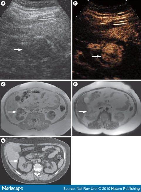
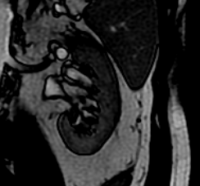
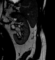
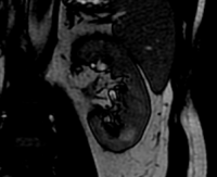
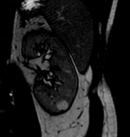
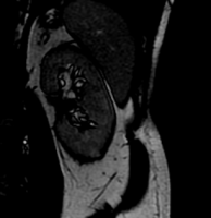
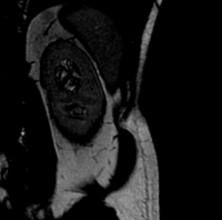
"Incidentally noted 10 mm T2 hypointense lesion in the lower pole of the left
kidney. Recommend evaluation with abdominal MRI without and with contrast."

after doing some research on the contrast agent, gadolinium, seems a bit sketchy, can collect in brain and also cause kidney problems plus who knows what else.

Anyone have any knowledge on the risks/benefits of the contrast agent with respect to a kidney lesion. BTW my mother, brother and cousin have all had kidney lesions found that turned out to be nothing dangerous.

My plan is to have the MRI but refuse the contrast.

@DrJ I appreciated your response on CT Scans and was wondering if you had anything else to add on MRI contrasting agents.
 

DrJ

Member
Joined
Jun 16, 2015
Messages
723
@x-ray peat If it were me, I would avoid them. I'd say I was allergic or refuse the waiver. They've been conducting MRIs successfully for a long time without the contrast agents. I suspect they are turning to this for the growing number of obese people. The skinnier you are, the better the image quality will be in MRI because there is less attenuation of the signal, both input and response. There's a similar problem in CT where they keep having to design them to fit wider and wider people, but the bigger design ends up being worse for a more average-sized body and especially kids.

That it's gadolinium is pretty ****88 up. A common material in x-ray detectors is gadolinium oxysulfide (Gd2O2S) or "GOS" for slang. I have frequently worked with it in building x-ray detectors. At my last job, I was working to build novel types of x-ray detectors, and often had to modify the GOS by cutting, grinding, etc. The dust particles are well-known to be damaging to the lungs. I received strict safety instruction on how to handle it from the senior detector scientist. I always had to wear a mask to prevent inhalation and wear gloves to protect my skin. I had to dispose of it in a hazardous waste container.

Also, when GOS combines with an acid it produces hydrogen sulfide gas, which is fairly toxic. I did an experiment in which I was trying to make "x-ray detector jello" using gelatin (thanks Ray Peat!) and GOS. Sounds stupid, but it didn't really occur to me that an amino acid was much of an acid. I made my mixture and all of the sudden it started bubbling up and had the distinct smell of rotten eggs - hydrogen sulfide. I had to clear everyone out of the lab for an while and let it vent off, even though it was a pretty small amount of GOS I had used to trigger the reaction - a few grams.

It's absolutely insane to me that they are injecting this stuff in people. I guess you could say that Gd2O2S and Gd are not the same, but there's plenty of oxygen and sulfur in the body, so I suspect it could be formed. But my understanding always was that the main toxic effects - like with the lungs - was due to the Gd. If it hurts the lung tissue, how can it not be hurting other tissues?! Soooo ****88 up.
 
OP
x-ray peat

x-ray peat

Member
Joined
Dec 8, 2016
Messages
2,343
@DrJ thanks for the reply and great info. Intravenously administering a rare earth metal doesn't seem like such a good idea on any level.

I did some more reading to see what is the difference in quality of the image due to the contrast and if I have it right, it looks like the contrast actually makes things worse. Image C is no contrast and D is post contrast. Could they have reversed the images?

MRI, Multidetector CT and Ultrasonography of the Same Kidney Lesion. The mass (arrow) was incidentally discovered on a | ultrasonography, as a hypoechoic exophytic lesion. b | Contrast‑enhanced ultrasonography highlighted bloodflow within this Fuhrman grade I renal cell carcinoma (RCC). MRI (c | precontrast and d | post‑contrast) revealed enhancement on T1‑weighted imaging. http://www.medscape.org/viewarticle/722370_3

722370-figure5.jpg
 

aguilaroja

Member
Joined
Jul 24, 2013
Messages
850
@x-ray peat.... They've been conducting MRIs successfully for a long time without the contrast agents. I suspect they are turning to this for the growing number of obese people. The skinnier you are, the better the image quality will be in MRI because there is less attenuation of the signal, both input and response. There's a similar problem in CT where they keep having to design them to fit wider and wider people, but the bigger design ends up being worse for a more average-sized body and especially kids.

That it's gadolinium is pretty ****88 up...

Thanks for the fascinating report.

I second the view that “plain MRI” gives very detailed, informative views, without contrast. There is also the added three family members with non-alarming kidney findings. Is there really a definite physician decision for this situation that is likely to change based on a second set of picture used gadolinium? Is the time course of the kidney finding known? If the hypointense finding may have there for months or longer, there is even less "urgency" to choose the riskier
(Of course, the hospital system has a financial incentive to do the more expensive test, including the use of contrast.)
 

Ras

Member
Joined
Sep 12, 2015
Messages
937
@DrJ thanks for the reply and great info. Intravenously administering a rare earth metal doesn't seem like such a good idea on any level.

I did some more reading to see what is the difference in quality of the image due to the contrast and if I have it right, it looks like the contrast actually makes things worse. Image C is no contrast and D is post contrast. Could they have reversed the images?

MRI, Multidetector CT and Ultrasonography of the Same Kidney Lesion. The mass (arrow) was incidentally discovered on a | ultrasonography, as a hypoechoic exophytic lesion. b | Contrast‑enhanced ultrasonography highlighted bloodflow within this Fuhrman grade I renal cell carcinoma (RCC). MRI (c | precontrast and d | post‑contrast) revealed enhancement on T1‑weighted imaging. http://www.medscape.org/viewarticle/722370_3

722370-figure5.jpg
MRI tech here.

I don't know what machine they used to acquire those images, but as someone that does MRIs of the abdomen every weekday, I think the quality of those images is very poor, insomuch that I suspect our Rads would refuse to read them.

I use a 3T Philips, and our non-cons look excellent. We acquire a variety of sequences with various weightings that give the Rads a lot to read, even without Gad. We use Gadavist almost exclusively, which, being macro-cyclic, is reputed to be safer if retained; we use Multihance in renal-compromised patients, as it will provide the same T1-relaxation at half the dose of gadolinium. Howbeit, gadolinium-based contrast of any brand has been found to be retained (Magnevist being especially bad), and they all will cause Nephrogenic Systemic Fibrosis and God knows what else when the gadolinium dissociates, so that's why I refuse to have an MRI with contrast of any kind for any reason, especially not for a renal cyst - even an amateur tech can identify a renal cyst and any associated pathology without contrast.
 
OP
x-ray peat

x-ray peat

Member
Joined
Dec 8, 2016
Messages
2,343
MRI tech here.

I don't know what machine they used to acquire those images, but as someone that does MRIs of the abdomen every weekday, I think the quality of those images is very poor, insomuch that I suspect our Rads would refuse to read them.

I use a 3T Philips, and our non-cons look excellent. We acquire a variety of sequences with various weightings that give the Rads a lot to read, even without Gad. We use Gadavist almost exclusively, which, being macro-cyclic, is reputed to be safer if retained; we use Multihance in renal-compromised patients, as it will provide the same T1-relaxation at half the dose of gadolinium. Howbeit, gadolinium-based contrast of any brand has been found to be retained (Magnevist being especially bad), and they all will cause Nephrogenic Systemic Fibrosis and God knows what else when the gadolinium dissociates, so that's why I refuse to have an MRI with contrast of any kind for any reason, especially not for a renal cyst - even an amateur tech can identify a renal cyst and any associated pathology without contrast.
wow this forum has got just about everything covered. Thanks for the inside scoop.
 

Fiver

New Member
Joined
Dec 12, 2016
Messages
1
Hello,
Radiologist here. The reason for the gadolinium in your case is to determine if your kidney lesion is vascular (enhancing). It sounds like it is not a simple cyst, because simple cysts are hyperintense on T2. It could be a complex cyst (no vascular component, not enhancing), meaning that there is more than just water inside (blood, protein), or it could be a tumor (solid tissue with vascularity). An MRI picture with gadolinium can be digitally "subtracted" from the picture without gadolinium, in order to tell if the whole lesion or any part of the lesion is vascular. If there is no enhancing component, you can rest assured that it is not cancer.

That said, you are correct that there is concern over residual gadolinium in the body. The gadolinium contrast agents that leave the least amount of gadolinium behind are Dotarem, Gadavist, and Prohance, and I would certainly tell you to request one of those. Since the lesion is so small, you could track its size with non-gadolinium MRIs to see if it grows over time. How long has it been since the lesion was initially found? Another alternative is ultrasound, but that depends on where the lesion is (how deep from the skin) in terms of how well it can be characterized (complex cyst vs tumor) that way. Ultrasound can also be used to track the size of the lesion over time, instead of the more expensive MRI. CT can also tell if it is vascular or not, but that involves iodinated contrast and radiation.

Finally, there is one kind of kidney tumor that has fat in it and that is usually a benign tumor. If they detect fat in the tumor by MRI (gadolinium is not needed for this), they do not need to give you gadolinium. You could ask the techs to have the radiologist check the images for fat in the tumor after the first part of the study, to see if you need the gadolinium or not. Good luck to you and let us know how it goes.
 
OP
x-ray peat

x-ray peat

Member
Joined
Dec 8, 2016
Messages
2,343
Hello,
Radiologist here. The reason for the gadolinium in your case is to determine if your kidney lesion is vascular (enhancing). It sounds like it is not a simple cyst, because simple cysts are hyperintense on T2. It could be a complex cyst (no vascular component, not enhancing), meaning that there is more than just water inside (blood, protein), or it could be a tumor (solid tissue with vascularity). An MRI picture with gadolinium can be digitally "subtracted" from the picture without gadolinium, in order to tell if the whole lesion or any part of the lesion is vascular. If there is no enhancing component, you can rest assured that it is not cancer.

That said, you are correct that there is concern over residual gadolinium in the body. The gadolinium contrast agents that leave the least amount of gadolinium behind are Dotarem, Gadavist, and Prohance, and I would certainly tell you to request one of those. Since the lesion is so small, you could track its size with non-gadolinium MRIs to see if it grows over time. How long has it been since the lesion was initially found? Another alternative is ultrasound, but that depends on where the lesion is (how deep from the skin) in terms of how well it can be characterized (complex cyst vs tumor) that way. Ultrasound can also be used to track the size of the lesion over time, instead of the more expensive MRI. CT can also tell if it is vascular or not, but that involves iodinated contrast and radiation.

Finally, there is one kind of kidney tumor that has fat in it and that is usually a benign tumor. If they detect fat in the tumor by MRI (gadolinium is not needed for this), they do not need to give you gadolinium. You could ask the techs to have the radiologist check the images for fat in the tumor after the first part of the study, to see if you need the gadolinium or not. Good luck to you and let us know how it goes.
Great info! Great to see an MD who has broken through their Operant Conditioning:)
I think I will go ahead and have the MRI w/o contrast and then see what they say once a Radiologist looks at the pics.
 
Last edited:

EndAllDisease

Member
Joined
Jul 6, 2014
Messages
195
If it's at all possible to avoid it I would avoid getting any kind of contrast agent.

Why do you need an MRI anyways?
It'll simply tell you if your cells are or are not oxidizing properly.

Hit whatever cells you're concerned about with some red light after eating a plate of liver and a jug of orange juice and some aspirin and you're on your way to healing whatever problems you may be experiencing.
 
OP
x-ray peat

x-ray peat

Member
Joined
Dec 8, 2016
Messages
2,343
@Fiver, @DrJ, @Ras So I had the follow up MRI without contrast and the radiologist said that the scan was inconclusive and wants to do another scan in a few months. I actually got a copy of the mri scans but its a bit too complicated to upload (comes with its own software to view).

I'm seeing a kidney specialist tomorrow. I am sure they will want to redo the scan with contrast. Any thoughts? Would the contrast make that much of a difference. Im not overweight so I would think I would have gotten a good scan as DRJ mentioned.
 

DrJ

Member
Joined
Jun 16, 2015
Messages
723
They may respect your wish to go without contrast. Ask if you can be scanned in a more powerful MRI in lieu of using contrast agent. Like 5T (tesla) or 7T (if they have those outside research facilities). Or ask if it can be found through ultrasound. If you're not overweight, it should not be so far from the surface of the skin that ultrasound wouldn't work. If they need to know the location of a potential lesion, then ultrasound isn't so good at that, but if they just need to know present/ not present then ultrasound fits that functionality.
 
OP
x-ray peat

x-ray peat

Member
Joined
Dec 8, 2016
Messages
2,343
They may respect your wish to go without contrast. Ask if you can be scanned in a more powerful MRI in lieu of using contrast agent. Like 5T (tesla) or 7T (if they have those outside research facilities). Or ask if it can be found through ultrasound. If you're not overweight, it should not be so far from the surface of the skin that ultrasound wouldn't work. If they need to know the location of a potential lesion, then ultrasound isn't so good at that, but if they just need to know present/ not present then ultrasound fits that functionality.
Thanks for the info. Just got back from the urologist and got an earful about how I should have taken the contrast. despite the FDA warning and several lawsuits etc. Anyway he agreed to go with an ultrasound as a next step.

I screen shot some of the kidney MRI pics if anyone can see something in them besides the grey ball I see.
pictures are at location =
14.62 mm
8.62 mm
2.62 mm
-3.37 mm
-9.37 mm
-15.37 mm
zoom out
 

Attachments

  • 1 kidney loc 14.62 mm.png
    1 kidney loc 14.62 mm.png
    13 KB · Views: 17
  • 2 kidney loc 8.62 mm.png
    2 kidney loc 8.62 mm.png
    16.2 KB · Views: 16
  • 3 kidney loc 2.62 mm.png
    3 kidney loc 2.62 mm.png
    13.2 KB · Views: 15
  • 4 kidney loc -3.37.png
    4 kidney loc -3.37.png
    12.5 KB · Views: 16
  • 5 kidney loc -9.37.png
    5 kidney loc -9.37.png
    13.8 KB · Views: 16
  • 6 kidney loc -15.37 mm.png
    6 kidney loc -15.37 mm.png
    14.5 KB · Views: 16
  • Kidney zoom out.png
    Kidney zoom out.png
    85.6 KB · Views: 16

Snowdrop

Member
Joined
Mar 3, 2014
Messages
45
I went for a brain MRI today. I only got told the day before that they would be using a Gadolinium contrast media, via injection.

After reading all I could, I had a little freak out about going through with the MRI. However, I did find the courage to go and thought having a chat with the radiologist would do no harm.

As it turns out, the Scotsman was very positive about the result the MRI would produce without the contrast media. Phew!

I received Ray Peat's reply after the MRI and as usual he was spot on.

Here is what he had to say about the Gadolinium contrast media...
From the animal studies, it damages all the essential organs. Competent workers can make fine MRI images without it. It’s a holdover from x-ray times, by people who don’t understand the principle of MRI. RP

I got lucky!!!
 

alywest

Member
Joined
Apr 19, 2017
Messages
1,028
In a strange moment of synchronicity I just happened to go from reading Snowdrop's post to reading this excerpt:

My Neuro ordered 6 MRIs ALL with Gadolinium in one day. I survived the first one and had to leave immediately. My reactions were immediate, as I assume they would be with anyone whose brain is filled with mercury. My entire mouthful of mercury fillings were dry- drilled out over 2 days. Hot mercury dripping into my gums leaving tattoos everywhere. I had mine removed for my health... I should have just left them. Now, people are hearing about the dangers and how to have them removed properly by a biological dentist. Back then, no one had ever heard of a biological dentist! Just think of injecting Gadolinium along the lines of having all your mercury removed improperly. It's just as dangerous as vaccines. WAY worse for an already vaccine injured kid. Please be advised that the imaging facility slipped it into my IV telling me it was Valium for claustrophobia. I would never have consented to this poison. My life is over now because of it. Once you "Consent to Treat", they can inject you with any poison they feel like. And they ARE doing so without ones' informed consent, ALL THE TIME !! I have strabismus now from one dose. I'll never drive again, or enjoy a movie. I want to make sure you understand that they are slipping it in to IVs WITHOUT CONSENT!! I was afraid the Neuro Opthomologist was going to tell me he had to surgically remove my eyeballs to correct the damage! No, not even, I'm too far gone for even that. I was pretty much a quadriplegic and still have much difficulty using my muscles, 2 years post Gadolinium. Brushing my teeth is way too difficult, I don't have any strength at all and also suffer from incontinence. - LS

From What NOT To Do
 
OP
x-ray peat

x-ray peat

Member
Joined
Dec 8, 2016
Messages
2,343
I went for a brain MRI today. I only got told the day before that they would be using a Gadolinium contrast media, via injection.

After reading all I could, I had a little freak out about going through with the MRI. However, I did find the courage to go and thought having a chat with the radiologist would do no harm.

As it turns out, the Scotsman was very positive about the result the MRI would produce without the contrast media. Phew!

I received Ray Peat's reply after the MRI and as usual he was spot on.

Here is what he had to say about the Gadolinium contrast media...


I got lucky!!!
omg between what you found and what @alywest reported I feel very lucky as well. The question is how do you find a competent MRI technician to run a proper scan or a competent radioligist who can read them without the contrast. My urologist was no help either. He seems ready to jump in and cut out a big chunk of my kidney. Anyway I hope my ultrasound tells them what they need. No way will I get the contrast after reading what you guys just posted
 

Ras

Member
Joined
Sep 12, 2015
Messages
937
Here is what he had to say about the Gadolinium contrast media...

I consider myself to be what Rays calls a "competent worker," yet I fail to see how I can duplicate, through imaging parameters alone, the T1 relaxation effects of gadolinium on the myriad tissues of the body. How could one capture without contrast the differing relaxivities of healthy and unhealthy tissues in the liver in a patient with HCC? It would be altogether impossible for a technologist using a GE scanner, the Playskool of MRI machines. At least Philips allows its tech to change manifold more parameters and tells him useful sequence characteristics like shot length and scan percentage. You can't even change refocusing control on a GE or Siemens, not to mention their lack of no-fail fat-suppressing techniques like mDIXON TSE. I doubt Ray knows MRI technology well enough to judge these matters.
 
OP
x-ray peat

x-ray peat

Member
Joined
Dec 8, 2016
Messages
2,343
I consider myself to be what Rays calls a "competent worker," yet I fail to see how I can duplicate, through imaging parameters alone, the T1 relaxation effects of gadolinium on the myriad tissues of the body. How could one capture without contrast the differing relaxivities of healthy and unhealthy tissues in the liver in a patient with HCC? It would be altogether impossible for a technologist using a GE scanner, the Playskool of MRI machines. At least Philips allows its tech to change manifold more parameters and tells him useful sequence characteristics like shot length and scan percentage. You can't even change refocusing control on a GE or Siemens, not to mention their lack of no-fail fat-suppressing techniques like mDIXON TSE. I doubt Ray knows MRI technology well enough to judge these matters.
thanks for the good info. so should we be looking for MRI centers that use phillips machines? Should we also try to get the highest Tesla machines or are lower tesla machines more appropriate for certain scans?

edit: what do you think of the quality of my scans I posted? You can see the lesion most clearly on pic number 4
 

Ras

Member
Joined
Sep 12, 2015
Messages
937
thanks for the good info. so should we be looking for MRI centers that use phillips machines? Should we also try to get the highest Tesla machines or are lower tesla machines more appropriate for certain scans?

edit: what do you think of the quality of my scans I posted? You can see the lesion most clearly on pic number 4
What machine produces the best images is a matter of subjective opinion. I have no doubt that some Radiologists prefer the images produced by a certain manufacturer's algorithms; some of this is due to their being used to reading the kind of images that their 3T GE produces with the shim it's had since its last complete field service, and some of it is more objective. As a technologist, I prefer Philips, having used GE, Siemens, Toshiba (CT), and Philips; I prefer Philips for many reasons, one of which is the quality of its images, howbeit I am no Radiologist. Your task is two-fold: find a center with admirable, skilled technologists who use a scanner that allows them to alter many parameters; secondly, find an honorable Radiologist whose knowledge, wisdom, ethic, and skills you admire, ask him which manufacturer he prefers and why, and get him to analyze your study. The latter is especially hard, given the corruption of our medical field, and the former is a trial also, since many technologists are button-pushers. But take hope: I'm not a button-ape, and I work with at least one Radiologist that I admire very much, and I know another.

With greater magnetic field strength, one finds better image quality, but also greater artifact. You'd not want to do an abdomen study on your 3T on a patient with ascites, but the images from your 1.5T will have more noise and less signal. It depends also on the patient's body habitus, with more rotund citizens being harder to image with excellence due to artifact and SAR limitations. In general, I prefer a 3T magnet, though they have greater field inhomogeneity.

The images you posted are fine enough to locate the lesion, but because I can't adjust the window and level and scroll through all of the slices, I can't give them a better assessment. And I am confident a Radiologist would agree. It happens sometimes that one does a CT stone hunt that shows the lesion incidentally, which leads to their having an US that was inconclusive, which caused the Radiologist to suggest an enhanced MRI in the report of which he - because of liability - did not state conclusively the nature of the lesion, only suggesting it could be this or that and saying that it should be correlated clinically, which necessitated the patient having a biopsy, which confirmed it was a benign growth that should be followed, first every ninety days, then every one-hundred and eighty, then annually.
 
EMF Mitigation - Flush Niacin - Big 5 Minerals

Similar threads

Back
Top Bottom