COVID-19 And Carbon Monoxide

OP
md_a

md_a

Member
Joined
Aug 31, 2015
Messages
468
Increased Carbon Monoxide in Exhaled Air of Subjects with Upper Respiratory Tract Infections
Received: November 14, 1997


Viral infection may induce the expression of heme oxygenase, resulting in increased carbon monoxide (CO) formation. CO may be produced by various cells of the upper and lower respiratory tract and may be detected in the exhaled air. Therefore, exhaled CO concentrations were measured on a CO monitor by vital capacity maneuver in subjects with upper respiratory tract infections (URTIs) and in nonsmoking and smoking healthy control subjects. At the time of symptoms of URTI, exhaled CO concentrations were 5.6 ± 0.4 ppm and decreased to 1.0 ± 0.1 ppm during recovery. Recovery values of exhaled CO were similar to those in age-matched nonsmoking healthy control subjects (1.2 ± 0.3 ppm). Smoking healthy control subjects had the highest levels of exhaled CO concentration among the groups (18.5 ± 2.5 ppm). These findings suggest that symptomatic URTIs increase the concentration of CO in exhaled air. This may reflect the induction of heme oxygenase that has an antiviral effect in the airways.

Carbon monoxide (CO) has been reported to have several biologic actions (1-4) and may play a role in pathophysiology of airway diseases (5). CO is made by an enzyme called heme oxygenase, and two forms of heme oxygenase have been characterized (6). Constitutive forms of the enzyme are widely distributed throughout the body with high concentrations in the brain (6), but another isoform is induced in several types of cells after exposure to inflammatory cytokines (7, 8), oxidants (9, 10) and NO (11). CO is detectable in the exhaled air of normal persons (5, 12) and exhaled CO is increased in asthma (5), which may reflect an expression of inducible heme oxygenase in airway epithelial cells (13). This is supported by the fact that inhaled corticosteroids inhibit the increase in exhaled CO in asthmatic patients (5).

DISCUSSION
The present study has shown that exhaled CO can be reliably measured in healthy control subjects and subjects during the acute phase of URTIs. The values of exhaled CO in nonsmoking and smoking control subjects were similar to those of previous studies (12, 21, 22). Because the carboxyhemoglobin level in the blood declines exponentially and becomes normal 24 h after the cessation of smoking (23), the present study should underestimate the exhaled CO concentration in smoking control subjects. However, we have demonstrated that URTIs are associated with an increase in exhaled CO in normal persons in the acute phase when symptoms are present, and that there is a reduction in exhaled CO after recovery to values that are similar to those in age-matched normal subjects. This suggests that URTI, presumably caused by influenza viral infection, increases the production of CO in the respiratory tract.

However, several factors might influence the exhaled CO concentration in the present study. First, some variations in the exhaled CO concentration among three sequential maneuvers were observed. Although the reason for observed variations is uncertain, the mean exhaled CO concentrations were similar among three sequential maneuvers in nonsmoking control subjects and the subjects during and after URTIs. Furthermore, the variation between the exhaled CO concentrations on separate days in nonsmoking control subjects was small. Second, the subjects exposed to cigarette smoke as passive smokers might have the same range as the subjects with URTIs. However, it seems unlikely because we selected the subjects with URTIs and none of them were smokers, ex-smokers, or passive smokers. Finally, the subjects might encounter much higher ambient CO levels than those at the place of the CO measurement on their way to the hospital. However, the route and means of transportation to the hospital were the same in the subjects with URTIs between the time of the acute phase and after 3 wk of recovery, and they visited the hospital at a similar time of day. Therefore, the ambient CO levels on the way to the hospital could little influence the difference in the exhaled CO concentration between the time of the acute phase and after 3 wk of recovery in the subjects with URTIs.

The exhaled CO concentration was higher without an expiratory resistance used to close the velum during expiration to exclude nasal CO that may leak throughout expiration in the presence of an open velum (20). Therefore, the increase in CO may be derived from both nasal tissues and the lower respiratory tract. It is of interest that a large proportion of subjects with URTIs in this study complained of lower respiratory tract symptoms, including cough and chest pain accentuated by coughing, indicating that the lower respiratory tract was likely to have been involved.

Heme oxygenase plays an important role in the resolution of inflammation in animals (24, 25) and pulmonary epithelial cells in vitro (26). Likewise, heme oxygenase protects against viral infection and replication in cultured human airway epithelial cells (13). Viral infections may induce heme oxygenase in a variety of cell types, including airway epithelial cells and macrophages (27) via the induction of proinflammatory cytokines (7, 8) and NO (11). The increased heme oxygenase activity may then serve to limit the virus infection by inhibiting viral replication (13) and airway inflammation by anti-inflammatory actions (24-26). Because inhaled corticosteroids inhibit the increase in exhaled CO in asthmatic patients (5), corticosteroids may impair host defenses against the viral infection through downregulation of heme oxygenase activity. However, Farr and colleagues (28) reported that the trend toward less increase in nasal obstruction, middle ear pressure, mucus production, and nasal mucus kinin and albumin concentrations during the first 2 d after rhinovirus inoculation was temporally related to the simultaneous administration of oral prednisone and intranasal beclomethasone. Furthermore, dexamethasone inhibits rhinovirus-induced production of cytokines and intercellular adhesion molecule-1 productions and replication of rhinovirus in the cultured human airway epithelial cells in vitro (29). Therefore, corticosteroids may have effects on upper respiratory tract infections through several mechanisms, and a further study is needed to clarify this issue. Furthermore, the administration of NO synthease inhibitors by nebulization to normal subjects and patients with asthma produces a fall in exhaled NO levels (30). Therefore, NO synthase inhibitors may downregulate heme oxygenase activity, thereby decreasing the exhaled CO.

Although we have shown an elevation of exhaled CO in URTIs that decreases after recovery, it is uncertain whether the level of CO in exhaled air is merely an indicator of airway inflammation or a causative link in the biology of airway viral infection. However, the demonstration that URTIs are associated with a high level of exhaled CO suggests that respiratory viral infections may induce the expression of heme oxygenase in the airway. Heme oxygenase therefore may act as a host defense against URTIs.
Because upper respiratory tract infections (URTIs) are reported to induce increases in interleukin (IL)-1β, IL-6, and tumor necrosis factor-α in nasal lavage fluid (14) as well as increases in exhaled NO (15), it is likely that viral respiratory tract infection increases CO production. We have, therefore, studied whether URTIs increase the concentration of CO in the exhaled air of normal persons.

https://www.atsjournals.org/doi/10.1164/ajrccm.158.1.9711066
 
OP
md_a

md_a

Member
Joined
Aug 31, 2015
Messages
468
Exhaled carbon monoxide in patients with lower respiratory tract infection

Abstract

The concentration of carbon monoxide (CO) in exhaled air is increased in patients with asthma, bronchiectasis and upper respiratory tract viral infections. However there is no information about the level of CO in patients with lower respiratory tract infection. We studied a group of 35 patients (22 males) aged 45 +_3 (SEM) years with cough productive of purulent phlegm and pyrexia in a general practice setting. All were non-smokers or ex-smokers and none had a previous history of respiratory problems or diabetes. We measured CO level in exhaled air before and after a course of antibiotics. Therapy was deemed successful when patient no longer complained of cough productive of purulent phlegm.

Twenty-eight of 35 patients had elevated CO level at their initial visit. Twenty-two out of 35 patients reported clinical improvement after antibiotic treatment and this was associated with a fall in exhaled CO level from 5.2 +_0.5 ppm to2.3+_0.3 ppm (P<0.0001).We suggest that simple CO measurements in exhaled air can detect the inflammatory process within the airways caused by infection and that a repeat measurement can be used to assess the nature of inflammation.

DISCUSSION

Our study showed that the majority of patients with clinical symptoms of lower respiratory tract infection had elevated exhaled CO levels. The clinical improvement after the treatment with antibiotics was associated with a reduction of exhaled CO. However, in 13 patients there was no clinical improvement after the treatment with antibiotic. In this group of patients the exhaled CO levels were lower, but statistically not significant, compared to the group who did respond. The reason for that could be that either they were treated with inappropriate antibiotic or they were suffering from a viral infection.

In the other similar study there was a significant reduction in exhaled CO level after the upper respiratory tract infection, but the observation period was much longer than in our study (6). Seven patients had ‘normal’ initial measurement of exhaled CO level (below 3 ppm).

However our ‘normal’ values differed from the other, similar study. If we accepted normal values our patients would have abnormal measurements. It is therefore necessary to establish and unify ‘normal’ values for the measurement of exhaled CO.

Although the exact cellular source of exhaled CO is not known, its elevation may reflect presence of inflammation and/or oxidative stress within airways (11).

An elevation in exhaled CO has been reported in patients with mild and acute asthma with a subsequent fall after the treatment with inhaled corticosteroids (4). CO levels were elevated in patients with asthma following allergen challenge (12). More recently, an elevation of exhaled CO has been found in patients with upper respiratory tract infection, which subsequently fell after the recovery (6). Some of the patients in this study also had symptoms of lower respiratory tract infection.

Similarly, nitric oxide (NO), another marker of airways inflammation, has been found to be elevated in subjects with upper respiratory tract infection. NO level subsequently fell after the recovery period (13).

Also, an elevated NO has been found in patients with asthma, which fell on treatment with inhaled steroids.

Our study showed an elevation of exhaled CO level in majority of patients with lower respiratory tract infection, which subsequently fell after the treatment with antibiotics. Bacterial infection may induce production of heme oxygenase, which has been shown to participate in resolution of inflammation in animals (17). Therefore, increased levels of exhaled CO in lower respiratory tract infection may reflect increased inflammation and oxidative stress in airways and could be a single, non-invasive tool for monitoring treatment with antibiotic. We have previously demonstrated an elevation of exhaled CO in patients with bronchiectasis (7) and cystic fibrosis (8), indicating that different types of inflammation may increase exhaled CO and that this measurement is not specific for any particular inflammatory disease of airways. Repeated measurements after treatment with either an inhaled corticosteroid (4,5) or an antibiotic provides information about the control of inflammation. Our study confirmed an elevation of exhaled CO in patients with symptoms of lower respiratory tract infection. The levels of CO subsequently fell after the treatment with antibiotic, which coincided with clinical improvement.

We suggest that simple CO measurement in exhaled air can detect increased inflammatory process within the airways, which has been caused by infection. The return of CO levels to normal after the treatment may be related to the reduction in inflammation. Elevated exhaled CO levels may provide an early warning signal for an acute infection episode.

Exhaled carbon monoxide in patients with lower respiratory tract infection - ScienceDirect
 
OP
md_a

md_a

Member
Joined
Aug 31, 2015
Messages
468
Host heme oxygenase‐1: Friend or foe in tackling pathogens?
First published:14 May 2018

Abstract

Infectious diseases are a major challenge in management of human health worldwide. Recent literature suggests that host immune system could be modulated to ameliorate the pathogenesis of infectious disease. Heme oxygenase (HMOX1) is a key regulator of cellular signaling and it could be modulated using pharmacological reagents. HMOX1 is a cytoprotective enzyme that degrades heme to generate carbon monoxide (CO), biliverdin, and molecular iron. CO and biliverdin (or bilirubin derived from it) can restrict the growth of a few pathogens. Both of these also induce antioxidant pathways and anti‐inflammatory pathways. On the other hand, molecular iron can induce proinflammatory pathway besides making the cellular environment oxidative in nature. Since microbial infections often induce oxidative stress in host cells/tissues, role of HMOX1 has been analyzed in the pathogenesis of number of infections. In this review, we have described the role of HMOX1 in pathogenesis of bacterial infections caused by Mycobacterium species, Salmonella and in microbial sepsis. We have also provided a succinct overview of the role of HMOX1 in parasitic infections such as malaria and leishmaniasis. In the end, we have also elaborated the role of HMOX1 in viral infections such as AIDS, hepatitis, dengue, and influenza.

CONCLUDING REMARKS
Research from last three decades has established that HMOX1 is a master regulator of stress response. Products of HMOX1 mediated degradation of heme namely CO and biliverdin are potent antioxidants and molecular iron acts as an oxidant if left unattended. Emerging literature suggests that HMOX1 plays an important role in several infections. Importantly CO and biliverdin (or its derivative bilirubin) can themselves kill several pathogens. A number of microbial infections induce oxidative stress in the infected cells. Thus, in many infections HMOX1 could protect the cells from the pathogen inflicted oxidative stress. On the contrary, oxidative stress could induce pathways that help the host cells in elimination of pathogens. Thus depending upon host cell type and the pathogen, the HMOX1 could act in protection of the host from infection or it may assist the growth of intracellular pathogen. Its protective role is clear in microbial sepsis and in several viral infections including HIV. On the hand, HMOX1 helps in the survival of intracellular leishmania. The role of HMOX1 is not clear in case of Malaria, TB and salmonella infection. In these infections, HMOX1 could perform both host protective function or may help intracellular growth of pathogen through downregulation of inflammatory pathways. We believe that further research will reveal the precise role of HMOX1 in these infections.

Error - Cookies Turned Off
 
OP
md_a

md_a

Member
Joined
Aug 31, 2015
Messages
468
Verné Saville
April 18 at 4:24 PM ·
Dear PM Scott Morrison

I'm a Veterinary Surgeon that graduated from Melbourne University with a double degree ( 1st: Veterinary Science, 2nd: pathology/toxicology), I have two Masters degrees one of which is in disease surveillance (epidemiology), and I've been doing my PhD on epidemiology and risk assesment. I was also awarded a recent alumni achievement award from Melbourne University. I've been watching this outbreak since it started, and have correctly predicted its course.

You are doing a fabulous job at slowing our curve, and I commend you on what you are doing as you have certainly bought us time.

I have two urgent matters to bring to your attention.
The first is it is unlikely a vaccination will work. Veterinarians are the only ones with a coronavirus vaccine, and what's been found in vaccinated animals that are subjected to infection with another coronavirus often results in worse pathology and they even have fatal consequences as demonstrated in a few studies.
This makes prevention very difficult, and vaccine efficacy will be questionable. This means the focus will need to be on treatment/cure.

The second matter is my current disease hypothesis that may result in successful treatment of critical cases.
I have a crazy but very plausible hypothesis, and there's a toxicologist in the USA that has released a similar hypothesis this week, so that's at least 2 of us that believe this is plausible.
My theory is that SARS-cov2 causes an increase in endogenous (produced in our body) carbon monoxide production in the body, resulting in carbon monoxide poisoning.

Carbon monoxide toxicity fits with everything we are seeing. It fits with the high fatalaties with comorbidities particularly diabetes, heart disease and obesity. It fits with cases overseas just dropping dead in the streets, it fits with the ground glass lung pathology seen. It fits with the symptoms, as carbon monoxide poisoning is often misdiagnosed as the flu, causes headaches, dizziness, fatigue, breathlessness. It fits with the lower than expected success with ventilated patients, as carbon monoxide actually increases in ventilated patients. It also explains the neurological signs seen in some patients, and it fits with the success seen with zinc.

This is 100% a hypothesis, but I can't physically test it. It's simple though, doctors need to test for Carbon monoxide which is simple to do if they are looking for it. We aren't looking for it, so no one is testing for it.
Up to now doctors have assumed this is a viral induced disease. I believe the virus does not mean to kill us, this is a mistake that has occurred in our bodies in response to the virus, causing a toxicity event. This is why most people are asymptomatic or have mild disease.

So my theory in a nut shell is the virus causes our bodies to produce more carbon monoxide than usual, which inadvertently causes carbon monoxide toxicity.

Treatment is relatively simple as its just in addition to what is already happening, with the addition of using a hyperbaric oxygen chamber.

How I came up with this hypothesis. I lived in bogota Colombia for 3 years and suffered from altitude sickness. To me this sounds like and looks exactly the same as what I suffered. Carbon monoxide poisoning and altitude sickness present very similar. Humans produce endogenous carbon monoxide, so I started looking for instances where this Carbon Monoxide production could be exaggerated, and it all started to make sense. In addition in bogota the young coped very well, and travelers new to the region suffered greatly the older they were, so the age fits too with what we are seeing in cases.

Whilst I plan to publish this in a formal paper this will take a month or 2, and I would like Australia to have access to this knowledge on the off chance I'm right and we can save lives.

It's easy to test for, and if it is Carbon Monoxide toxicity it's easy to treat.
Carbon monoxide has been found to Increase in pathological conditions, it's not that hard a jump to think that somehow the virus induces certain people to get their carbon monoxide production into a slight overdrive. It doesn't take much to result in carbon monoxide toxicity.

Thank you for everything you are doing, I know where we would be without the measures you have put in place, and you have done an outstanding job.

Sincerely
Dr Verné Dove
BVSc Hons BAnimSc (Research: Veterinary Pathology) MVS (Veterinary Conservation Med.)
MVS ( Veterinary Disease Surveillance) Dip. Conservation
PhD candidate (Dolphin Health Assessment)
Murdoch University, Australia
Universidad de los Andes, Colombia

18th April 2020.
 

gaze

Member
Joined
Jun 13, 2019
Messages
2,270
“Porphyrins are the pigment molecules that include the heme of hemoglobin, of the respiratory cytochromes, the peroxidases including thyroid peroxidase, and of the P450 enzymes which, among other things, produce steroid hormones.” RP

“The heme group (of hemoglobin and the respiratory enzymes, for example) is the iron-binding oily molecule that interacts with oxygen, and it is called a porphyrin.”RP


“Besides the frequently discussed interactions of excessively accumulated iron with the unsaturated fatty acids, producing lipid peroxides and other toxins, the accumulated calcium very probably forms some insoluble soaps with the free fatty acids which are released even from intracellular fats during stress. The growth of new mitochondria probably occasionally leaves behind such useless materials, combining soaps, iron, and porphyrins remaining from damaged respiratory enzymes.

When the background of carbon dioxide is high, circulation and oxygenation tend to prevent the anaerobic glycolysis that produces toxic lactic acid, so that a given level of activity will be harmful or helpful, depending on the level of carbon dioxide being produced at rest.” Mitonchondria and mortality

“Sensitivity to the harmful effects of light can be increased by some drugs and by excess porphyrins produced in the body (and by the porphyrin precursor, delta-amino levulinic acid), leading to rosacea, so those factors should be considered, but too often alcohol (which can cause porphyrin to increase) is blamed for rosacea and rhinophyma, without justification. There are many ways in which poor health can increase light sensitivity. Some types of excitation produced by metabolites (or by the failure of inhibitory metabolites) can produce vasodilation, involving the release of nitric oxide (Cardenas, et al., 2000), setting off a series of potentially pathological reactions, including fibrosis. The nitric oxide increases glycolysis while lowering energy production. The excitatory metabolite glutamate, and nitric oxide, are both inhibited by aspirin (Moro, et al., 2000).


Estrogen's most immediate effect on cells is to alter their oxidative metabolism. It promotes the formation of lactic acid. In the long run, it increases the nutritional requirements for the B vitamins, as well as for other vitamins. It also increases the formation of aminolevulinic acid, a precursor of porphyrin, and increases the risk of excess porphyrin increasing light sensitivity. Both aminolevulinic acid and excess porphyrins are toxic to mitochondria, apart from their photosensitizing actions. Nitric oxide, glutamate, and cortisol all tend to be increased by estrogen.


Lactate, glutamate, ammonium, nitric oxide, quinolinate, estrogen, histamine, aminolevulinate, porphyrin, ultraviolet light, polyunsaturated fatty acids and endotoxin contribute to excitatory and excitotoxic processes, vasodilation, angioneogenesis, and fibrosis.” Rosacea, inflammation, and aging: The inefficiency of stress

“My results, in which estrogen interfered with respiration, made me think of Warburg's description of cancer metabolism. He saw cancer's,respiratory defect’; as depriving it of the energy it needed to function as a useful tissue, leaving it only the primitive function of growth. I considered many ways in which estrogen might be a cancer hormone, including its promotion of the oxygen-wasting age pigment, and its stimulation of porphyrin metabolism, since some researchers had seen an association between cancer and porphyrins. At that time, it wasn't known that the breakdown of the porphyrin, heme, produced carbon monoxide.

But beyond the possibility that estrogen was deeply involved in the nature of cancer, I felt that its biological role had to do with its interference with oxidative metabolism. Selye had characterized estrogen's effect as ‘like the shock phase of the stress reaction’. Estrogen does act in conjunction with histamine, and histamine alone tends to cause circulatory collapse by allowing fluid to leak out of blood vessels. Lack of oxygen probably relates more generally to the shock reaction than does histamine.The reduction of cellular energy is probably estrogen's central action, and in Warburg's scheme, this would be the way to turn on cell division and growth. In the absence of oxygen, cells take up water, and when water-logged (even from being placed in a hypotonic fluid), they begin to divide.” Ray Peat - From PMS To Menopause Females Hormones In Context

“The increased energy deficiency produced by accumulated toxins and fibrosis can stimulate the production of porphyrins for the heme enzymes that control respiration and detoxification, and this can lead to the production of carbon monoxide and other anti-respiratory factors. Increasing amounts of ammonia and lactic acid circulate through the tissues.” Ray Peat - FIBROSIS

...

“Almost any sort of liver disease increases the systemic ammonia level. Estrogen can cause a large variety of liver diseases, including fatty liver and cholestasis; ammonia, which is toxic in itself, also links into another potentially toxic system, the porphyrin synthetic system.

Porphyria (acute intermittent porphyria, hepatic porphyria) is a disease that can cause nerve damage, hypertension, and connective tissue damage. It typically involves an excess of two precursors of heme, and sometimes a deficiency of heme (needed for respiratory enzymes), and it is often triggered by hypoglycemia, by exercise, and by estrogen or by certain poisons. Although it is usually described as a strictly genetic disease, it is higly susceptible to environmental influences, and a proper reevaluation of the evidence might show that it is more often environmental than genetic. Most of the evidence for a genetic cause consists of measurements that show low activity of certain enzymes. Since the conditions prevailing when a protein is synthesized can affect its structure and functions, the simple measurement of enzyme activity is hardly an appropriate argument.

The two substances that accumulate happen to be in a synthetic sequence, subsequent to a step in which C02 is removed, and before a step in which NH3, ammonia, is removed. The principle of mass action indicates that a reaction will slow or stop when there is a certain concentration of the product of the reaction. High C02 and low NH3 will prevent an accumulation of these chemicals, one of which is a potent neurotoxin. The opposite situation, low C02 and high NH3 (ammonia), will tend to cause an accumulation of these substances. Therefore, a simple metabolic shift that predictably happens in stress and malnutrition, can explain the main type of porphyria, independently of specific genetic problems. Everyone's genetic constitution is unique, and in a metabolically complex condition such as porphyria, there will be a spectrum of susceptibility. To draw a line across the spectrum, dividing people with "genetic defects" from the normal, is a purely arbitary and illogical procedure. It is much more important to identify and eliminate "porphyriogenic" environments.

Since porphyria attacks commonly occur premenstrually or after skipping a meal, the food cravings caused by increased estrogen ·and lowered blood sugar, are probably reinforced in many people by dread of the terrible symptoms that can be produced by not eating enough, resulting from the increased ammonia and porphyrins or porphyrin precursors. Calorie restriction can be dangerous when porphyria is developing. The presence of porphyrin poisoning, with its associated free radical toxins, can lead to the activation of heme oxygenase, the enzyme which produces carbon monoxide, which I have discussed elsewhere as a cause of the respiratory defect that characterizes cancer. Both ammonia and porphyria have been implicated in the production of cancer.

Acetazolamide, a drug that causes carbon dioxide to be retained in the tissues, tends to block the formation of ammonia. This is probably a confirmation of the importance of carbon dioxide as an anti-ammonia factor. High altitude also causes increased retention of carbon dioxide in the tissues, because of the Haldane-Bohr effect, and the reduction of ammonia (production, serum concentration, and excretion) at high altitude is probably even greater than the reduction of lactate production.” Ray Peat -Estrogen, Aging, Radiation, Migraine & Energy

...

“THE ORIGINAL ARGUMENT:

Cancer is the result of ordinary physiological processes which become autonomous because of regulatory weaknesses in the organism. Respiration is essential for the maintenance of the higher forms of life, and it is a respiratory 'defect, on both the cellular and the organismic levels, which allows cancer to persist and develop.

The heme group, because it serves many respiratory functions--hemoglobin, mitochondrial respiratory enzymes, steroid synthesizing enzymes, formation of thyroid hormone, detoxifying enzymes--is regulated in relatively primitive ways within each cell, and in more complex ways at higher organismic levels.

When the cell needs more respiratory energy, some fuel is diverted into the production of porphyrin, which is then turned into heme, which would normally provide for the efficient production of energy and protective factors.

When the efficient energy-producing systems are blocked, by injury, oxygen deficiency, toxins, or by the lack of one or more essential nutritional factors, heme production is activated.

Excess heme is destroyed by the enzyme heme oxygenase, which converts heme into biliverdin and carbon monoxide. Both of these factors have effects on the cell which are characteristic of cancer.

Estrogen, radiation, chemical carcinogens, and other forms of stress, activate the heme oxygenase enzyme.

Estrogen causes both porphyria and jaundice and is associated with increased formation of carbon monoxide. It inhibits many types of liver function, including detoxification.

The production of carbon monoxide by cancer cells can account for cancer's self-sustaining, "hereditary," property, without invoking genetic mutations which are now known to be consequences, rather than causes of cancer.

The production of carbon monoxide and biliverdin can account for many of the structural and biochemical abnormalities of cancer cells, and for their induction of abnormalities in adjacent cells.

"Genetic" theories of cancer have now reached a dead end, and the epigenetic, developmental physiological approach remains as the only plausible description of cancer.” Carbon monoxide, estrogen, and the medical cancer cult

...

“THE ARGUMENT:

Cancer is the result of ordinary physiological processes which become autonomous because of regulatory weaknesses in the organism. Respiration is essential for the maintenance of the higher forms of life, and it is a respiratory defect, on both the cellular and the organismic levels, which allows cancer to persist and develop. The heme group, because it serves many respiratory functions--hemoglobin, mitochondrial respiratory enzymes, steroid synthesizing enzymes, formation of thyroid hormone, detoxifying enzymes--is regulated in relatively primitive ways within each cell, and in more complex ways at higher organismic levels. When the cell needs more respiratory energy, some fuel is diverted the production of porphyrin which is then turned i which would normal provide for the efficient production of energy and protective factors.

When the efficient energy-producing systems are blocked by injury, oxygen deficiency, toxins, or by the lack of one or more nutritional factors, heme production is activated. Excess heme is destroyed by the enzyme heme oxygenase which converts heme into biliverdin and monoxide. Both of these factors have effects on the cell wich are characteristic of cancer. Estrogen, radiation, chemical carcinogens and other forms of stress, activate the heme oxygenase enzyme.

Estrogen causes both porphyria and jaundice and is associated with increased formation of carbon monoxide. It inhibits types of liver function, including detoxification.

The production of carbon monoxide by cancer cells can account for cancer's self-sustaining, "hereditary," property, without invoking genetic mutations which are now known to be consequences, rather than causes of cancer.

The production of carbon monoxide and biliverdin can account for many of the structural and biochemical abnormalities of cancer cells, and for their induction of abnormalities in adjacent cells. "Genetic" theories of cancer have now reached a dead end, and the epigenetic, developmental physiological approach remains as the only plausible description of cancer.


Strong discovered that and poor nutrition lead to overproduction of the porphyrin pigment, and contribute to the development of cancer. He showed that the liver was involved in the control of cancer. The Shutes, the Biskinds, and Alexander Lipshutz were at this time revealing other effects of estrogen that illuminated Strong's discoveries.” Ray Peat ‘Carbon Monoxide, Stress, and Cancer: 1999 status’

...

“Estrogen promotes lactic acid formation, and promotes porphyrin synthesis, providing the material for forming heme and carbon monoxide. Besides causing porphyria, estrogen causes many other liver diseases, including chole"stasis, the failure to release bile; the inhibition of carbon monoxide formation has been found to promote bile flow. Estrogen promotes the formation of the enzyme heme oxygenase, which forms carbon monoxide from heme. Carbon monoxide increases the formation of cortisol, by stimulating ACTH release from the pituitary.

The stress response is self-sustaining on several levels.

For example, stress increases the absorption of bacterial endotoxin from the intestine, which increases the estrogen level and synergizes with biliverdin and cortisol.

While estrogen does cause direct DNA damage, its clearest effect in carcinogenesis is on the cytoskeleton which regulates cell division, and defective cell division, rather than "gene mutation," is one of the important steps in the progression of cancer (Rubin, Duesberg).

Cancer cells are protected against the body's ability to destroy them, by the antioxidant functions of carbon dioxide and biliverdin.


This would include antiestrogen regimes, antiinflammatory and antihistamine factors (histamine interacts closely with nitric oxide and carbon monoxide), adequate nutrition, carbon dioxide, and specific anti-carbon monoxide therapies (such as light, alcohol, and possibly the minerals which convert porphyrin into compounds that inhibit the production of carbon monoxide), and methods to decrease nitric oxide formation and to restrain cortisol production, since these promote the formation of carbon monoxide. One of the most interesting approaches to inhibiting carbon monoxide production is to use vitamin B12, as hydroxocobalamin, as an antidote to nitric oxide, preventing the nitric oxide from stimulating the formation of heme oxygenase.

Wherever carbon monoxide mediates a biological malfunction, as in aquired immunodeficiency, Alzheimer's disease, and cancer, vitamin B12 seems to have a place as a detoxicant.

Progesterone and thyroid have several desirable properties that make them generally useful in cancer prevention and therapy. The shorter-chain saturated fatty acids contained in coconut oil have several beneficial effects. Aspirin and vitamin E and simultaneously destabilizing normal cellular phenotypes.” Ray Peat

...

When something interferes with the normal, productive use of oxygen, there is a great increase in the destructive forms of oxidation, such as lipid peroxidation, and the antioxidative reserves become crucial. That is, decreased respiration of the productive sort tends to increase the destructive use of oxygen.

In 1895 Magnus-Levy demonstrated that hypothyroid people have abnormally low heat production,

and that their heat production could be brought up to normal by giving them thyroid substance.

In 1926, Otto Warburg showed that the respiratory enzyme, containing the heme group, is inhibited by carbon monoxide, which binds to that enzyme, as it does to hemoglobin. Warburg also showed that visible light restores the activity of the respiratory enzyme by dissociating it from the carbon monoxide.

The heme group (of hemoglobin and the respiratory enzymes, for example) is the iron-binding oily molecule that interacts with oxygen, and it is called a porphyrin. There is a long history of investigating the interactions of porphyrin metabolism with estrogen with diet, and with excess iron. Estrogens are known to cause porphyria and to exacerbate the symptoms and biochemical disturbances in· people with subclinical porphyria.

Sometimes symptoms occur premenstrually, during the time of increased estrogen production—the term "ovulocyclic porphyria" has been in use for a long time. Puberty therefore increases the susceptibility to symptomatic episodes. Jaundice in pregnancy and in oral contraceptive users is probably a closely related phenomenon.

Porphyrin synthesis begins at an important cross-over point of protein and carbohydrate metabolism.

Succinyl CoA and amino levulinic acid can enter the Krebs cycle or the porphyrin pathway.

Protein catabolism feeds into these pathways. Increased protein catabolism or blockage of oxidative consumption of Krebs cycle fuel--for example by poisoning--makes these precursors available to enter the porphyrin pathway. Stress-induced oxidation of heme can eliminate feedback control, but the specific outcome can be modified in many ways...

Low blood sugar, most often caused by hypothyroidism, and diabetes--which involves poor absorption of sugar by cells--both tend to lower the respiratory quotient, the amount of carbon dioxide produced in relation to the amount of oxygen used.

High carbohydrate diets, sometimes with insulin have been used to treat porphyria. The use of carbon dioxide inhalation in psychiatry has many metabolic justifications, one of which might be the importance of carbon dioxide in glucose regeneration. It is also essential for detoxifying ammonia.

Whenever a symptom is relieved by glucose, I think we should suspect that thyroid and carbon dioxide might be deficient.

Many serious long-range consequences of excess heme/porphyrin production and metabolism are currently being investigated, suggesting that the criterion of "twice the upper limit of normal" excretion that was recently proposed by a government agency, for recognizing that a problem exists, could allow far more serious problems to develop over time, that on the surface might seem unrelated to porphyria. I consider any porphyrin excess to be a serious indicator of physiological stress. The ramifications of disturbed heme metabolism, resulting from exogenous factors, are far-reaching. For example, G. Y. Kennedy, at the Cancer Research Laboratory, University of Sheffield, observed that a porphyrin shortened the time required to induce tumors, and porphyrin derivatives have been proposed to be “cancer hormones."

The carbon monoxide produced in the breakdown of heme inhibits many enzymes. The consequences of slight excesses in porphyrin metabolism just haven't been investigated, because of the genetic dogmatism that denies that the person's environment could be at fault.

The synthesis of heme/porphyrin, and the production of red blood cells, are stimulated by a lack of oxygen, or by toxins such as arsenic and iron, which cause oxidative stress. Emphysema, high elevation, sluggish circulation, and nocturnal breathing problems can cause enough oxygen deficiency to stimulate the formation of new red blood cells. Newborn babies often have polycythemia, as a result of limited prenatal oxygen supply. At a certain point, the continued production of red blood cells can make the blood so viscous that this viscosity impairs circulation through capillaries, and creates a vicious circle, stimulating the formatjon of more red blood cells. Men are more likely than women to have polycythemia rubra vera, possibly because testosterone is anabolic to the bone marrow, and estrogen tends to slow blood cell formation (females of all species are relatively "anemic" compared to males, partly because their blood is more dilute), but I think the greater ability of men's marrow to respond proliferatively to hypoxia is influenced by many factors.” Ray Peat – ‘Optimizing respiration’

...


“Oxygen deprivation causes tissues to retain calcium (and iron), as does estrogen in many cases, being similar to aging in promoting cellular uptake of calcium. Since the porphyrins strongly bind metals, it has been suggested that they may have a role in mediating the deposition of metals in stressed tissues. Paroxysmal vasospasm occurs in about 90% of scleroderma patients, and estrogen and adrenalin are known to synergize in producing vascular spasm; hypothyroidism normally involves elevations of both estrogen and adrenalin.

The porphyrins break down into bilirubin, which also poisons mitochondrial respiration (Zetterstrom and Ernster).”


“When extracts of cancers were injected into healthy animals, some of them became sick, and were inclined to develop cancer. Several lines of investigation led to the belief that pyrroles and porphyrins, related to heme (the iron-binding pigment in hemoglobin and various oxidative enzymes), might be a "cancer hormone," but the idea lacked charm, and didn't catch on.

I made extracts from aged uterine tissue, thinking it might contain estrogen, and found that when I injected it into a hamster, it seemed to cause secretion of porphyrin from the animal's eye. This led me to get interested in the hormonal significance of the porphyrin pigment, and it was known to be related to estrogen excess and cancer susceptibility. Two of cancer's most mysterious features are its respiratory defect, identified by Otto Warburg, in which it converts glucose to lactic acid even in the presence of oxygen, and its resistance to lipid peroxidation. Lipid peroxidation is intimately involved in the control of cell division and aging, and in susceptibility of cells to elimination by the immune system, so cancer's antioxidative capacity seems to be closely related to its "immortal" nature. Iron (either free or bound to heine) is known to catalyze lipid peroxidation, but its presence in cancer cells simply supports their growth, rather than causing peroxidation.

Warburg discovered that light desorbs carbon monoxide and cyanide from respiratory pigments. In trying to understand light's effects on respiration, it occurred to me that it might be desorbing those, or other toxins that bind to and inhibit the respiratory enzymes. Cancer cells lack the ability to detoxify cyanide, so it has seemed possible that cyanide might contribute to the respiratory defect of cancer; bowel bacteria can produce small amounts.

But carbon monoxide is always being produced in the body, by the enzyme heme oxygenase, which is involved in the breakdown of hemoglobin. Carbon monoxide, by binding to heme-iron, inhibits lipid peroxidation, as well as inhibiting the respiratory pigments in the mitochondria.

Warburg observed that depriving growing cells of oxygen was sufficient to cause some of them to turn into cancer cells.

Anything which causes oxygen deprivation stimulates the formation of heme.

If the breakdown of heme occurs in cancer cells, that is, if heme oxygenase can be demonstrated in them, then the conditions exist for a stable, heritable but non-genetic state which, as a result of the carbon monoxide which is produced in heme metabolism, combines a respiratory defect with resistance to lipid peroxidation. Heme oxygenase is induced by a variety of stresses, especially oxidative stress, and is known to exist in at least some cancers. I think it will turn out to be a universal feature of cancer. Heme could function as a systemic toxin, if produced in cancer cells in abundance, since it would be metabolized in the liver, with production there of abnormally large amounts of carbon monoxide. Liver abnormalities have long been recognized as an important feature of cancer. And carbon monoxide, produced by a large tumor, would certainly be a systemic toxin. It could also account for the "regional cancerization" which has been reported to occur in the area immediately around a tumor, in which normal cells seem to be modified by the cancer as if by an inductive agent. These observations have always been discounted by the genetic dogmatists.

The relation between estrogen and porphyrin (which can be seen in some types of porphyria), and their association with cancer susceptibility, probably is a consequence of estrogen's interference with blood oxygenation, which would tend to cause exaggerated production of heine in various tissues. A sensitive instrument is now available, which can measure carbon monoxide in the breath; this could become an important diagnostic instrument.

Besides using light to desorb toxins from the heme group, there are probably various ways to directly inhibit the formation of heme. For example, ethyl alcohol inhibits heme formation (the "hemeless" ring sideroblast is often considered to be a sign of alcoholism). Alcohol is superior in many ways to morphine for pain control in cancer patients, and if carbon monoxide produced by heme breakdown turns out to be a factor in cancer's persistence, alcohol might become an important factor in the prevention or treatment of cancer. It would be necessary to use a highly purified form of vodka, free of estrogen and other carcinogens. Except for bowel and liver cancer, the alcohol should be taken transdermally or intravenously. Anti-inflammatory and antihistamine agents, magnesium, progesterone, pregnenolone and other substances could be used to support oxidative metabolism.

The material used in heme synthesis is diverted from energy production. Useless heme production would contribute to cancer's energy-depleting effect on the organism.

Although carbon monoxide production by cancer cells will seem merely an incidental feature to the genetic dogmatists, I think it offers the opportunity for a unifying perspective on cancer, explaining both its systemic effects (immune suppression, wasting, and adrenal activation, for example) and its cellular features, including the respiratory defect, dedifferentiation, resistance to killing by lipid peroxidation, and – in some ways the most important feature - its stability, which has led so many people to call it a "genetic disease." Metabolic stress does cause chromosomal damage and mutations, but without the intrinsic resistance to lipid peroxidation, these defects would lead to the cells' death.” - Ray Peat ‘Carbon Monoxide: Cancer Hormone?’

this was a great post.
Thanks for collecting all the passages.
 

aliml

Member
Joined
Apr 17, 2017
Messages
692
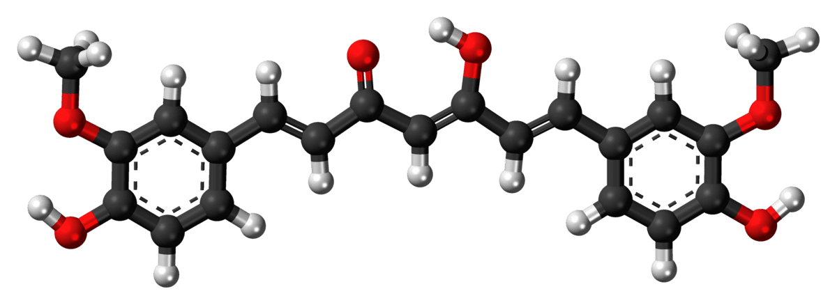
Boron enhances the antiviral activity of the curcumin against SARS-CoV-2


In order to help elucidating future treatment decisions for COVID-19, we are proposing that the HBT model to protect the blood against SARS-CoV-2 infection. Thus, we propose to utilize the following two classes of natural compounds suited for defending the blood against SARS-CoV-2: hemin and boron–curcumin complexes. The advantages of the HBT model in identifying prophylactic and therapeutic strategies against COVID-19 are related to the following physiological aspects:
(i) HO-1 has a significant activity in the spleen of HBT patients in the degradation of hemoglobin during red blood cells recycling (0.8% of the red blood cells fund per day), which represents approximately 80% of the production of endogenous carbon monoxide (CO)-derived heme [25].
(ii) HBT patients have hemin concentration in the blood, while normal people have a total lack of free hemin [23].
(iii) In HBT patients, erythropoietin (EPO), the hormone that regulates erythrocyte production, is also dramatically increased in response to anemia and hypoxia. EPO-induced HO-1 expression is likely to provide cytoprotection against oxidative stress [26]. These effects are actually a link between EPO and HO-1. In addition to HO-1 induction, EPO can exert its antioxidant effects by inducing antioxidant enzymes, such as superoxide dismutase, catalase, and glutathione peroxidase [27,28].
(iv) Two platelet function markers belonging to the group of membrane glycoproteins – selectin P and serum soluble cluster of differentiation 40 ligand (sCD40L) – were present at lower levels in individuals with HBT compared to controls [29]. Subjects with HBT have a lower frequency of cardiac and cerebral ischemic events than controls in some studies and this difference was supported by a meta-analysis and recently multiple sclerosis is very low in thalassemics [30].
HO-1 is a metabolic enzyme that catalyzes the degradation of heme into CO, biliverdin and free iron [25]. This enzyme has anti-inflammatory and antioxidant properties, which modulate innate and adaptive immune responses. In addition, recent studies have reported that HO-1 may exert significant antiviral activity against a wide variety of viruses, including HIV, hepatitis C virus, hepatitis B virus, enterovirus 71, influenza virus, respiratory syncytial virus, dengue virus, and Ebola virus, among others [31]. The activation of HO-1 by its substrate hemin ensured the protection against HIV infection. Treatments against viral infections that enhance HO-1 include various anesthetics (isoflurane or sevoflurane), hemin, estrogen, statins, curcumin, resveratrol, and melatonin [32]. Therefore, HO-1 expression can be strongly induced by heme analogues, such as hemin and curcumin derivatives, such as boron–curcumin complexes [33,34].
Lymphocytopenia is a common feature of SARS-Cov-2 infection, which can be used as an early marker in patients with SARS-CoV-2 infections, and is more pronounced in patients infected with SARS-CoV-2. Current data show that hemin can ameliorate lung damage resulting from virus infection. In addition, reports indicate that hemin may alleviate lung damage caused by other factors, such as lipopolysaccharide, mechanical ventilation, complement activation, and transfusion. All these results suggest that the effect of hemin in protecting the lungs has a broad spectrum and potentially involves against several diseases in the clinical setting [35].
Do rosocyanine (boron–curcumin) and hemin [iron(III)–porphyrin chloride] act against SARS-CoV-2? The following candidate molecules could be able to ensure very good blood protection against SARS-CoV-2:
(i) Rosocyanine is a promising and inexpensive alternative used in the photodynamic inactivation of viruses. Rosocyanine has been shown to be an extremely effective inhibitor of viral proteases (HIV-1 and HIV-2) and in addition a potent chelator of free iron. Taking into consideration the above-presented scientific arguments and also the capacity of curcumin to form boron adducts, several boron-based curcumin nutraceuticals could be prepared for viral disease (COVID-19) prevention [36].
(ii) Hemin, a commercial heme compound, used as an inducer of HO-1 has been shown to have antiviral activity. Hemin has been approved as a drug by the Food and Drug Administration (FDA) for use in patients with acute porphyria since the 1970s. Scientific data show that protoporphyrin, hemin and other natural porphyrins, as well as related metalloporphyrins, have activity against ribonucleic acid (RNA) viruses through an inhibition mechanism of viral enveloped proteases. In addition, it is also known that porphyrins can be a remarkable viral trap (for SARS-CoV-2 too), as they can be intercalated in the host cell membranes affecting the fusion of the virus with the human body. Considering the present demand of medication, it could be worth testing the antiviral properties and safety of hemin against COVID-19 [37]. Various compelling evidence indicate a possible role for hemin-induced HO-1 as a therapy strategy against the SARS-CoV-2-induced cytokine storm syndrome. Hemin-induced HO-1 might be a harmless, novel, and promising approach for controlling SARS-CoV-2 infection and limiting cytokine [38].
Recently, it has been hypothesized that heterozygous patients with HBT would develop resistance against SARS-CoV-2 due to the absence of the beta chain in hemoglobin (potential target of the virus). This hypothesis has been studied starting from the high percentages of the beta-thalassemia population present in four areas of Italy: Sardinia, Sicily, Puglia, and Emilia Romagna [39,40]. To this end, there are two classes of natural compounds suitable for protecting the blood against SARS-CoV-2: exogenous porphyrins [41] and boron–curcumin compounds [3]. Our current approaches for prophylaxis and treatment for COVID-19, based on HBT model and on using iron as a “bargaining chip” for blood protection and boron–curcumin complex as well, should be taken into consideration by specialized agencies – FDA, World Health Organization (WHO), European Medicines Agency (EMA). We believe that new decisions for COVID-19 prophylaxis and treatment are being actually imperative due to the spread of SARS-CoV-2 infections at the present moment.


cedric

Member
Joined
Jul 26, 2018
Messages
156
CO-rna-"virus"- "great mimicker"
The name of coronavirus could be carbon monoxide -
Low zinc/melatonin, respirators, hypoxia, hyperoxia -all generate excessive carbon monoxide.
Now we have time of geosolar disturbances, low Earth magnetic field which causes excessive heme degradation through heme oxygenase-1 producing iron, carbon monoxide, biliverdin/bilirubin.
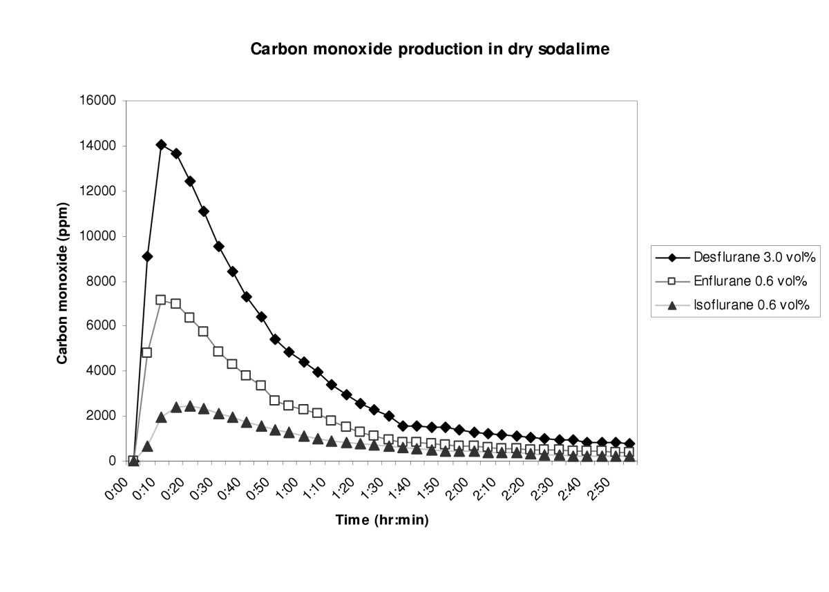
Halomethanes could decompose to carbon monoxide.
So called long covid could be carbon monoxide toxicity.
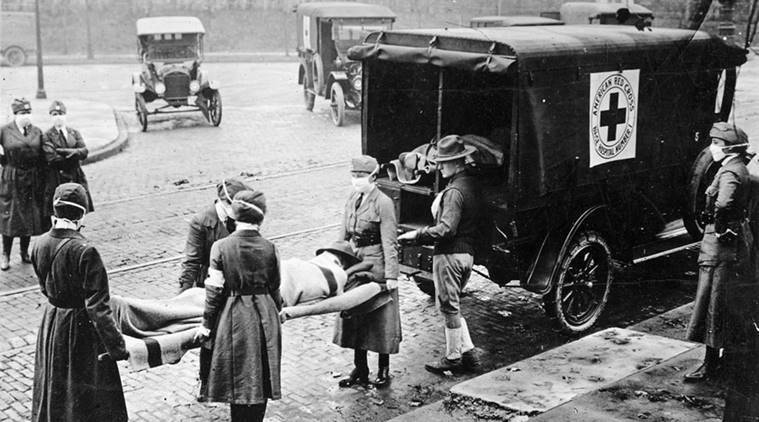
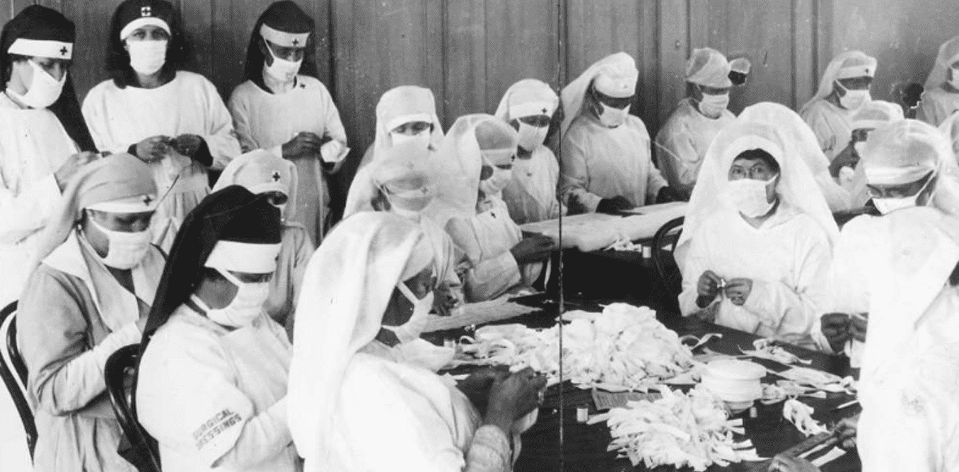
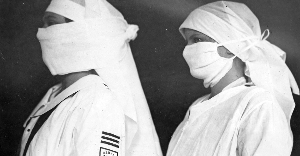
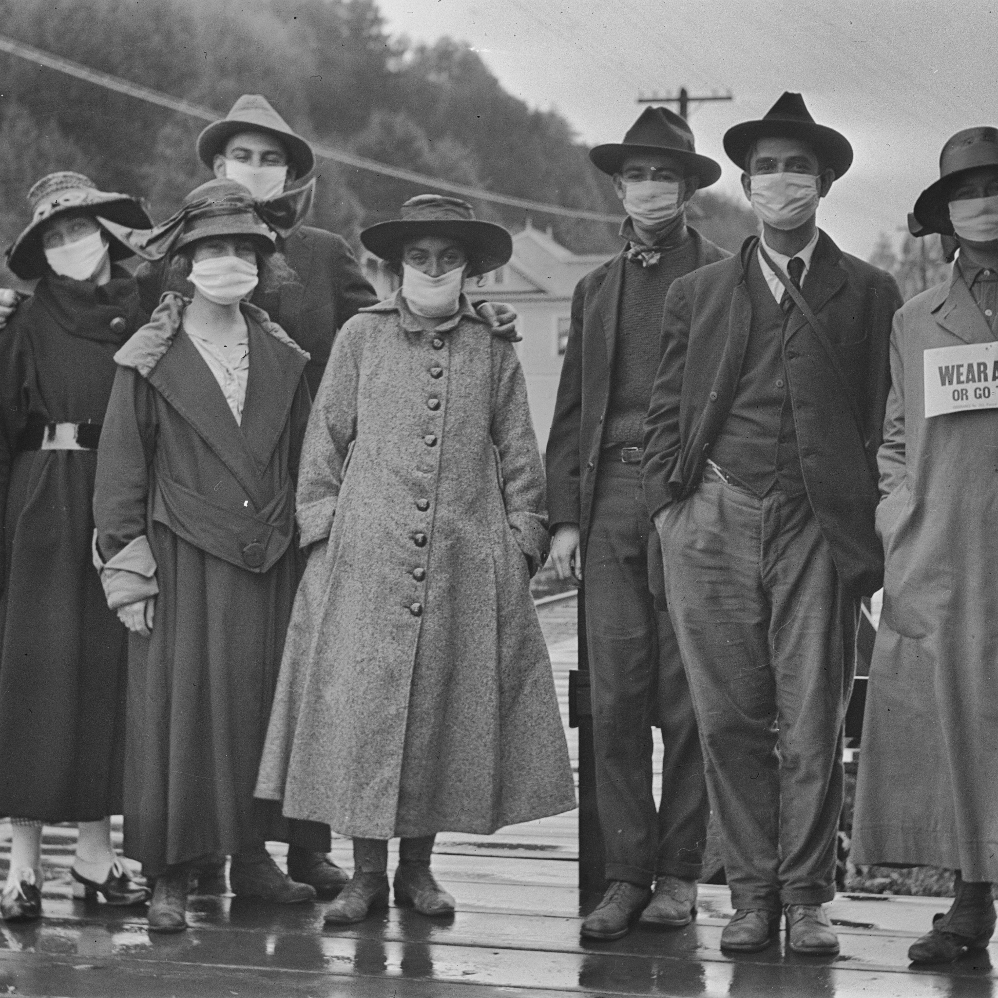
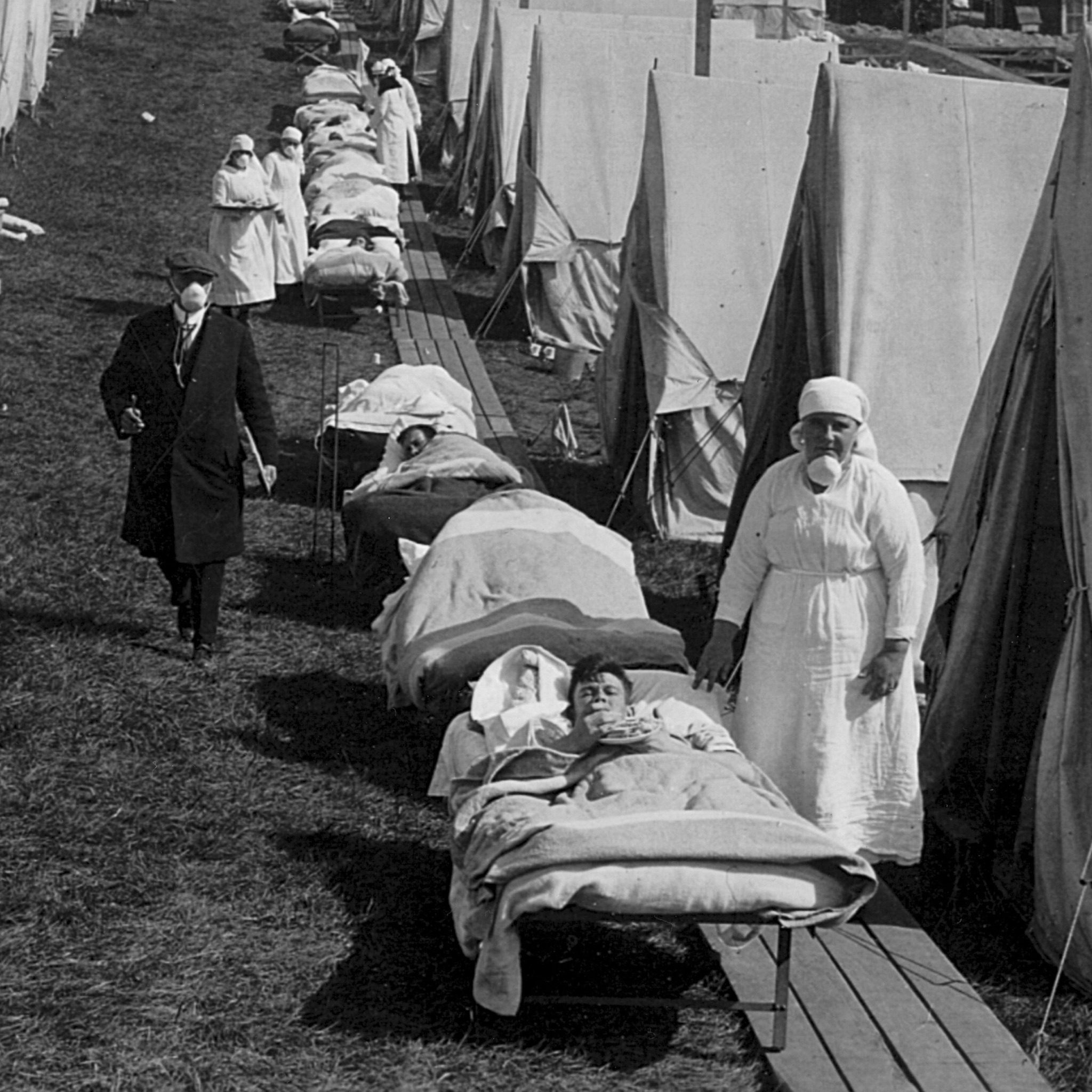
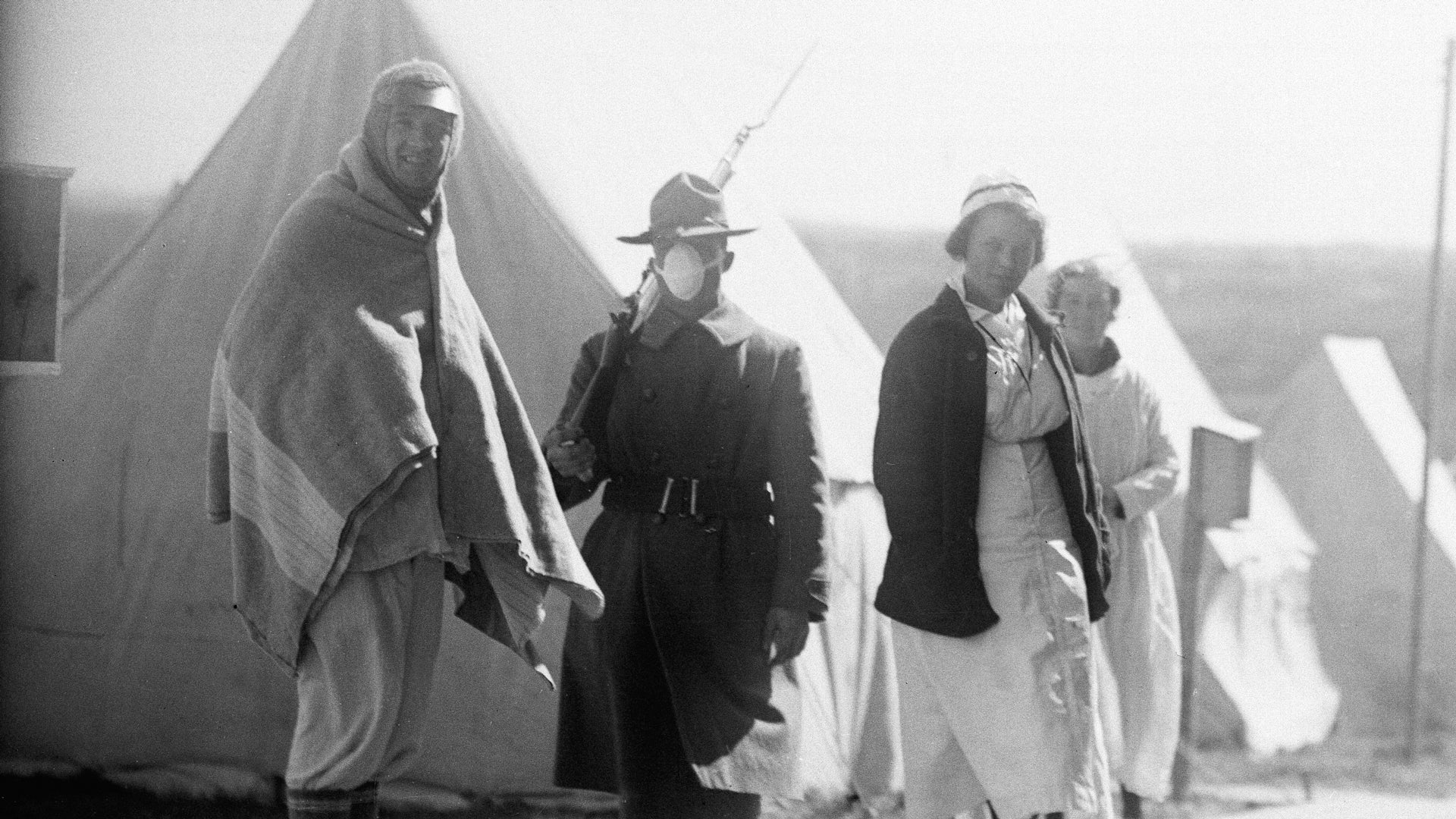
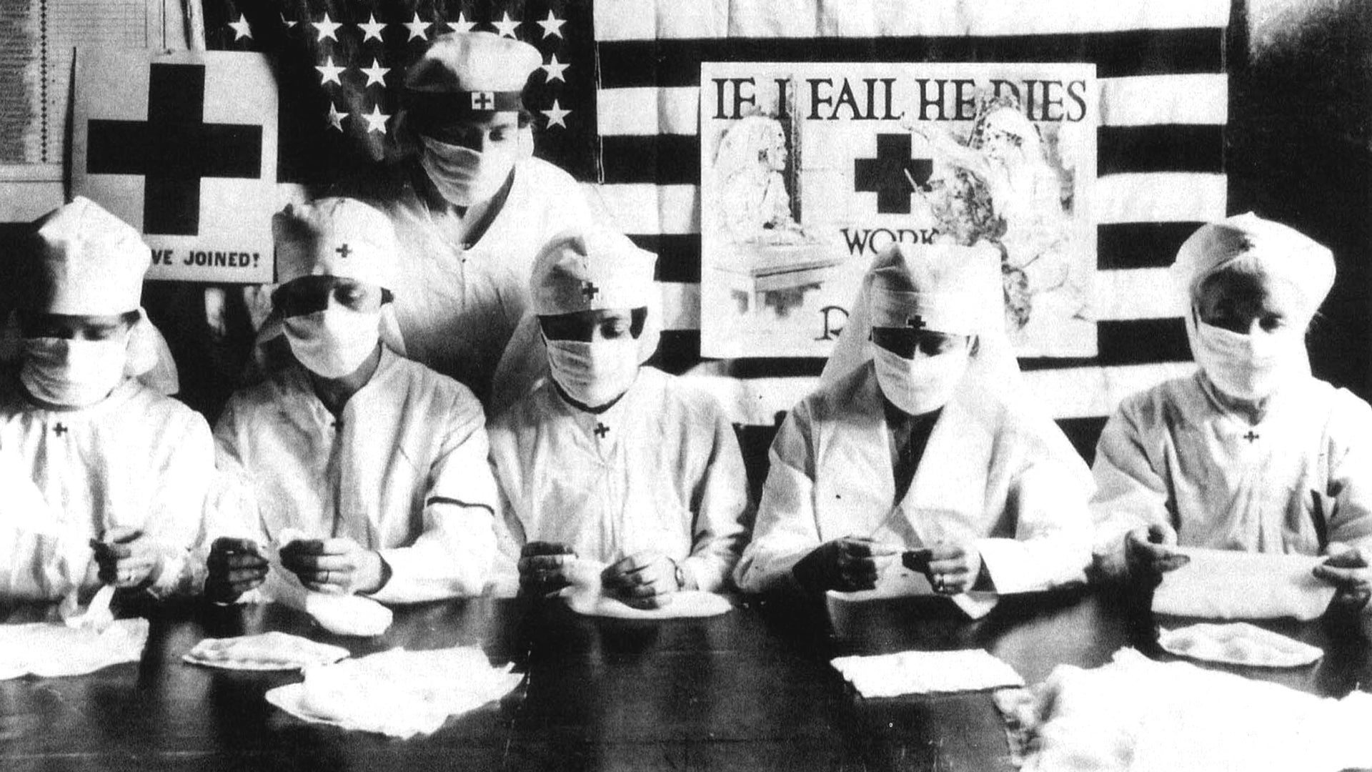
During so called “spanish flu” there were also mask mandates.
Soldiers used anti-gas masks.
Small endogenous amounts of CO are protective, but after mask mandates they become exogenous and chronically toxic
"Bag breathing" has a burden of carbon monoxide

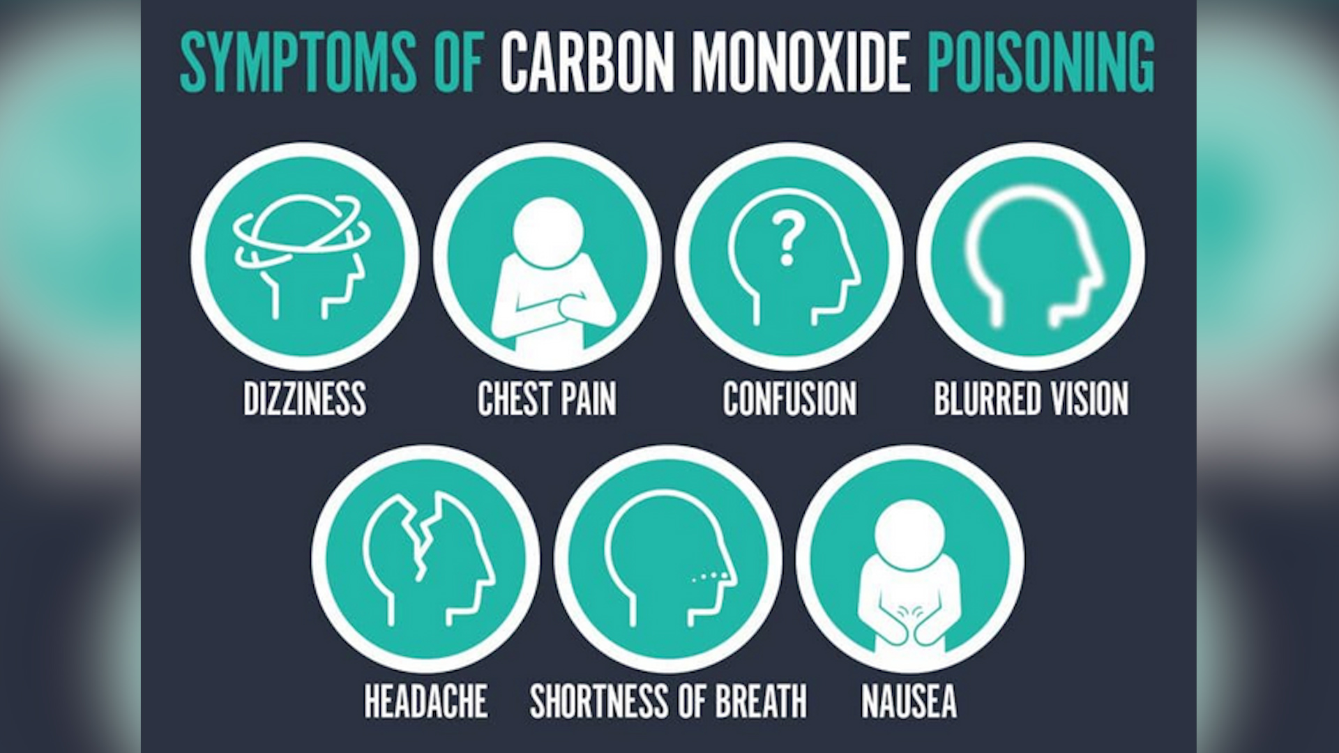
" Carbon monoxide poisoning typically occurs from breathing in carbon monoxide (CO) at excessive levels.[3] Symptoms are often described as "flu-like" and commonly include headache, dizziness, weakness, vomiting, chest pain, and confusion.[1] Large exposures can result in loss of consciousness, arrhythmias, seizures, or death.[1][2] The classically described "cherry red skin" rarely occurs.[2] Long-term complications may include chronic fatigue, trouble with memory, and movement problems.[5][...]

Diagnosis is typically based on a HbCO level of more than 3% among nonsmokers and more than 10% among smokers.[2] The biological threshold for carboxyhemoglobin tolerance is typically accepted to be 15% COHb, meaning toxicity is consistently observed at levels in excess of this concentration.[7] The FDA has previously set a threshold of 14% COHb in certain clinical trials evaluating the therapeutic potential of carbon monoxide.[8] In general, 30% COHb is considered severe carbon monoxide poisoning.[9] The highest reported non-fatal carboxyhemoglobin level was 73% COHb.[9][...]
The discovery that hemoglobin is affected by CO emerged with an investigation by James Watt and Thomas Beddoes into the therapeutic potential of hydrocarbonate in 1793, and later confirmed by Claude Bernard between 1846 and 1857.[9][...]
The carbon monoxide tolerance level for any person is altered by several factors, including genetics (hemoglobin mutations), behavior such as activity level, rate of ventilation, a pre-existing cerebral or cardiovascular disease, cardiac output, anemia, sickle cell disease and other hematological disorders, geography and barometric pressure, and metabolic rate.[17][18][19][9][...]
The main manifestations of carbon monoxide poisoning develop in the organ systems most dependent on oxygen use, the central nervous system and the heart.[31] The initial symptoms of acute carbon monoxide poisoning include headache, nausea, malaise, and fatigue.[36] These symptoms are often mistaken for a virus such as influenza or other illnesses such as food poisoning or gastroenteritis.[28]
Headache is the most common symptom of acute carbon monoxide poisoning; it is often described as dull, frontal, and continuous.[37] Increasing exposure produces cardiac abnormalities including fast heart rate, low blood pressure, and cardiac arrhythmia;[38][39] central nervous system symptoms include delirium, hallucinations, dizziness, unsteady gait, confusion, seizures, central nervous system depression, unconsciousness, respiratory arrest, and death.[40][41] Less common symptoms of acute carbon monoxide poisoning include myocardial ischemia, atrial fibrillation, pneumonia, pulmonary edema, high blood sugar, lactic acidosis, muscle necrosis, acute kidney failure, skin lesions, and visual and auditory problems.[38][42][43][44] Carbon monoxide exposure may lead to a significantly shorter life span due to heart damage.[45]

One of the major concerns following acute carbon monoxide poisoning is the severe delayed neurological manifestations that may occur. Problems may include difficulty with higher intellectual functions, short-term memory loss, dementia, amnesia, psychosis, irritability, a strange gait, speech disturbances, Parkinson's disease-like syndromes, cortical blindness, and a depressed mood.[28][46] Depression may occur in those who did not have pre-existing depression.[47] These delayed neurological sequelae may occur in up to 50% of poisoned people after 2 to 40 days.[...]

Chronic poisoning
Chronic exposure to relatively low levels of carbon monoxide may cause persistent headaches, lightheadedness, depression, confusion, memory loss, nausea, hearing disorders and vomiting.[49][50] It is unknown whether low-level chronic exposure may cause permanent neurological damage.[28] Typically, upon removal from exposure to carbon monoxide, symptoms usually resolve themselves, unless there has been an episode of severe acute poisoning.[49] However, one case noted permanent memory loss and learning problems after a three-year exposure to relatively low levels of carbon monoxide from a faulty furnace.[51] Chronic exposure may worsen cardiovascular symptoms in some people.[49] Chronic carbon monoxide exposure might increase the risk of developing atherosclerosis.[52][53] Long-term exposures to carbon monoxide present the greatest risk to persons with coronary heart disease and in females who are pregnant.[54] In experimental animals, carbon monoxide appears to worsen noise-induced hearing loss at noise exposure conditions that would have limited effects on hearing otherwise.[55] In humans, hearing loss has been reported following carbon monoxide poisoning.[50] Unlike the findings in animal studies, noise exposure was not a necessary factor for the auditory problems to occur.[...]
"covid" w kopalniach?
"In caves carbon monoxide can build up in enclosed chambers due to the presence of decomposing organic matter.[94] In coal mines incomplete combustion may occur during explosions resulting in the production of afterdamp. The gas is up to 3% CO and may be fatal after just a single breath.[82] Following an explosion in a colliery, adjacent interconnected mines may become dangerous due to the afterdamp leaking from mine to mine. Such an incident followed the Trimdon Grange explosion which killed men in the Kelloe mine.[95][...]

Central nervous system effects
The mechanism that is thought to have a significant influence on delayed effects involves formed blood cells and chemical mediators, which cause brain lipid peroxidation (degradation of unsaturated fatty acids). Carbon monoxide causes endothelial cell and platelet release of nitric oxide, and the formation of oxygen free radicals including peroxynitrite.[36] In the brain this causes further mitochondrial dysfunction, capillary leakage, leukocyte sequestration, and apoptosis.[156] The result of these effects is lipid peroxidation, which causes delayed reversible demyelination of white matter in the central nervous system known as Grinker myelinopathy, which can lead to edema and necrosis within the brain.[150] This brain damage occurs mainly during the recovery period. This may result in cognitive defects, especially affecting memory and learning, and movement disorders. These disorders are typically related to damage to the cerebral white matter and basal ganglia.[156][157] Hallmark pathological changes following poisoning are bilateral necrosis of the white matter, globus pallidus, cerebellum, hippocampus and the cerebral cortex.[30][28][158]

Pregnancy
Carbon monoxide poisoning in pregnant women may cause severe adverse fetal effects. Poisoning causes fetal tissue hypoxia by decreasing the release of maternal oxygen to the fetus. Carbon monoxide also crosses the placenta and combines with fetal hemoglobin, causing more direct fetal tissue hypoxia. Additionally, fetal hemoglobin has a 10 to 15% higher affinity for carbon monoxide than adult hemoglobin, causing more severe poisoning in the fetus than in the adult.[11] Elimination of carbon monoxide is slower in the fetus, leading to an accumulation of the toxic chemical.[159] The level of fetal morbidity and mortality in acute carbon monoxide poisoning is significant, so despite mild maternal poisoning or following maternal recovery, severe fetal poisoning or death may still occur.[160]"

hydrocarbonate in 1793
Water gas - Wikipedia
"Diseases treated by hydrocarbonate included: tuberculosis, inflammation, asthma, expectoration, hemoptysis, pneumonia, hydrothorax, spasm and other indications.[2][3] Many of the diseases treated with hydrocarbonate, whose active ingredient was carbon monoxide, are now being investigated using modern biomedical research methods to determine the therapeutic potential of carbon monoxide. For example, James Lind recognized hydrocarbonate to effectively treat lung inflammation;[2] delivery of carbon monoxide via inhalation protocol or carbon monoxide-releasing molecules has significant preclinical data indicating an effective treatment for inflammation.[5] "

Grinker myelinopathy - Wikipedia
" Grinker's myelinopathy, also known as anoxic leukoencephalopathy,[2] is a rare disease of the central nervous system. The disease is characterized by a delayed leukoencephalopathy after a hypoxic episode.[2] It is typically, though not necessarily, related to carbon monoxide poisoning or heroin overdose. It occurs in roughly 2.8% of those who experience an acute hypoxic/anoxic episode.[3] Because of the wide range of symptoms and the delay in onset, it is often misdiagnosed as other neuropathologies. Grinker's myelinopathy was originally characterized by Roy R. Grinker in 1925[4][5] or 1926,[3] depending on the source.

Categorization
Following an apparent rehabilitation from a severe episode of prolonged cerebral oxygen deprivation, patients with Grinker's myelinopathy begin to experience massive white matter death that leads to a wide range of neurological dysfunctions ranging from confusion and apathy to Parkinson-like symptoms.[3]

Symptoms
The symptoms have been known to include apathy, dementia, Parkinsonism, agitation, urinary incontinence, and pseudobulbar palsy, among many other neuropsychiatric symptoms. Microscopically, extensive hemispheric demyelination and the degeneration of basal ganglia are observed.[1]

Onset
The onset of the symptoms usually occurs several weeks after the initial hypoxic episode. The hypoxic episode is necessarily severe, usually with an arterial oxygen partial pressure less than 40mmHg.[3] Following the severe hypoxia, the patient typically falls unconscious or into a coma, with the exception of cases of carbon monoxide poisoning.[2][1] If the patient recovers from this unconscious state, usually within 24 hours, it is typically followed by a successful recovery over a few days (generally 4 to 5). After the short recovery, a lucid period is observed, lasting anywhere from 1 to 4 weeks, in which the patient exhibits no symptoms related to the anoxic episode. It is after this period that the degenerative symptoms begin to appear and rapidly grow in severity.[6]"
Heme oxygenase - Wikipedia
Carboxyhemoglobin - Wikipedia
" Overview
The average red blood cell contains 250 million hemoglobin molecules.[7] Hemoglobin contains a globin protein unit with four prosthetic heme groups (hence the name heme -o- globin); each heme is capable of reversibly binding with one gaseous molecule (oxygen, carbon monoxide, cyanide, etc.),[8] therefore a typical red blood cell may carry up to one billion gas molecules. As the binding of carbon monoxide with hemoglobin is reversible, certain models have estimated that 20% of the carbon monoxide carried as carboxyhemoglobin may dissociate in remote tissues.[7]

COVID-19 and heme oxygenase: novel insight into the disease and potential therapies

"COVID-19 and heme oxygenase: novel insight into the disease and potential therapies"

https://link.springer.com/article/10.10 ... 21-01246-w

"Heme oxygenase agonists—fluvoxamine, melatonin—are efficacious therapy for Covid-19"


" The spike protein of the causative COVID-19 virus induces heme oxygenase-1: Pathophysiologic implications"
Carbon monoxide is PUFA metabolite
warm CO2 - infrared


1654886767451.jpeg


loss of potassium
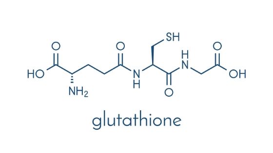
light and glutathione
low Earth geomagnetic field/geomagnetic disturbances/solar flares like now and melatonin/heme oxygenase-1/carbon monoxide, mass shootings, depressions, suicides

 
Last edited:

Jam

Member
Joined
Aug 10, 2018
Messages
2,212
Age
52
Location
Piedmont
 

cedric

Member
Joined
Jul 26, 2018
Messages
156
Exhaled carbon dioxide correlates with exhaled carbon monoxide/CO-rna"virus"
Bloomberg - Are you a robot?
” Carbon-dioxide monitors can assess how Covid-risky a space is because they help tell you whether you’re breathing in clean air. They measure the concentration of carbon dioxide, which people exhale when they breathe, along with other things like, potentially, virus particles. The more well-ventilated a space, the lower the reading on my monitor’s screen — meaning not only less carbon dioxide but also less of the stuff like Covid that might make people sick. “
 

cedric

Member
Joined
Jul 26, 2018
Messages
156
CO2 can protect against CO-rna"virus"/carbon monoxide poisoning
War against CO2 started many years ago in medicine. Now we have "mad"icine.
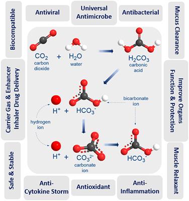
Humidified Warmed CO2 Treatment Therapy Strategies Can Save Lives With Mitigation and Suppression of SARS-CoV-2 Infection: An Evidence Review " Humidified Warmed CO2 Treatment Therapy Strategies Can Save Lives With Mitigation and Suppression of SARS-CoV-2 Infection: An Evidence Review" Rapid elimination of CO through the lungs: coming full circle 100 years on " At the start of the 20th century, CO poisoning was treated by administering a combination of CO2 and O2 (carbogen) to stimulate ventilation. This treatment was reported to be highly effective, even reversing the deep coma of severe CO poisoning before patients arrived at the hospital. "
 
EMF Mitigation - Flush Niacin - Big 5 Minerals

Similar threads

Back
Top Bottom