Be Wary Of Vitamin D Supplementation

Amazoniac

Member
Joined
Sep 10, 2014
Messages
8,583
Location
Not Uganda
They decided to test the hair of malnourished fetuses to relate nutrients. One of them was zinc, it was pointed out that its content in hair is stable when compared to other tissues, but I guess that they decided to do it anyway because it's practical, non-invasive and more sensitive than blood (which was also tested).

- Hair Zinc Level Analysis and Correlative Micronutrients in Children Presenting with Malnutrition and Poor Growth

"Zinc concentration remains constant in hair, skin, heart and skeletal muscle, whereas that in plasma, liver, bone and testis easily fluctuates [7]."

"Vitamins can affect zinc status. Vitamin-induced suppression of zinc can occur either directly or indirectly. Vitamin D can suppress zinc absorption through increased absorption of calcium. Also, by decreasing thyroid activity and increasing parathyroid activity, vitamin D contributes to zinc deficiency by decreasing its absorption [33]. Similarly, in our study hair zinc level had negative correlationship to serum 25-hydroxy vitamin D."

upload_2020-1-18_20-20-18.png

It's challenging to find meaning when there's no intervention, but it's interesting that zinc tended to be higher in hair as levels of the rubbish were lower, the opposite would be expected. If it was due to malnourishment, how could it explain greater killcidiol levels? For example, (assuming that the more you have available, the more it can overflow) zinc being used as a proxy for nutrient intake such as killcium, it would be found decreased along with the level of killcidiol.

For the hair analysts to comment on the interaction, it must have been observed that dosing killciol decreases its content in hair and the situation is improved with extra.
 
Last edited:

Amazoniac

Member
Joined
Sep 10, 2014
Messages
8,583
Location
Not Uganda
With the spikes described below you prime the body for excretion or storage as an attempt to ease the intoxication.

- Methods for controlled release oral dosage of a vitamin D compound (US8778373B2) | Google Patents

"Cholecalciferol and ergocalciferol are normally present at stable, low concentrations in human blood. Slight, if any increases in blood Vitamin D levels occur after meals since unsupplemented diets have low Vitamin D content, even those containing foods fortified with Vitamin D. Almost all human Vitamin D supply comes from fortified foods, exposure to sunlight or from dietary supplements, with the latter source becoming increasingly important. Blood Vitamin D levels rise only gradually, if at all, after sunlight exposure since cutaneous 7-dehydroxycholesterol is modified by UV radiation to pre-Vitamin D3, which undergoes thermal conversion in the skin to cholecalciferol over a period of several days before circulating in the blood. In contrast, supplements such as those currently available, do cause marked increases in intraluminal, blood and intracellular levels of Vitamin D proportional to the dose administered."

"Surges in the blood levels of cholecalciferol and/or ergocalciferol (“cholecalciferol/ergocalciferol”) can transiently raise intracellular Vitamin D concentrations, accelerating prohormone production and elevating intracellular and blood prohormone concentrations. Surges in the blood levels of cholecalciferol and/or ergocalciferol also can saturate the enzymes which produce the prohormones, causing the excess Vitamin D to be catabolized or shunted to long-term storage in adipose tissue. Vitamin D stored in adipose tissue is less available for future conversion to prohormones. Surges in intraluminal levels of Vitamin D after ingestion of current oral supplements can directly boost Vitamin D and prohormone concentrations in the local enterocytes, thereby exerting “first pass” effects on calcium and phosphorus metabolism in the small intestine."

"Surges in blood or intracellular prohormone concentrations can promote excessive extrarenal hormone production, leading to local adverse effects on calcium and phosphorus metabolism. Such surges also can inhibit hepatic prohormone production from subsequent supplemental Vitamin D and promote catabolism of both Vitamin D and 25-hydroxyvitamin D in the kidney and other tissues."

"Currently available oral Vitamin D supplements are far from ideal for achieving and maintaining optimal blood 25-hydroxyvitamin D levels. These preparations typically contain 400 IU to 5,000 IU of Vitamin D3 or 50,000 IU of Vitamin D2 and are formulated for quick or immediate release in the gastrointestinal tract. When administered at chronically high doses, as is often required for Vitamin D repletion, these products have significant and, often, severe limitations which are summarized below."

Killciol

"High doses of immediate release Vitamin D supplements produce marked surges in blood Vitamin D levels, thereby promoting: (a) storage of Vitamin D in adipose tissue, which is undesirable because stored Vitamin D is less available for later hepatic conversion to 25-hydroxyvitamin D; (b) hepatic catabolism of Vitamin D to metabolites, which are less useful or no longer useful for boosting blood 25-hydroxyvitamin D levels, via 24- and/or 26-hydroxylation; and, (c) excessive intracellular 24- or 25-hydroxylation of Vitamin D, which leads to increased risk of hypercalciuria, hypercalcemia and hyperphosphatemia."

"Furthermore, high doses of immediate release Vitamin D supplements produce supraphysiologic pharmacological concentrations of Vitamin D, e.g., in the lumen of the duodenum, promoting: (a) 25-hydroxylation in the enterocytes and local stimulation of intestinal absorption of calcium and phosphorus, leading to increased risk of hypercalciuria, hypercalcemia and hyperphosphatemia; (b) catabolism of Vitamin D by 24- and/or 26-hydroxylation in the local enterocytes, causing decreased systemic bioavailability; and (c) absorption primarily via chylomicrons, leading to increased hepatic catabolism."​
Killcidiol

"High doses of immediate release Vitamin D supplements also produce surges or spikes in blood and intracellular 25-hydroxyvitamin D levels, thereby promoting: (a) excessive extrarenal production of Vitamin D hormones, and leading to local aberrations in calcium and phosphorus homeostasis and increased risk of hypercalciuria, hypercalcemia and hyperphosphatemia; (b) accelerated catabolism of both Vitamin D and 25-hydroxyvitamin D by 24- and/or 26-hydroxylation in the kidney and other tissues; (c) down-regulation of hepatic production of Vitamin D prohormones, unnecessarily impeding the efficient repletion of Vitamin D insufficiency or deficiency; and, (d) local aberrations in calcium and phosphorus homeostasis mediated by direct binding to VDR."​
 
Joined
Apr 24, 2017
Messages
625
By supplement, because histamine issues affect what I can eat. It is interesting that you mention Vitamin K2, I had problems with that as well, and it was manganese that was needed (2-5mg/meal and it helped with tolerating K2, D3, iodine, selenium). I wish I knew why that was.
Wow! I'm dealing with histamine issues too. Also vitamin D and K2 supplementation made me worse. I also remember eating blue mussels regularly years ago and didn't have these issues back then even though I was using vitamin D supplements. Looking into their nutritional data and it says 100gs of them has 3,4mg manganese (148% RDA). Gonna start eating them again. Has plenty of other vitamins and cheap too.
 

LLight

Member
Joined
May 30, 2018
Messages
1,411
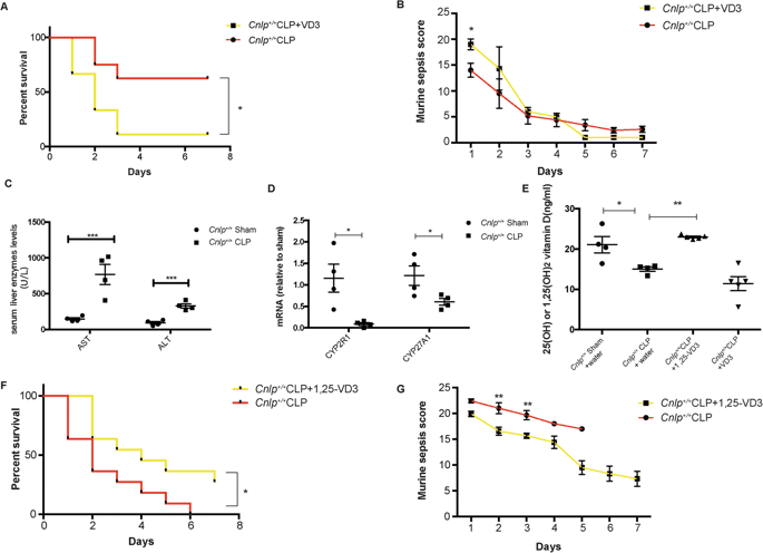
Cathelicidin preserves intestinal barrier function in polymicrobial sepsis | Critical Care | Full Text

"Our data suggested that 1alpha, 25-dihydroxyvitamin D3 but not cholecalciferol is a potential therapeutic agent for treating sepsis."

13054_2020_2754_Fig10_HTML.png


"Furthermore, we assessed the therapeutic use of the active form and the inactive form of VD3 in our CLP model. We observed that administration of calcitriol (an active form of VD3) but not cholecalciferol (an inactive form of VD3) after the onset of sepsis led to a better survival outcome in CLP mice. In line with recent publications, high-dose VD3 (cholecalciferol, inactive form of VD3) did not improve the survival outcomes of critically ill patients in terms of 90-day mortality [50]. Since hepatic cytochrome P450 (CYPs) play an essential role in the conversion of VD3 into 25-hydroxyVD3 together with additional evidence showing that hepatic CYPs dysfunctions are linked to sepsis [51,52,53], we further examined the functions of the liver after the onset of sepsis. Our results demonstrated that CLP induced hepatic damage and the associated downregulations of hepatic CYPs at mRNA level, resulting in decreased serum intermediate and active VD3. Fortunately, the administration of calcitriol (an active form of VD3) can bypass hepatic biotransformation of cholecalciferol into 25-hydroxyVD3 mediated by CYP system, directly entering the circulatory system and exerting the beneficial effects. Taken together, we confirmed that the active form of VD3 but not the inactive form of VD3 is a therapeutic drug in our CLP model. Noticeably, the latter worsened 7-day mortality and the associated symptoms in CLP-operated mice, the mechanism of which remains unclear."
 

Amazoniac

Member
Joined
Sep 10, 2014
Messages
8,583
Location
Not Uganda
- Glutathione deficiency induces epigenetic alterations of vitamin D metabolism genes in the livers of high-fat diet-fed obese mice

"We recently demonstrated that GSH has a positive relationship with 25(OH)vitamin D3 (25(OH)VD3) in the blood of type 2 diabetic and obese subjects[6–9]. Also, supplementation with L‐cysteine (LC), a rate-limiting precursor of GSH[5], boosts the levels of GSH, reduces oxidative stress, and improves circulating 25(OH)VD3 levels[7–12]."

"This study examined the hypothesis that GSH-deficiency induces epigenetic alterations of VD metabolism genes, which can reduce the circulating 25(OH)VD3 levels in obesity."

"The animals were fed either a standard chow diet (Harlan TD.08485, providing 5.2% calories as fat; Control) or a high-fat diet (Harlan TD.88137, containing 42% calories as fat; HFD) for 16 weeks. The detailed composition of these diets appears in a recent publication[46]."

"The expression of the VD metabolism genes CYP2R1 and CYP27A1 (25-hydroxylase), CYP27B1 (1-α-hydroxylase), and vitamin D receptor (VDR) were downregulated in the livers of mice fed an HFD (GSH-deficient) compared with control diet-fed group. The expression of CYP24A1 (24-hydroxylase) was significantly increased, which catabolizes both 25(OH)VD3 and 1α,25-hydroxyvitaminD3."

upload_2020-2-20_21-21-23.png

"Gene-specific hypermethylation of 25-hydroxylase, 1-α-hydroxylase, and VDR, and hypomethylation of CYP24A1 was observed in HFD-fed mice."

upload_2020-2-20_21-21-31.png

"To rule out the non-specific effect of HFD conditions and other confounding variables in animals, we carried out in vitro experiments to determine the direct impact of glutathione deficiency status on VD metabolism genes in hepatocytes. GSH deficiency was induced by GCLC siRNA (Fig. 4a) or treatment with the pharmacological inhibitor BSO (Fig. 5a); in both approaches, cell viability was not affected in the present study." "Under GSH-deficient conditions, treatment with siRNA (Fig. 4b,c) and the inhibitor BSO (Fig. 5b) resulted in decreased expression of CYP27A1, CYP27B1, and VDR. Both 25-hydroxylases (CYP2R1 and CYP27A1) were expressed in the liver, while CYP27A1 expression was seen only in cultured hepatocytes (FL83B mouse hepatocyte cell line). Additionally, CYP24A1 showed it upregulated mRNA and protein expression in hepatocytes treated with GCLC siRNA or its inhibitor. These results suggest that GSH may have a direct effect on VD metabolism."

"GSH deficiency induced in cultured hepatocytes caused an increase in oxidative stress and alterations in VD regulatory genes."

"Supplementation of hepatocytes with GSH or its precursors [did not] affect cell viability. The levels of GSH significantly increased after treatment with LC, NAC, and GSHee [described below] (Fig. 8a). There was a significant loss of Dnmt activity and rise in Tet activity in the treatment group. Data from DNA dot-blots and mRNA levels of Dnmt and Tet support the activity data (Fig. 8b–d). The levels of CYP27A1 and CYP27B1 were significantly upregulated along with those of VDR mRNA, but CYP24A1 was downregulated in groups supplemented with GSH or its precursor (Fig. 8e)."

upload_2020-2-20_21-21-40.png


I haven't considered if the amounts are sane.

"GSH replenishment in human tissues is difficult due to its shorter half-life, and not easy to penetrate the plasma membrane hence use of prodrugs/precursors such as cysteine, and glycine is the most promising approach[4,45]. This provides evidence for the positive effect of glutathione on VD metabolism and vitamin D receptor gene expression."​


--
It's rarely taken into account the rate of utilization when attempting push what's acceptable as physiological dose higher: it takes a few days for realization of intoxication if synthesized on skin; when ingested, it's much faster.

Also, if the last step before turning into venom D depends on temperature, what happens in cold climates where the exposed parts that get sunlight are the ones that will be cold for most of the time?

In case of sun exposure..

- Physiological Amputation of Limb (Limb Gangrene) after Tourniquet Application for 12 days following Snake Bite

"Do not use tourniquets because they obstruct arterial flow and cause ischemia. Constriction bands may be useful, especially when immediate medical care is not available. A constriction band is an elastic bandage or Penrose drain, thick rope, or piece of clothing wrapped circumferentially above the bite, applied with enough tension to restrict superficial venous and lymphatic flow while maintaining distal pulses and capillary filling. Apply the band snugly but loose enough to avoid arterial compromise. A constriction band can delay venom absorption without causing increased swelling.[11]"​

Don't expect a light publication (now that you will click on it).
 
Last edited:

Amazoniac

Member
Joined
Sep 10, 2014
Messages
8,583
Location
Not Uganda
- Hector F. DeLuca | ResearchGate
Vitamin D binding protein is required to utilize skin-generated vitamin D

"Vitamin D binding protein (DBP), also known as GC-globulin, is the primary transport protein for vitamin D metabolites in the blood (5, 6). However, the important finding that DBP−/− mice are clearly responsive to orally administered vitamin D did not support an essential role of DBP in vitamin D metabolism and function (7, 8). Although the lack of DBP did not alter tissue distribution, uptake, metabolism, or biological potency of vitamin D, it did result in diminished levels of circulating 25- hydroxyvitamin D (25(OH)D) and 1,25(OH)2D (7, 8)."

"Our results demonstrate that DBP is absolutely required for the utilization of vitamin D produced in the epidermis by UV light. Most convincing is that in the absence of DBP, UVB exposure had no effect on vitamin D deficiency or the resultant hypocalcemia while UVB treatment of vitamin D-deficient DBP+/+ mice restored serum calcium and 25(OH)D to normal. Direct measurement of vitamin D in skin confirmed that UV successfully produces vitamin D in both DBP+/+ and DBP−/− mice. In agreement with a previous report, levels of UV-generated vitamin D decreased in the skin of DBP+/+ mice over a 2-d period following UV irradiation (9); however, the vitamin D levels were unchanged in the skin of DBP−/− mice. Of particular significance is the fact that injection of recombinant DBP restored serum calcium of the irradiated DBP−/− mice, thus recapitulating the DBP+/+ phenotype. Taken together, these results demonstrate that DBP is required for the utilization of cutaneous produced vitamin D."

upload_2020-2-22_10-35-52.png

"The observation that these DBP knockout mice are phenotypically normal with detectable 25(OH)D and 1,25(OH)2D levels when maintained on vitamin D containing diets indicates that DBP is not required for the absorption of vitamin D from the gut (7, 8). Indeed, absorption and transport of vitamin D from the gut on chylomicrons has been repeatedly demonstrated (5, 10, 11). As animals in previous studies were maintained on diets containing vitamin D, this likely explains why this epidermal function of DBP has been overlooked. The presence of 25(OH) D and 1,25(OH)2D also suggests DBP is not required for their release from the liver or kidney."

"In the circulation, virtually 100% of vitamin D metabolites are bound with ∼90% being associated with DBP and the remaining 10% associated with albumin (5, 10, 12). Although the ability to bind alternative carriers creates the possibility of a compensatory transport mechanism for cutaneous vitamin D in the absence of DBP, the data presented here show that the transport is DBP specific."

"While it is now possible to replace cutaneous synthesis using oral supplements, cutaneous-derived vitamin D is more biologically efficient and serves as a more sustained source without the risk of toxicity (4, 20–22)."​
 

Amazoniac

Member
Joined
Sep 10, 2014
Messages
8,583
Location
Not Uganda
Only if you're struggling to make supplemental venom D work:

- Oxalate: From the Environment to Kidney Stones

You can try to concentrate supplementation away from meals that contain significant amounts of oxalate or precursors (not underestimating these; such as extra collagen, ascourgic acid, creatide, and so on; read article).

Killcium carbonate decreases the acidity of some body compartments, it's conceivable that it makes them prone to the formation of crystals. Here it's claimed that venom D can do something similar, but in this case I don't think that it's concerning because its metabolism is better regulated than the reaction of carbonate salts with stomach acid, which inedivatbgaobly happen, so you might benefit from distributing it by applying on skin, should ease the punch. There must be a period of the day when the internal metabolites that are potential problems are consistently at their lowest, another good period to take it. And finally, killcium citrate should be the safest in this regard if you want to take both at the same time.
 

Terma

Member
Joined
May 8, 2017
Messages
1,063
- Glutathione deficiency induces epigenetic alterations of vitamin D metabolism genes in the livers of high-fat diet-fed obese mice

"We recently demonstrated that GSH has a positive relationship with 25(OH)vitamin D3 (25(OH)VD3) in the blood of type 2 diabetic and obese subjects[6–9]. Also, supplementation with L‐cysteine (LC), a rate-limiting precursor of GSH[5], boosts the levels of GSH, reduces oxidative stress, and improves circulating 25(OH)VD3 levels[7–12]."

"This study examined the hypothesis that GSH-deficiency induces epigenetic alterations of VD metabolism genes, which can reduce the circulating 25(OH)VD3 levels in obesity."

"The animals were fed either a standard chow diet (Harlan TD.08485, providing 5.2% calories as fat; Control) or a high-fat diet (Harlan TD.88137, containing 42% calories as fat; HFD) for 16 weeks. The detailed composition of these diets appears in a recent publication[46]."

"The expression of the VD metabolism genes CYP2R1 and CYP27A1 (25-hydroxylase), CYP27B1 (1-α-hydroxylase), and vitamin D receptor (VDR) were downregulated in the livers of mice fed an HFD (GSH-deficient) compared with control diet-fed group. The expression of CYP24A1 (24-hydroxylase) was significantly increased, which catabolizes both 25(OH)VD3 and 1α,25-hydroxyvitaminD3."

"Gene-specific hypermethylation of 25-hydroxylase, 1-α-hydroxylase, and VDR, and hypomethylation of CYP24A1 was observed in HFD-fed mice."

"To rule out the non-specific effect of HFD conditions and other confounding variables in animals, we carried out in vitro experiments to determine the direct impact of glutathione deficiency status on VD metabolism genes in hepatocytes. GSH deficiency was induced by GCLC siRNA (Fig. 4a) or treatment with the pharmacological inhibitor BSO (Fig. 5a); in both approaches, cell viability was not affected in the present study." "Under GSH-deficient conditions, treatment with siRNA (Fig. 4b,c) and the inhibitor BSO (Fig. 5b) resulted in decreased expression of CYP27A1, CYP27B1, and VDR. Both 25-hydroxylases (CYP2R1 and CYP27A1) were expressed in the liver, while CYP27A1 expression was seen only in cultured hepatocytes (FL83B mouse hepatocyte cell line). Additionally, CYP24A1 showed it upregulated mRNA and protein expression in hepatocytes treated with GCLC siRNA or its inhibitor. These results suggest that GSH may have a direct effect on VD metabolism."

"GSH deficiency induced in cultured hepatocytes caused an increase in oxidative stress and alterations in VD regulatory genes."

"Supplementation of hepatocytes with GSH or its precursors [did not] affect cell viability. The levels of GSH significantly increased after treatment with LC, NAC, and GSHee [described below] (Fig. 8a). There was a significant loss of Dnmt activity and rise in Tet activity in the treatment group. Data from DNA dot-blots and mRNA levels of Dnmt and Tet support the activity data (Fig. 8b–d). The levels of CYP27A1 and CYP27B1 were significantly upregulated along with those of VDR mRNA, but CYP24A1 was downregulated in groups supplemented with GSH or its precursor (Fig. 8e)."

View attachment 16733

I haven't considered if the amounts are sane.
"GSH replenishment in human tissues is difficult due to its shorter half-life, and not easy to penetrate the plasma membrane hence use of prodrugs/precursors such as cysteine, and glycine is the most promising approach[4,45]. This provides evidence for the positive effect of glutathione on VD metabolism and vitamin D receptor gene expression."​
People or not, this seems like a logical role for the liver. It has role in GSH synthesis due to the transsulfuration pathway, and if it cells can't defend against oxidative stress, they couldn't handle more calcium either, which would be more likely to go toward oxalates, too. So if you increase D to fight virus, reasoning via ACE2, this suggests more protection is at least prudent (seems a logical therapy overall: Role of Combined Lipoic Acid and Vitamin D3 on Astrocytes as a Way to Prevent Brain Ageing by Induced Oxidative Stress and Iron Accumulation). Good to bump this in the circumstances. If vit D is a reaction to UVB and it triggers cellular defense via things like FoxO it's tightly linked to the antioxidant system by nature, and increasing FoxO too far in nutrient deficiency sounds like a recipe for apoptosis as well as insulin resistance by liver. All in all I do fairly poorly on vit D but don't regularly supplement the stronger antioxidants anymore so this is interesting.
 
Joined
Dec 18, 2018
Messages
2,206
People or not, this seems like a logical role for the liver. It has role in GSH synthesis due to the transsulfuration pathway, and if it cells can't defend against oxidative stress, they couldn't handle more calcium either, which would be more likely to go toward oxalates, too. So if you increase D to fight virus, reasoning via ACE2, this suggests more protection is at least prudent (seems a logical therapy overall: Role of Combined Lipoic Acid and Vitamin D3 on Astrocytes as a Way to Prevent Brain Ageing by Induced Oxidative Stress and Iron Accumulation). Good to bump this in the circumstances. If vit D is a reaction to UVB and it triggers cellular defense via things like FoxO it's tightly linked to the antioxidant system by nature, and increasing FoxO too far in nutrient deficiency sounds like a recipe for apoptosis as well as insulin resistance by liver. All in all I do fairly poorly on vit D but don't regularly supplement the stronger antioxidants anymore so this is interesting.

What do you experience from taking D?How much?
I have goodwill towards it,cured me from recurrent
low grade sore throat and tonsilitis.
Individuals who dont test for it seem to be underdosing according to the
numerous dose-ranging studies which got publishment.
 

Terma

Member
Joined
May 8, 2017
Messages
1,063
It aggravated mental stress and sometimes energy levels, and some extra bodily sensations I won't try to describe. In my estimate some of my problems relate to calcium channels like TRP (even related to oxalates: Thermo-Sensitive TRP Channels: Novel Targets for Treating Chemotherapy-Induced Peripheral Pain) although many stress signals use calcium, and I rely often on brute force solutions to that like phenibut and THC. I had the 5,000IU/drop supplement so no worries about dosage (40,000+) plus have a UVB light and take tons of magnesium all the time. And yes I had problems when consuming too much calcium, repeatedly.
 
Last edited:

SOMO

Member
Joined
Mar 27, 2018
Messages
1,094

Vitamin D gave me HYPERCALCEMIA.
A calcium level of 16 which is kidney-failure levels.

I was taking 50,000 IU orally.


That study Amazoniac posted states:
"Some find all of these recommendatiosn too conservative and suggest much higher dosing regiments. While the concern of hypervitmainosis D is legitimate, HYPERCALCEMIA has only been reported with serum concentrations of 25(OH)D exceeding 150 ng/mL. Models have suggested that to achieve that concentration would require daily oral supplementation of 40,000 IU of vitamin D."
 
Joined
Dec 18, 2018
Messages
2,206
Vitamin D gave me HYPERCALCEMIA.
A calcium level of 16 which is kidney-failure levels.

I was taking 50,000 IU orally.


That study Amazoniac posted states:

What was the treatment for it,how long did it last.
How long did you take that dose,what was the total dose?How high was your Calcium intake and Salt intake?Do you have labs for that,had you an measurable PTH?
 

Amazoniac

Member
Joined
Sep 10, 2014
Messages
8,583
Location
Not Uganda
So, there's no cure to low levels of Vitamin D ?
Inflammation suffices to drop them, but the challenge is knowing if or up to when it's productive to force the elevation and address directly. Below they had to recruit wealthy people that was deemed deficient to serve as reference, which makes us wonder: is they wealthy in spite of low levels?

- Vitamin D administration leads to a shift of the intestinal bacterial composition in Crohn's disease patients, but not in healthy controls (vitad.org)
 
Last edited:

Amazoniac

Member
Joined
Sep 10, 2014
Messages
8,583
Location
Not Uganda
"There are nevertheless several lines of evidence suggesting that hypercalcemia is of secondary importance to vitamin D toxicity. First, both vitamin A [5,6] and the bone resorption inhibitor ibandronate [7] reduce or eliminate soft tissue calcification, anorexia, weight loss, lethargy and death induced in animals by toxic doses of vitamin D without reducing the concomitant hypercalcemia. Second, Warfarin, a coumadin derivative that inhibits the recycling of vitamin K, produces a toxicity profile almost identical to that of vitamin D but does not increase serum calcium levels [8]. Third, vitamin D can induce renal calcification in chickens [9] and possibly bone resorption in humans [10] at doses that do not result in hypercalcemia. These observations show that vitamin D can induce hypercalcemia in the absence of toxicity and can exert toxicity in the absence of hypercalcemia, and therefore demand an alternative explanation for the mechanism of this toxicity."​

Each curve is a different formulation..

75 mcg/d (tablets):
upload_2020-8-31_10-16-46.png
upload_2020-8-31_10-17-6.png
x2?

180 mcg/d (oil capsules, peaking earlier):
upload_2020-8-31_10-16-53.png
 
Last edited:

Amazoniac

Member
Joined
Sep 10, 2014
Messages
8,583
Location
Not Uganda
"We recently demonstrated that the increment in serum 25(OH)D after a vitamin D-3 supplement was greater when the supplement was taken with a meal than in the fasting state.[3] However, that work did not investigate the specific meal components responsible for the apparently improved absorption."

"Serum 25(OH)D is neither the most direct nor optimal measure of vitamin D absorption. Serum 25(OH)D levels are influenced by several factors that are at least partially independent of vitamin D supplementation, including genetic regulators of vitamin D binding proteins in the circulation,[4-6] 25(OH)D metabolism,[6,7] and the state of inflammation (increased inflammation is associated with lower 25(OH)D levels,[8,9] possibly as a result of increased urinary losses).[10] The most direct way to assess vitamin D-3 absorption is to measure the peak circulating parent vitamin D-3 level 12 hours after ingestion of the vitamin D dose.[11]"

"The objectives of this study were to determine whether vitamin D-3 absorption from a supplement taken with a meal was influenced by the presence or absence of fat in that meal and, if so, whether MUFA:PUFA in the meal influenced vitamin D-3 absorption."

upload_2020-11-12_10-3-36.png

"This study extends the earlier finding by determining that taking vitamin D-3 with a meal containing a typically consumed amount of fat, when compared with a fat-free meal, significantly improved its absorption by an average of 32% at the standard peak absorption time 12 hours after the dose. The variability around this estimate is consistent with a difference between groups of 11% to 52%. Even an 11% difference in our view is clinically meaningful." "Absorption at 10 and 14 hours was also greater, by means of 40% and 25%, respectively. This study provides the rationale for recommending that vitamin D supplements be taken with a meal containing fat. MUFA:PUFA in the meal does not appear to be important."

- Effect of fat on serum 25-hydroxyvitamin D levels after a single oral dose of vitamin D in young healthy adults: a double-blind randomized placebo-controlled study

upload_2020-11-12_10-3-45.png

"In these healthy older men and women with no evidence of malabsorption, vitamin D3 absorption occurred in the no‐meal group, clearly indicating that a meal is not essential for vitamin D absorption. The presence of a meal did improve absorption, however, at least when the meal had a low‐fat content. Absorption after the low‐fat meal was 20% higher when compared with no meal; it was 16% higher when compared with the high‐fat meal."

upload_2020-11-12_10-3-53.png

"The basis for lower absorption in the presence of the high‐fat compared with the low‐fat meal is speculative. Hollander and colleagues.[7] offered several potential explanations. First, more fat in the intestinal lumen may have increased the solubility of vitamin D in the micelles and changed the partition coefficient such that the vitamin D stayed in the micelle. Alternatively, it may have increased the size of the micelle and thereby reduced its diffusion rate and increased its difficulty in crossing the unstirred water layer lining the intestinal mucosa.[8]"

"Several factors known or thought to influence vitamin D absorption should be considered in interpreting our findings. Drugs known to alter gastric acid production were exclusions because of the possibility that gastric acid production would influence the absorption of fats and fat‐soluble vitamins by reducing bile secretion.[16,17] Naturally occurring hypochlorhydria or achlorhydria may impair absorption by a similar mechanism. However, the subjects in our meal groups appeared to be balanced in gastric acid production, at least as indicated by their similar pepsinogen 1 levels.[18] We have previously observed that healthy older men and women who reported consuming diets with a low monounsaturated fatty acid (MUFA)/PUFA ratio had smaller increments in 25(OH)D in response to supplementation with 700 IU of D3 per day than subjects consuming diets with a high MUFA/PUFA ratio.[19] The reduced increment in 25(OH)D could reflect reduced absorption or altered metabolism of the vitamin. In the current study, the MUFA/PUFA ratio was actually lower in the low‐fat meal than in the high‐fat meal and thus may possibly have masked some of the favorable effect of the low‐fat meal on absorption."

"The importance of the impact of a meal and its fat content on vitamin D homeostasis may be questioned by our second finding, which was that the increment in plasma 25(OH)D after 1 and 3 months of supplementation did not differ in the three groups. We cannot exclude the possibility that this null finding was related to the relatively high vitamin D dose we used, and that a favorable effect of higher absorption would have been evident in the 25(OH)D level had a lower dose of vitamin D been used."

upload_2020-11-12_10-4-3.png

"In conclusion, a meal is not necessary for vitamin D absorption; however, absorption of vitamin D after a 50,000 IU dose was greater when the supplement was taken with a low‐fat meal than when taken with no meal (after a 12‐hour fast) or with a high‐fat meal. Improved absorption from the 50,000 IU dose, however, did not translate to greater increases in plasma 25(OH)D levels from monthly dosing at either 30 or 90 days. The impact of the meal condition on vitamin D absorption and increment in plasma 25(OH)D after lower doses of vitamin D remains to be determined."

The difference in how much it's obtained from this factor alone can be marked. Given how levels are similar with time, favoring the correction through lower doses (without attempting to boost absorption) and relying on the contamination to run its natural course is preferable, ingesting in wasteful amounts might only serve to use up nutrients while being equally effective in the end, yet there's no way to get there (Strokes, 2003).

- Vehicles for the Absorption of Vitamin D In Cystic Fibrosis: Comparison of Powder Vs. Oil
 
Last edited:

Amazoniac

Member
Joined
Sep 10, 2014
Messages
8,583
Location
Not Uganda
- The association between ketoacidosis and 25(OH)-vitamin D3 levels at presentation in children with type 1 diabetes mellitus

"The association between reduced vitamin D levels and acidosis may result from direct effects of acidosis on vitamin D physiology. A number of studies have demonstrated impaired 1-alpha-hydroxylase activity and conversion of 25(OH)-vitamin D3 to 1,25(OH)2-vitamin D3 with induced chronic metabolic acidosis (24–26), while others have shown increased levels of 1,25(OH)2-vitamin D3 with chronic metabolic acidosis (27, 28). It is also possible that the acidotic state results in a reduction of vitamin D-binding proteins and a corresponding reduction in measured 25(OH)-vitamin D3 levels in accordance with the Michaelis–Menten equilibrium. A direct negative effect of acidosis on 25(OH)-vitamin D3 levels is consistent with the normalization of 25(OH) vitamin D3 levels with resolution of the acidosis in 10 of the 11 children who were followed up. This normalization of the 25(OH)-vitamin D3 levels occurred without any supplementation or advice regarding increasing sun exposure." "A causal relationship cannot be deduced from this study."​
 

iso1

Member
Joined
Jul 3, 2020
Messages
78
I've been trying to fix my deficiency several times, and every time I am getting severe anhedonia/ brain fog and hair loss from it. Have no idea why. My calcium is on the higher upper level. I was supplementing 2000 iu of D3 and also added magnesium and k2. The vitamin d I tried had some additives like titanium dioxide and mag stearate, maybe i am very sensitive to it ? seems it can be toxic ? Maybe i should try supplementing vit d2 from mushrooms ? any ideas what to do ?
 
Last edited:
EMF Mitigation - Flush Niacin - Big 5 Minerals

Similar threads

Back
Top Bottom