DHA And Visual Acuity Development In Infants

noordinary

Member
Joined
Jun 1, 2016
Messages
209
@Travis could you please check this one out:
The DIAMOND (DHA Intake And Measurement Of Neural Development) Study: a double-masked, randomized controlled clinical trial of the maturation of infant visual acuity as a function of the dietary level of docosahexaenoic acid
They supplemented DHA and arachidonic acid.
See fatty acids profile of the formulas: http://ajcn.nutrition.org/content/suppl/2010/03/19/ajcn.2009.28557.DC1/ajcn_28557_sd1.pdf
I checked the Visual Evoked Potential test they used and it seems alright.
How did they get better vision equity results in supplemented groups? Am i missing something?
 

Travis

Member
Joined
Jul 14, 2016
Messages
3,189
I think this makes sense. It would appear that these changes are consequent of the DHA—not the arachindonic acid. The ingestion of DHA has been shown to increase myelination, and the increased myelination should increase nerve velocity.

'Myelination also proceeds rapidly after birth and in this process neuroglial cells envelop the axons of cortical neurones with sheaths of myelin which speed the rate of transmission of electrical messages between neurones, other central nervous system cells, and end organs such as muscle and skin.' ―Farquharson⁽¹⁾

Using DHA to promote myelination has been shown in children with Zellweger's syndrome. This condition is not to be confused with the nerve condition, of the same name, resulting from watching Bridget Jones' Diary for the first time. This version here is charaterized by the absence of peroxisomes and their associated enzymes, including those which desaturate and/or oxidize long‐chain fatty acids. There is a bit of dispute as to what enzymes are deleted as the process of DHA synthesis itself is under dispute: some biochemists assume the necessity of a Δ⁴‐desaturase; others assume the combination of Δ⁶‐desaturase followed by a two‐carbon deletion.⁽²⁾ Regardless of which particular enzymes are involved, the lack of DHA in the brain of children classified with Zellweger's syndrome is undeniable:

zellweger.png


Although not shown in the above graphic: arachidonic and linoleic acids are often found increased in Zellweger's syndrome, so this condition cannot be explained by a lack of the eicosanoid precursors (ω−6). When all lines of evidence are considered, I think it must be admitted that the cause of Zellweger's is the lack of 22:5 (DPA) and 22:6 (DHA) combined; but similar studies on subjects with related pathologies—having normal brain levels of 22:5 but decreased 22:6—highlight the necessity of just docosahexaenoic acid. This is not made by elongation of linoleic acid (18:2), but by the similar‐sounding linolenic acid (18:3); this is done through the same elongase and desaturase enyzmes the Zellwegers are deficient in. Linolenic acid can be found in grass and grass‐fed milk, and leaf‐eating herbivores such as deer and rabbits have plenty of myelin. Consumption of fish is unnecessary for myelin synthesis in most people.

Docosahexaenoic acid is requisite for normal myelin formation as the MRI images below indicate:⁽³⁾

mri.png


The administration of DHA to subjects having Zellweger's improves myelination, muscle control, and eyesight.

What is known for certain is that myelin consists mainly of progesterone, pregnenolone, and sphingomyelin. Triglycerides exist in smaller amounts in myelin than in the grey matter (GM), and the triglycerides in myelin have a lower prevalence of this fatty acid:⁽⁴⁾

myelin.png


So paradoxically, myelin itself has a relatively low concentration of DHA. Perhaps the DHA is needed by the grey matter to repel and displace saturated lipids and sterols towards the myelin, where they then nucleate like a lipid crystal over the microtubule bundles? I don't know, but it's apparent that DHA will improve myelination regardless of the fact that its not a very significant constituent of it (by mass).

So with increased myelination, you'd expect a quicker visual evoked response (VEP)—the metric used for 'visual acuity' in the study in question. The phenomenon of the visual evoked response was discovered sometime in the '50s, perhaps accidentally through normal EEG readings. Historically, one electrode had been placed on the back of the skull and one on the ear (as 'ground') with the subject viewing a screen onto which a strobe light had been directed. As soon as it had become clear that the sharpness of the projected image could effect the magnitude of the spike detected by the EEG trace, it had become immediately apparent VEP could be used as a measure of visual acuity.⁽⁵⁾

vision.png


But this was still somewhat crude. Superimposing multiple traces led to greater accuracy—as seen below—and in the late '60s this addition of waveforms started to go electronic. Millisecond‐long electrical feeds were split and fed into banks of capacitors and/or triode amplifiers while the subjects viewed a strobe projection—serially sampling a corresponding part of each wave and averaging them to produce one waveform. This was found necessary because the evoked response will change upon lens accommodation, there is also noise from something as simple as the subject shifting in his seat (nerve potentials from elsewhere can be picked‐up). Further improvements were made in the '70s with the use of an oscillator, which could freeze one waveform on a screen with the use of a 'frequency sweep' (we are all familiar with this: this is where you seen a flickering sine wave slowly moving across the oscillator screen, and after the knob is adjusted it appears stationary).

potential.png


The waveform is highly variable from person to person (see directly above), so the most useful data appears to be the maximum velocity and the amplitude (electrical potential). It had been later discovered that white on a black background, as in bars or checkerboards, could illicit a response of varying character dependent upon the bar spacing or checkerboard pattern. The visual test in the study under question—the sweep visual evoked potential (with DHA)—had been first described in 1979.⁽⁶⁾ This test has the user view a series of black bars, the width of which are progressively decreased and concomitantly alternated with white until the viewer is left staring at a blank page—either all black or all white; this is the bars per degree visual angle (c/deg) on the x‐axis seen below. The point in which the user can distinguish a fine line, and respond to it, is taken to represent visual acuity; this is numerically given by the intersection of a tangent line extrapolated across the x‐axis. Since these scientist types were familiar with the oscilloscope, they understandably had used the word 'sweep' to describe the progressive increase or decrease in the image bar size—analogous to the frequency sweep on the monitor. This redundancy of terms can perhaps add a bit of confusion to the technique since there are two actually 'sweeps' used in such articles (spatial and time frequency), but it's firmly solidified in the lexicon.

sweep.png


Acuity measured in the way can probably be taken to measure the coherence of light through the optic nerve, which is dependent on both the lens and the cornea. But since DHA in small amounts probably cannot be said to greatly influence lens accommodation or methylglyoxal metabolism—or even osmolarity if you're one on those people—the slight increase in visual acuity was most likely the result of myelination, a process which it has been shown to accelerate.⁽³⁾ I think this could be for two reasons, both dependent on the same thing: the 10‧Å fenestrations on the microtubule wall.⁽⁷⁾⁽⁸⁾

fenestration.png


These fenestrations could perhaps distrupt Förster resonance energy transfer in two ways: (1) they could cause cause fluorescence to escape (leak), or (2) perhaps they could allow ions to diffuse into the microtubule lumen—quenching fluorescence directly. I think the second possibility is more realistic since any energy that would normally be directed towards the lumen wall would likely be lost anyway, being absorbed by the peptide backbone and diverse non‐resonant amino acid side‐chains. And besides: Förster resonance energy transfer is a radiationless process that has more to do with dipole–dipole coupling than direct fluorescent transfer, though important.

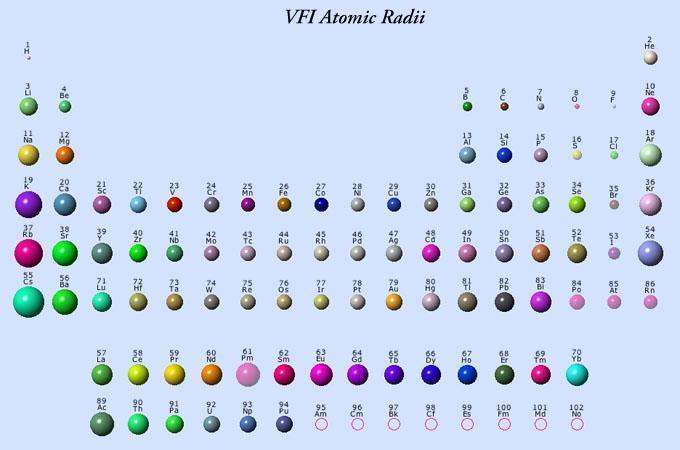
All atomic ions—such as calcium (Ca²⁺), lithium (Li⁺), and fluoride (F⁻)—are much smaller than microtubule fenestrations. The largest atomic ion (cesium) has a radius of only 2.65‧Å:

VFI_Atomic_Radii_sm.jpg


Small molecular ions such as phosphate, sulfate, and borate are also smaller than ten angstroms; these small molecular ions aren't much bigger than cesium.* (However! molecular ions such as phosphate and sulfate are doubly charged and would likely adhere to something before gaining entry.) I think this is really the best explanation for why small unreactive species like methane, halothane, and xenon and general anesthetics: they diffuse into the microtubule lumen and quench fluorescence. The mechanism of action of general anesthetics have always been a matter of great interest because the don't even have 'receptors;' they are nonpolar and can't be said to associate with anything besides lipids. The anesthetic ions fluoride and lithium can perhaps be explained as such based on their size: lithium is the smallest group IA ion and fluoride the smallest halogen.

And non‐myelinated microtubules are also less stable, being more easily depolymerized

[1] Farquharson, James. "Effect of diet on the fatty acid composition of the major phospholipids of infant cerebral cortex." Archives of disease in childhood (1995)
[2] Qiu, Xiao. "Biosynthesis of docosahexaenoic acid: two distinct pathways." Prostaglandins, Leukotrienes and Essential Fatty Acids (2003)
[3] Martinez, Manuela. "MRI evidence that docosahexaenoic acid ethyl ester improves myelination in generalized peroxisomal disorders." Neurology (1998)
[4] O'Brien, John S. "Fatty acid and fatty aldehyde composition of the major brain lipids in normal human gray matter, white matter, and myelin." Journal of lipid research (1965)
[5] Duffy, Frank H. "Ametropia Measurements from the Visual Evoked Response." Optometry and Vision Science (1971)
[6] Tyler, Christopher W. "Rapid assessment of visual function: an electronic sweep technique for the pattern visual evoked potential." Investigative Ophthalmology & Visual Science (1979)
[7] Nogales, Eva. "High-resolution model of the microtubule." Cell (1999)
[8] Li, Huilin. "Microtubule structure at 8 Å resolution." Structure (2002)
 
Last edited:

Travis

Member
Joined
Jul 14, 2016
Messages
3,189
Explaining how DHA increases myelination appears to present difficulty once it's realized that myelin contains only ~1% of this particular fatty acid, yet the fact that it does so becomes undeniable after considering the clinical evidence. I had previously hypothesized that DHA catalyzed myelination through the exclusion of cholesterol from the grey matter's cell membranes towards the microtubules, increasing MRI phase contrast in the same process. This idea, as it turns out, appears to have a considerable amount of evidence to support it; the experimental evidence is really too vast to list in detail (there are literally dozens of articles showing this) so I'll just provide a litany of quotes in this regard taken from a meta‐article:

Stillwell, William. "Docosahexaenoic acid: membrane properties of a unique fatty acid." Chemistry and physics of lipids (2003)

The lipid number for DHA is 22:6 and most other acronyms are expanded for readability:


'There is a strong correlation between various membrane properties and the degree of unsaturation and cholesterol present.' ―Stillwell & Wassel

'In the plasma membrane of most animal cells, the major polar lipid is not a phospholipid but instead is cholesterol.' ―Stillwell & Wassel

'These results showed that cholesterol demonstrated little monolayer “condensation” and bilayer permeability with DHA-containing phosphotidylcholines and suggested that cholesterol may exhibit poor solubility in these membranes. Arriving at a similar conclusion by a completely different method, Dusserre reported cholesterol efflux from plasma membranes remained the same after incorporation of oleate, linoleate or arachidonate but increased with EPA and DHA.' ―Stillwell & Wassel
There must be a physical distinction between DPA (22:5) and DHA (22:6) because children having normal levels of DPA yet lacking DHA still have nervous pathologies, although more mild than those seen in Zellweger's.

'High conformational disorder of the multiple double bond-containing chain deters close contact with the rigid steroid moiety. Experimental evidence establishing preferential affinity of cholesterol for saturated over polyunsaturated chains has accumulated, supporting recent hypotheses of a lipid driven mechanism for lateral phase separation into cholesterol-rich and -poor microdomains.' ―Stillwell & Wassel

'The first quantitative demonstration that cholesterol has low affinity for polyunsaturated acyl chains came from solid state ²H NMR spectroscopy.' ―Stillwell & Wassel

'DHA produces a similar response. A solubility of ∼10 mol% was measured by ²H NMR of 22:6-22:6 phosphotidylcholine [contrast this to 18:0-20:4 phosphotidycholine, which will will absorb 49 mol% cholesterol].' ―Stillwell & Wassel

'Weaker association of cholesterol with DHA than saturated chains is confirmed by Langmuir trough measurements.' ―Stillwell & Wassel

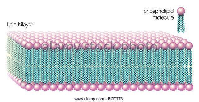
The unsaturated lipids exclude cholesterol the more double bonds they have, and also the closer these bonds are the Δ carbon, or carboxyl end of the molecule. This end would be closer to the extracellular fluid (outer boundry) and cytosol (inner boundary) in lipid bilayer orientation:

in-water-solutions-phospholipids-form-a-lipid-bilayer-the-fat-soluble-bce773.jpg


The six double bonds of DHA confer a rapid wiggling of the molecule, which increases volume and appears to repel cholesterol based on kinetic energy alone. Molecules such as DPA (22:5) and arachidonic acid (20:4) take up nearly the same space and have similar melting points, yet these do not exclude cholesterol to the extent that DHA does.

'Poor affinity between sterol and polyunsaturated acyl chain is again in evidence. Whereas the solubility of cholesterol in 16:0-18:1 phosphotidylethanolamine bilayers is ∼51 mol%, in 16:0-22:6 phosphotidylethanolamine the value is reduced to 32 mol%. These results demonstrate that unlike in phosphotidylcholine bilayers where a marked reduction in solubility requires polyunsaturation at sn-1 and sn-2 positions, DHA at the sn-2 position with a saturated sn-1 chain is sufficient in phosphotidylethanolamine to trigger exclusion of the sterol.' ―Stillwell & Wassel

'Differential affinities involving cholesterol, in particular, provide the basis of proposals for lateral segregation into DHA-rich/cholesterol-poor and DHA-poor/cholesterol-rich microdomains.' ―Stillwell & Wassel

'The presumption is that within mixed membranes the sterol separates into regions minimizing contact with polyunsaturated acyl chains.' ―Stillwell & Wassel

'Surface elasticity measurements on monolayers, and differential scanning calorimeter and solid state ²H NMR measurements on bilayers supported the idea of phase separation into a cholesterol-rich/sphingomyelin-rich liquid ordered phase and a cholesterol-poor/18:0-22:6‧phosphotidylethanolamine-rich liquid disordered phase.' ―Stillwell & Wassel

'Addition of PUFAs to membranes has also been shown to alter transbilayer sterol localization. While the transmembrane distribution of cholesterol is not known with absolute certainty, the outer leaflet of plasma membranes may contain more cholesterol and therefore be more rigid than the inner leaflet.' ―Stillwell & Wassel

'Upon addition of PUFAs in culture, a decrease in molecular order and a redistribution of cholesterol with more than 70% in the outer leaflet was reported. The DHA-induced cholesterol efflux from the plasma membrane of smooth muscle cells reported by Dusserre supports the notion that DHA may be incorporated into the inner leaflet which then forces more cholesterol into the outer leaflet where it is readily lost from membrane.' ―Stillwell & Wassel

'Conclusions: Is there anything truly unique about DHA or are its effects on membranes similar but perhaps slightly augmented over other polyunsaturated fatty acid? The involvement of DHA in so many unrelated diseases and the highly heterogeneous distribution of this fatty acid imply that DHA may indeed have a special place in biological systems. There are an increasing number of reports demonstrating that DHA increases the area/headgroup, increases permeability to water and other solutes, increases acyl chain flexibility, dynamics and disorder, increases packing free volume, drives HII phase, induces negative curvature strain, interacts poorly with cholesterol, and may enhance formation of lateral domains. Many reports now demonstrate that DHA-containing bilayers behave differently from bilayers containing the other major membrane PUFA, arachidonic acid. Further supporting the uniqueness of DHA is a remarkable observation from the nervous system. A simple elimination of one double bond from DHA producing DPA (22:5, omega-6) results in the loss of several behavioural features in animals. Clearly additional investigations on the uniqueness of DHA as well as a search for unique properties for other PUFAs will likely produce a plethora of unexpected and profound results.' ―Stillwell & Wassel

So I think you have a situation where the loss of de novo DHA biosynthetic capacity, as seen in Zellweger's syndrome (the other one), will cause a greater solubility of cholesterol in the grey matter. This would likely reduce cell membrane permeability, glucose influx, cholesterol synthesis, cholesterol efflux, and most noticeably MRI phase contrast—increased after DHA supplementation.

mri.png


The sterol redistribution above is noticeable: Images 'a' and 'c' represent before images, and you can distinctly see what appears to be misplaced cholesterol—or white matter in the grey matter. This disorder is reversed by DHA, apparently by simple displacement towards the 'nerves'.


dha.png


The regular double bonds of this long molecule give it the 3‐dimensional shape reminiscent of a crankshaft (albeit a very long one, like those found in submarine engines). Although molecules are always drawn static, they are all wiggling at every temperature above absolute zero (0 K)—and docosahexaenoic acid takes‐up more space than any other when it does so (it requires more 'wiggle room' in the cell membrane.)

'Rapid reorientation about its long molecular axis that wobbles relative to the bilayer normal characterizes motion...' ―Stillwell & Wassel

'Order parameters derived on the basis of ¹H–¹H and ¹H–¹³C dipolar couplings measured in NMR experiments on [²H]18:0-22:6 phosphotidylcholine are extremely low throughout the DHA chain and can only be reconciled with a high degree of motional freedom.' ―Stillwell & Wassel

 
Last edited:

Mito

Member
Joined
Dec 10, 2016
Messages
2,554
Explaining how DHA increases myelination appears to present difficulty once it's realized that myelin contains only ~1% of this particular fatty acid, yet the fact that it does so becomes undeniable after considering the clinical evidence.
Any idea how much DHA is needed in the diet for heathy myelination (assuming no Zellweger's syndrome)?
 

Travis

Member
Joined
Jul 14, 2016
Messages
3,189
Any idea how much DHA is needed in the diet for heathy myelination (assuming no Zellweger's syndrome)?
None, it can be made from α-linolenic acid (18:3) found in grass‐fed dairy and the grass itself. I think perhaps eating the precursor could be a better idea for adults, since α-linolenic acid is less prone to oxidation and would be expected to more readily penetrate the blood–brain barrier. But there exists a point where any more will do little good, since the brain has a finite volume.

This study is interesting because it shows a plateau in brain DHA ratio after α-linolenic acid feeding accompanied by a dose‐dependent enrichment in peripheral cell membranes:

plateu.png

[Showing ratio of DHA (22:6) to the similar DPA (22:5). The ratio of DHA in myelin is shown above, and not the total amount (which is only about 1%).]

So any DHA consumption exceeding what can be contained within the grey matter will be incorporated elsewhere. I think perhaps it could be a better idea to avoid linoleic acid, as arachidonic acid (20:4) can displace DHA from the membranes (sn‐2). In Zellweger's syndrome, an increase of brain arachidonic acid is seen as it's recruited to take the place of insufficient DHA and DPA. Perhaps just as important as ensuring adequate α-linolenic acid (or DHA) could be the avoidance of linoleic acid (18:2). Too much cholesterol could also limit brain contrast a bit, and necessitate adequate DHA levels to exclude it from the grey matter. I have read indications of the phase contrast being reduced upon aging but I don't know the mechanism of this yet. What is certain is that a sharp separation of white and grey areas on the MRI image corresponds to greater nerve velocity and intelligence.

I think its safe to say that α-linolenic acid (18:3) is an essential* amino acid yet linoleic acid (18:2) is not, and I would feel better about consuming extra α-linolenic acid than DHA—and especially if consuming a high-iron diet (lipofuscin).

[*] In the absence of DHA of course, but how many infants do you know or that eat fish?
 
Last edited:
OP
noordinary

noordinary

Member
Joined
Jun 1, 2016
Messages
209
Thank you @Travis for the detailed response.
arachidonic acid (20:4) can displace DHA from the membranes (sn‐2)
But in the study they supplemented arachidonic acid as well:
"fed 1 of the following 4 infant formulas containing equivalent nutrient amounts, except for long-chain polyunsaturated fatty acids: control (0% DHA), 0.32% DHA, 0.64% DHA, or 0.96% DHA; DHA-supplemented formulas also provided 0.64% arachidonic acid"
Not only they supplemented arachidonic acid but in quantity (0.64%) more (for 0.32% DHA group), equal (for 0.74% DHA group) and less (for 0.96% DHA group) and yet got similar results for DHA supplemented groups on visual equity test.
 
Last edited:

Travis

Member
Joined
Jul 14, 2016
Messages
3,189
Thank you @Travis for the detailed response.

But in the study they supplemented arachidonic acid as well:
"fed 1 of the following 4 infant formulas containing equivalent nutrient amounts, except for long-chain polyunsaturated fatty acids: control (0% DHA), 0.32% DHA, 0.64% DHA, or 0.96% DHA; DHA-supplemented formulas also provided 0.64% arachidonic acid"
Not only they supplemented arachidonic acid but in quantity (0.64%) more (for 0.32% DHA group), equal (for 0.74% DHA group) and less (for 0.96% DHA group) and yet got similar results for DHA supplemented groups on visual equity test.
This is just one study, and there are about seven such studies:

Birch EE. Dietary essential fatty acid supply and visual acuity development. 1992
Carlson SE. Visual-acuity development in healthy preterm infants: effect of marine-oil supplementation 1993
Carlson SE. A randomized trial of visual attention of preterm infants fed docosahexaenoic acid until two months. 1996
Carlson SE. Effect of long-chain n-3 fatty acid supplementatio n on visual acuity and growth of preterm infants with and without bronchopulmonary dysplasia. 1996
Uauy R. Visual and brain function measurements in studies of n-3 fatty acid requirements of infants. 1992
Faldella G. Visual evoked potentials and dietary long chain poyunsaturated fatty acids in preterm infants. 1996

Which I had found in the footnotes of the one that I had read:

Wezel‐Meijler, G. "Dietary supplementation of long‐chain polyunsaturated fatty acids in preterm infants: effects on cerebral maturation." Acta Paediatrica (2002)

Which had shown no significant change, as had at least one other:

'This failure correlates with the failure to show a positive influence on myelination of the cerebral visual system. It also conŽfirms the study results of Bougle who did not Ž find differences in VEP latencies between healthy preterm infants fed a formula devoid of L-C PUFAs or preterm infants fed either mothers’ milk or formula with L-C PUFAs. However, our Ž findings do not correlate with the results of Faldella who found differences in  flash-VEP wave form and latencies between premature infants fed a standard formula and those fed human milk or formula supplemented with L-C PUFAs.' ―Wezel‐Meijler

This study was conducted in The Netherlands. After reading this study, and a few others, I was left with the impression that the country could have something to do with this. I think it might be interesting to go over a few more VEP–DHA studies and look for differences. I do know that formula-fed infants have lower DHA because formula has less α-linolenic acid, and a child already getting enough α-linolenic acid cannot improve simply on account of finite intercranial volume. It certainly makes sense to me that increased DHA would increase VEP but only when the control group has a mild deficiency of α-linolenic acid.

I don't think arachidonic acid can help with visual acuity because I don't think it can increase myelination: I don't think it can increase myelination because it seems relatively ineffective at excluding cholesterol from the grey matter.


So it might seem that arachidonic acid can displace DHA only when DHA is present at low levels (DHA and DPA being more selectively incorporated into the grey matter).
 
Last edited:
OP
noordinary

noordinary

Member
Joined
Jun 1, 2016
Messages
209
It certainly makes sense to me that increased DHA would increase VEP but only when the control group has a mild deficiency of α-linolenic acid.
Would you consider the control group mildly deficient in ALA?
“The content of other major fatty acids, including LA (16.9–17.5% fatty acids) and ALA (1.61–1.68% fatty acids), were similar between all 4 formulas. ”
 
OP
noordinary

noordinary

Member
Joined
Jun 1, 2016
Messages
209
This study was conducted in The Netherlands. After reading this study, and a few others, I was left with the impression that the country could have something to do with this.
That would be interesting, considering that 3 of them plus the original one i posted were conducted by (or with partisipatiin of) the same auther Carlson SE (Susan E Carlson). I will read all studies you linked as well.
 
OP
noordinary

noordinary

Member
Joined
Jun 1, 2016
Messages
209
@Travis also should be considered:
- The original study I posted was “Supported by Mead Johnson Nutrition.”
- “JM and DAD-S are employees of Mead Johnson Nutrition. SEC and DRH have served on speakers’ panels at scientific and educational conferences on behalf of Mead Johnson.” (Abbreviations stand for authors names: SEC for Susan E Carlson)
- Mead Johnson Nutrition is a leading manufacturer of infant formula both domestically and globally with its flagship product Enfamil.

And the study actually concluded:
“Infants fed control formula had significantly poorer VEP visual acuity at 12 mo of age than did infants fed any of the DHA-supplemented formulas (P < 0.001).”
and at the same time:
“Whereas differences between the control and supplemented-formula groups in sweep VEP visual acuity are subtle (ie, 1–2 lines on an eye chart), they are important because they suggest diet-related modifications in the developmental course of structure and function in the brain and/or retina.”
 

Travis

Member
Joined
Jul 14, 2016
Messages
3,189
Would you consider the control group mildly deficient in ALA?
“The content of other major fatty acids, including LA (16.9–17.5% fatty acids) and ALA (1.61–1.68% fatty acids), were similar between all 4 formulas. ”
Okay, it now looks as if DHA itself might accelerate myelination over an equimolar amount of α-linolenic acid:

breast.png

breast 2.png


In phosphotidylethanolamine and phosphotidylserine, these breast‐fed infants had higher DHA and lower DPA and arachidonic acid concentrations—exemplifying the competition between the types while displaying a more favourable profile in the 'breast‐fed' group. Phosphotidylcholine fatty acid ratios were more stable.

So one might then wonder what the milk was composed of, the fatty acids specifically:

lipid.png


The two brands of formula lacked DHA completely and also had more linoleic acid, helping to explain the higher brain arachidonic acid in the formula‐fed groups. The 'breastfed' group had slightly edged‐out the 'SMA' (Wyeth) group despite having a lower α-linolenic acid concentration, ostensibly because it also had ~.4% DHA itself. And despite the fact that the DHA concentration was not enough to make up the difference, the 'breastfed' group had a greater DHA brain concentration on average; I think you could expect enhanced myelination and nerve velocity from this.

So you might think each DHA molecule is equal to about three α-linolenic acid molecules in myelination potential . . . or there could be other factors involved. Human milk is more complex than formulas sold to replace it, and DHA synthesis ➞ incorporation also could require brain energy (which needs vitamins.) But you do see a trend with increasing α-linolenic acid vs myelination in the two formula-fed groups, with DHA itself perhaps being a bit more potent in rapidly growing humans.. .

Farquharson, James. "Effect of diet on the fatty acid composition of the major phospholipids of infant cerebral cortex." Archives of disease in childhood (1995)
 
Last edited:
OP
noordinary

noordinary

Member
Joined
Jun 1, 2016
Messages
209
Hmm, interesting:
Susan E. Carlson
Ph.D. Nutrition
"By way of Carlson’s pioneering work and that of others who followed her in the field, the FDA approved the use of DHA and arachidonic acid (ARA) in infant formula. U.S. infant formulas with DHA have gone from zero in 2002 to over 90 percent in 2008."
 
Joined
Jul 6, 2016
Messages
550
Explaining how DHA increases myelination appears to present difficulty once it's realized that myelin contains only ~1% of this particular fatty acid, yet the fact that it does so becomes undeniable after considering the clinical evidence. I had previously hypothesized that DHA catalyzed myelination through the exclusion of cholesterol from the grey matter's cell membranes towards the microtubules, increasing MRI phase contrast in the same process. This idea, as it turns out, appears to have a considerable amount of evidence to support it; the experimental evidence is really too vast to list in detail (there are literally dozens of articles showing this) so I'll just provide a litany of quotes in this regard taken from a meta‐article:

Stillwell, William. "Docosahexaenoic acid: membrane properties of a unique fatty acid." Chemistry and physics of lipids (2003)

The lipid number for DHA is 22:6 and most other acronyms are expanded for readability:


'There is a strong correlation between various membrane properties and the degree of unsaturation and cholesterol present.' ―Stillwell & Wassel

'In the plasma membrane of most animal cells, the major polar lipid is not a phospholipid but instead is cholesterol.' ―Stillwell & Wassel

'These results showed that cholesterol demonstrated little monolayer “condensation” and bilayer permeability with DHA-containing phosphotidylcholines and suggested that cholesterol may exhibit poor solubility in these membranes. Arriving at a similar conclusion by a completely different method, Dusserre reported cholesterol efflux from plasma membranes remained the same after incorporation of oleate, linoleate or arachidonate but increased with EPA and DHA.' ―Stillwell & Wassel
There must be a physical distinction between DPA (22:5) and DHA (22:6) because children having normal levels of DPA yet lacking DHA still have nervous pathologies, although more mild than those seen in Zellweger's.

'High conformational disorder of the multiple double bond-containing chain deters close contact with the rigid steroid moiety. Experimental evidence establishing preferential affinity of cholesterol for saturated over polyunsaturated chains has accumulated, supporting recent hypotheses of a lipid driven mechanism for lateral phase separation into cholesterol-rich and -poor microdomains.' ―Stillwell & Wassel

'The first quantitative demonstration that cholesterol has low affinity for polyunsaturated acyl chains came from solid state ²H NMR spectroscopy.' ―Stillwell & Wassel

'DHA produces a similar response. A solubility of ∼10 mol% was measured by ²H NMR of 22:6-22:6 phosphotidylcholine [contrast this to 18:0-20:4 phosphotidycholine, which will will absorb 49 mol% cholesterol].' ―Stillwell & Wassel

'Weaker association of cholesterol with DHA than saturated chains is confirmed by Langmuir trough measurements.' ―Stillwell & Wassel

The unsaturated lipids exclude cholesterol the more double bonds they have, and also the closer these bonds are the Δ carbon, or carboxyl end of the molecule. This end would be closer to the extracellular fluid (outer boundry) and cytosol (inner boundary) in lipid bilayer orientation:

in-water-solutions-phospholipids-form-a-lipid-bilayer-the-fat-soluble-bce773.jpg


The six double bonds of DHA confer a rapid wiggling of the molecule, which increases volume and appears to repel cholesterol based on kinetic energy alone. Molecules such as DPA (22:5) and arachidonic acid (20:4) take up nearly the same space and have similar melting points, yet these do not exclude cholesterol to the extent that DHA does.

'Poor affinity between sterol and polyunsaturated acyl chain is again in evidence. Whereas the solubility of cholesterol in 16:0-18:1 phosphotidylethanolamine bilayers is ∼51 mol%, in 16:0-22:6 phosphotidylethanolamine the value is reduced to 32 mol%. These results demonstrate that unlike in phosphotidylcholine bilayers where a marked reduction in solubility requires polyunsaturation at sn-1 and sn-2 positions, DHA at the sn-2 position with a saturated sn-1 chain is sufficient in phosphotidylethanolamine to trigger exclusion of the sterol.' ―Stillwell & Wassel

'Differential affinities involving cholesterol, in particular, provide the basis of proposals for lateral segregation into DHA-rich/cholesterol-poor and DHA-poor/cholesterol-rich microdomains.' ―Stillwell & Wassel

'The presumption is that within mixed membranes the sterol separates into regions minimizing contact with polyunsaturated acyl chains.' ―Stillwell & Wassel

'Surface elasticity measurements on monolayers, and differential scanning calorimeter and solid state ²H NMR measurements on bilayers supported the idea of phase separation into a cholesterol-rich/sphingomyelin-rich liquid ordered phase and a cholesterol-poor/18:0-22:6‧phosphotidylethanolamine-rich liquid disordered phase.' ―Stillwell & Wassel

'Addition of PUFAs to membranes has also been shown to alter transbilayer sterol localization. While the transmembrane distribution of cholesterol is not known with absolute certainty, the outer leaflet of plasma membranes may contain more cholesterol and therefore be more rigid than the inner leaflet.' ―Stillwell & Wassel

'Upon addition of PUFAs in culture, a decrease in molecular order and a redistribution of cholesterol with more than 70% in the outer leaflet was reported. The DHA-induced cholesterol efflux from the plasma membrane of smooth muscle cells reported by Dusserre supports the notion that DHA may be incorporated into the inner leaflet which then forces more cholesterol into the outer leaflet where it is readily lost from membrane.' ―Stillwell & Wassel

'Conclusions: Is there anything truly unique about DHA or are its effects on membranes similar but perhaps slightly augmented over other polyunsaturated fatty acid? The involvement of DHA in so many unrelated diseases and the highly heterogeneous distribution of this fatty acid imply that DHA may indeed have a special place in biological systems. There are an increasing number of reports demonstrating that DHA increases the area/headgroup, increases permeability to water and other solutes, increases acyl chain flexibility, dynamics and disorder, increases packing free volume, drives HII phase, induces negative curvature strain, interacts poorly with cholesterol, and may enhance formation of lateral domains. Many reports now demonstrate that DHA-containing bilayers behave differently from bilayers containing the other major membrane PUFA, arachidonic acid. Further supporting the uniqueness of DHA is a remarkable observation from the nervous system. A simple elimination of one double bond from DHA producing DPA (22:5, omega-6) results in the loss of several behavioural features in animals. Clearly additional investigations on the uniqueness of DHA as well as a search for unique properties for other PUFAs will likely produce a plethora of unexpected and profound results.' ―Stillwell & Wassel

So I think you have a situation where the loss of de novo DHA biosynthetic capacity, as seen in Zellweger's syndrome (the other one), will cause a greater solubility of cholesterol in the grey matter. This would likely reduce cell membrane permeability, glucose influx, cholesterol synthesis, cholesterol efflux, and most noticeably MRI phase contrast—increased after DHA supplementation.

View attachment 8242

The sterol redistribution above is noticeable: Images 'a' and 'c' represent before images, and you can distinctly see what appears to be misplaced cholesterol—or white matter in the grey matter. This disorder is reversed by DHA, apparently by simple displacement towards the 'nerves'.


View attachment 8243

The regular double bonds of this long molecule give it the 3‐dimensional shape reminiscent of a crankshaft (albeit a very long one, like those found in submarine engines). Although molecules are always drawn static, they are all wiggling at every temperature above absolute zero (0 K)—and docosahexaenoic acid takes‐up more space than any other when it does so (it requires more 'wiggle room' in the cell membrane.)

'Rapid reorientation about its long molecular axis that wobbles relative to the bilayer normal characterizes motion...' ―Stillwell & Wassel

'Order parameters derived on the basis of ¹H–¹H and ¹H–¹³C dipolar couplings measured in NMR experiments on [²H]18:0-22:6 phosphotidylcholine are extremely low throughout the DHA chain and can only be reconciled with a high degree of motional freedom.' ―Stillwell & Wassel


I remember Tyw saying something similar in the past about DHA and cholesterol exclusion, the addition you have added to the theory is that DHEA/progesterone/pregnenolone are a major component of the white matter.

I had an idea. My friend has said after supplementing DHA his blood cholesterol dropped a ton. Could it be as simple as more DHA in blood -> cholesterol exclusion into tissues? That would actually explain all of the talk of PUFA decreasing blood cholesterol.

I wonder what's really going on in the brain. DHA seems to act to exclude sterols and place them in the right direction, but there's gotta be more. DHA is an insanely mobile molecule, capable of extreme molecular motion (which is why it's used with rhodsopin... it's like an extremely sensitive and fast antenna).

For instance the synaptic vesicles of neurons contain high amounts of both DHA and cholesterol.

Here's something you should read, I had no idea it was free online until just a second ago: Human Longevity

If you haven't read it before, you'll find some very interesting things about PUFA, and especially HUFA.

Apparently PUFA enriched membranes are highly permeable to things like H+, Na+, Ca+, etc etc. Could neurons use this permeability of DHA enriched membranes to allow rapid cycling of polarization? Like, all that matters is that the message is sent, that the polarization occurs (either hyper- or de-polarization). It doesn't matter really how strong or how weak the polarization is, it just matters that the message is sent, fast, and that it can occur at a high rate.

Really what's going on in the brain? What is the point of polarization of neurons if microtubules are the source of information transfer?
 
Joined
Nov 26, 2013
Messages
7,370
"By way of Carlson’s pioneering work and that of others who followed her in the field, the FDA approved the use of DHA and arachidonic acid (ARA) in infant formula. U.S. infant formulas with DHA have gone from zero in 2002 to over 90 percent in 2008."
31152423.jpg
 

Runenight201

Member
Joined
Feb 18, 2018
Messages
1,942
None, it can be made from α-linolenic acid (18:3) found in grass‐fed dairy and the grass itself. I think perhaps eating the precursor could be a better idea for adults, since α-linolenic acid is less prone to oxidation and would be expected to more readily penetrate the blood–brain barrier. But there exists a point where any more will do little good, since the brain has a finite volume.

This study is interesting because it shows a plateau in brain DHA ratio after α-linolenic acid feeding accompanied by a dose‐dependent enrichment in peripheral cell membranes:

View attachment 8245
[Showing ratio of DHA (22:6) to the similar DPA (22:5). The ratio of DHA in myelin is shown above, and not the total amount (which is only about 1%).]

So any DHA consumption exceeding what can be contained within the grey matter will be incorporated elsewhere. I think perhaps it could be a better idea to avoid linoleic acid, as arachidonic acid (20:4) can displace DHA from the membranes (sn‐2). In Zellweger's syndrome, an increase of brain arachidonic acid is seen as it's recruited to take the place of insufficient DHA and DPA. Perhaps just as important as ensuring adequate α-linolenic acid (or DHA) could be the avoidance of linoleic acid (18:2). Too much cholesterol could also limit brain contrast a bit, and necessitate adequate DHA levels to exclude it from the grey matter. I have read indications of the phase contrast being reduced upon aging but I don't know the mechanism of this yet. What is certain is that a sharp separation of white and grey areas on the MRI image corresponds to greater nerve velocity and intelligence.

I think its safe to say that α-linolenic acid (18:3) is an essential* amino acid yet linoleic acid (18:2) is not, and I would feel better about consuming extra α-linolenic acid than DHA—and especially if consuming a high-iron diet (lipofuscin).

[*] In the absence of DHA of course, but how many infants do you know or that eat fish?

Docosahexaenoic Acid (DHA): An Ancient Nutrient for the Modern Human Brain

@Travis I think your input on this whole study would be rather interesting, especially given the claim challenging the essentiality of ALA and the claim that in humans, preformed DHA is "conditionally essential".

They also hypothesize that the accelerated brain development and brain to body ratio that is unique to humans is solely because we started consuming sea food with preformed DHA.

In regards to your infant comment, human breast milk contains preformed DHA, and as the study points out, DHA is necessary for proper neurodevelopment (whether preformed or through the ALA pathway).

That review elucidated a lot of problems I've had with land meat and why perhaps consuming sea food is not only superior but perhaps optimal. Whatever the case, it is very clear that the increase in arachidonic acid at the expense of DHA incorporation into the cellular membrane is problematic and contributes to a number of inflammatory pathologies, and at the least arachidonic acid should be minimized.
 

magnesiumania

Member
Joined
Jul 17, 2018
Messages
607
DHA from supplements dont make it to the brain typically because its not in the SN2 position. To my knowlegde the primary function of DHA is to make a DC electric current. This 22 carbon fat can convert light energy to electrical energy and is what it primarily does. However it makes sense that DC current help stimulate the formation of myelin even if DHA is not an integral component in the myelin substance itself.
 

dabdabdab

Member
Joined
May 28, 2020
Messages
250
DHA from supplements dont make it to the brain typically because its not in the SN2 position. To my knowlegde the primary function of DHA is to make a DC electric current. This 22 carbon fat can convert light energy to electrical energy and is what it primarily does. However it makes sense that DC current help stimulate the formation of myelin even if DHA is not an integral component in the myelin substance itself.
are you saying that dha is beneficial if it's created inside the body, just like cholesterol which peat is against dietery form but favors its synthesis from sugar inside the body?
 
EMF Mitigation - Flush Niacin - Big 5 Minerals
Back
Top Bottom